Online First
Articles in press have been peer-reviewed and accepted, which are not yet assigned to volumes/issues, but are citable by Digital Object Identifier (DOI).
Display Method:
, Available online , doi: 10.3760/cma.j.cn121381-202304023-00397
 [PDF 1822KB]
Abstract:
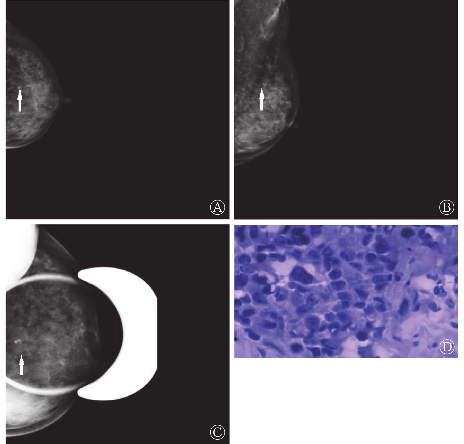
Objective To explore the value of low-dose digital breast tomosynthesis (DBT) and full-field digital mammography (FFDM) in screening early breast cancer. Methods 246 female patients (age (47.3±6.2) years old, ranging from 28 to 65 years old) with breast lumps and breast swelling pain treated in the Second People's Hospital of Liaocheng from January 2020 to April 2022 were prospectively selected. All patients underwent routine FFDM and low-dose DBT examinations, and underwent ultrasound-guided puncture biopsy. The pathological examination results of biopsy tissue was taken as the "gold standard", and the sensitivity, specificity, accuracy, positive predictive value and negative predictive value of FFDM, low-dose DBT and combination of the two in screening early breast cancer were compared and analyzed, and the consistency (Kappa value) between FFDM, low-dose DBT, alone and combination examination of the two and histopathological examination results were analyzed and compared. The average glandular dose and examination time of different examination methods were compared. The intergroup comparison of quantitative data adopted t test or one-way analysis of variance, while the intergroup comparison of counting data adopted χ2 test, and analysis of consistency between different examination methods and histopathological examination results adoptedKappa test. Results Of the 246 patients included in the study, 192 cases were diagnosed as early breast cancer and 54 cases were diagnosed as benign breast lesions by histopathological examination. 154 cases of early breast cancer and 92 cases of benign breast lesions were diagnosed by FFDM. 169 cases of early breast cancer and 77 cases of benign breast lesions were diagnosed by low-dose DBT. 177 cases of early breast cancer and 69 cases of benign breast lesions were diagnosed by FFDM combined with low-dose DBT. The sensitivity (86.98%), specificity (96.30%), accuracy (89.02%), positive predictive value (98.82%) and negative predictive value (67.53%) of low-dose DBT in the diagnosis of early breast cancer were higher than those of FFDM (75.00%, 81.48%, 76.42%, 93.51% and 47.83%), and the differences were statistically significant (χ2=6.000~13.677, all P<0.05). The sensitivity (90.63%), specificity (94.44%), accuracy (91.46%), positive predictive value (98.31%) and negative predictive value (73.91%) of FFDM combined with low-dose DBT in the diagnosis of early breast cancer were higher than those of FFDM, and the differences were statistically significant (χ2 =4.285~20.644, all P<0.05). There was no significant difference in the sensitivity, specificity, accuracy, positive predictive value and negative predictive value between low-dose DBT alone and FFDM combined with low-dose DBT in diagnosing early breast cancer (χ2=0.159~1.283, all P>0.05). The results of FFDM in the diagnosis of early breast cancer has good consistency with the histopathological examination results (Kappa value=0.655), and the results of low-dose DBT in the diagnosis of early breast cancer has good consistency with the histopathological examination results (Kappa value=0.722), and the combination diagnostic results of the two has good consistency with the histopathological examination results (Kappa value=0.792).. There was no statistically significant difference in the average glandular dose among FFDM [(1.03±0.18)mGy], low-dose DBT [(1.04±0.19) mGy]and combined examination of the two [(1.06±0.21) mGy] (F=1.529, P>0.05), and there was no statistically significant difference between FFDM examination time [(6.25±0.52) min] and low-dose DBT examination time [(6.33±0.57) min] (t=1.626, P>0.05). Conclusions Compared with FFDM, low-dose DBT has higher application value in screening early breast cancer, and it has good consistency with histopathological examination results, which can be used as an important examination method for clinical screening of early breast cancer.

, Available online , doi: 10.3760/cma.j.cn121381-202309017-00393
Abstract:
Langerhans cell histiocytosis (LCH) is a rare neoplastic disease with abnormal proliferation of immature dendritic cells, and histopathological examination is the "gold standard" for its diagnosis. The author reports a case of multisystem LCH in an adolescent with CT, MRI, and 18F-fluorodeoxyglucose(FDG) PET/CT imaging, and analyzes the characteristics of the disease from the clinical, histopathological, imaging, and therapeutic perspectives, and deepens the understanding of the disease by reviewing the literature to provide more references for the diagnosis of the disease.
Langerhans cell histiocytosis (LCH) is a rare neoplastic disease with abnormal proliferation of immature dendritic cells, and histopathological examination is the "gold standard" for its diagnosis. The author reports a case of multisystem LCH in an adolescent with CT, MRI, and 18F-fluorodeoxyglucose(FDG) PET/CT imaging, and analyzes the characteristics of the disease from the clinical, histopathological, imaging, and therapeutic perspectives, and deepens the understanding of the disease by reviewing the literature to provide more references for the diagnosis of the disease.


, Available online , doi: 10.3760/cma.j.cn121381-202303006-00378
Abstract:
Objective To investigate the predictive value of 18F-fluorodeoxyglucose(FDG) PET/CT primary lesions metabolic heterogeneity index for occult lymph node metastasis(OLM) in gastric cancer. Methods A retrospective analysis was performed on 79 patients [62 males, 17 females, age (63.8±9.0) years] with gastric cancer who underwent 18F-FDG PET/CT imaging and were diagnosed as clinical (c)N0 stage before surgery from January 2016 to December 2022 in the First Affiliated Hospital of Zhengzhou University. All patients underwent radical gastrectomy in our hospital within 1 month after imaging, and were divided into OLM-positive group and OLM-negative group according to postoperative pathology to determine whether there was lymph node metastasis. The following PET/CT parameters were measured: The maximum, mean and peak normalized uptake values (SUVmax, SUVmean, SUVpeak) , tumor metabolic volume (MTV) and total focal glycolysis (TLG)of the primary lesions.And TLR (tumor - liver ratio), heterogeneity index -1 (HI-1) and heterogeneity index -2 (HI-2) were calculated. The t test and Mann-Whitney U test of two independent samples were used to compare the parameters between groups. The independent risk factors of OLM were analyzed by logistic regression. The diagnostic efficacy of heterogeneity index on OLM was analyzed by receiver operating characteristic (ROC) curve. Results A total of 39 (49.4%, 39/79) of the 79 patients were pathologically confirmed to have OLM. HI-2 in OLM positive group was higher than that in OLM negative group [4.98 (2.68, 8.44) vs 2.61 (1.84, 4.23), z=−3.178, P < 0.05], while SUVmax in OLM negative group was higher than that in OLM negative group [5.59 (4.46, 7.51) vs 6.91 (5.11, 10.64). z=−2.000, P < 0.05], SUVmean[3.33 (3.06, 3.85) vs 3.65 (3.25, 4.64), z=−2.001, P < 0.05], HI-1[0.23±0.12 vs 0.29±0.14, t=2.096, P < 0.05] were significantly higher than those in OLM positive group. Multivariate logistic regression analysis showed that HI-2 was an independent risk factor for OLM [odds ratio (OR) =6.893, 95%CI: 1.922-24.718, P < 0.05]. The area under ROC curve (AUC) of HI-2 for OLM prediction was 0.708 (95%CI: 0.237-0.483, P=0.001), and the sensitivity and specificity for OLM diagnosis were 51.3% (20/39) and 87.5% (35/40), respectively, when the threshold was 4.962. Conclusion 18F-FDG PET/CT tumor metabolic heterogeneity index has predictive value for OLM in gastric cancer, and heterogeneity index -2 is an independent risk factor for OLM.


, Available online , doi: 10.3760/cma.j.cn121381-202211020-00395
Abstract:
Objective To explore the application of enhanced CT combined with 18F-FDG PET/CT in the diagnosis of pulmonary sequestration (PS) and analyze the image characteristics. Methods The clinical data of 6 patients with PS, including 2 males and 4 females, aged (49.8±17.5) years, who were surgically confirmed to be accompanied by elevated levels of tumor markers at the Affiliated Qingdao Central Hospital of Qingdao University from October 2007 to December 2020 were retrospectively analyzed.18F-FDG PET/CT imaging and enhanced CT scanning were performed in the 6 patients, and the location of the lesions, morphology, density, CT enhancement characteristics and 18F-FDG metabolism were observed. Results The lesions in the six patients were all located in the posterior basal segment of the lower lobe of the lung, including four cases in the right lung and two cases in the left lung. The maximum diameter of the lesion was (4.3±2.0) cm, and the CT value on plain scan was (27.2±13.9) HU. 2 patients had oval, rounded, or triangular-shaped lesions, and 1 patient had calcified foci within the lesion; 1 patient had marked enhancement on the CT image, 4 had moderate enhancement, and 1 had no marked enhancement. 6 patients were found to have an abnormal arterial blood supply originating from the thoracic aorta. Two patients had cystic masses, three patients had solid masses, and one patient had a cystic-solid mass. There were 3 patients with localized vascularization, coarsening, and disorganization in the lobes of the lungs, 2 patients with moderately increased 18F-FDG metabolism, 3 patients with mildly increased metabolism, and 1 patient with no metabolism increased metabolism. Conclusion The possibility of PS should be considered when patients have different degrees of elevated levels of tumour markers, round-like, oval-like, triangular-like nodules or masses found adjacent to the spine in the lower lobes of both lungs, increased 18F-FDG metabolism and abnormal blood-supplying arteries detected on CT-enhanced scans with mild-to-moderate enhancement, or when the lesion has no increased 18F-FDG metabolism and there is no significant enhancement on CT-enhanced scans, but abnormal blood-supplying arteries were detected. CT enhancement combined with 18F-FDG PET/CT imaging improves the accuracy of PS diagnosis.


, Available online , doi: 10.3760/cma.j.cn121381-202307002-00396
Abstract:
Objective To explore the value of derived parameters based on DCE-MRI in the evaluation of epilepsy recurrence caused by cerebral cysticercosis (CC). Methods 40 patients with acute epilepsy caused by CC treated in the second people's Hospital of Baoshan City, Yunnan Province from January to December 2020 were analyzed retrospectively, including 22 males and 18 females, aged (35.6 ±11.0) years. According to the recurrence of epilepsy within half a year, all patients were divided into recurrent group and non-recurrent group. The dynamic contrast enhanced MRI (DCE-MRI) derivative parameters, such as rate constant (Kep), volume transfer constant (Ktrans) and extracellular space volume fraction (Ve), were observed and recorded in all patients at first admission and half a year after follow-up, respectively. The permeability of blood-brain barrier (BBB) was evaluated in two groups. Independent sample t-test or χ2 test was used for inter-group comparison. Results There was no significant difference in general data such as gender, age, first onset time, and epilepsy types between the two groups(χ2=0.020, t=0.692, t=0.902, χ2=0.030, all P >0.05). At the first admission, the levels of Kep, Ve and K trans in the non-recurrent group were (30.17±5.32)×10−2/min, (102.32±6.58)×10−2 and (19.98±2.64) × 10−2/min, respectively, which were significantly lower than those in the recurrent group [(36.32±4.36)×10−2/min, (110.35±7.12)×10−2, (23.21±3.21)×10−2/min] (t=3.839、3.660、3.477, all P<0.001). After half a year of follow-up, the levels of Kep, Ve and Ktrans in the non-recurrent group were (12.57±3.29)×10−2/min, (78.02±4.36)×10−2 and (17.96±3.01)×10−2/min, respectively, which were also significantly lower than those in the recurrent group (24.25±3.58)×10−2/min, (90.37±8.27)×10−2, (23.32±3.98)×10−2/min, and the differences were statistically significant (t=10.620、10.161、4.848, all P<0.001). Conclusion Using DCE-MRI derived parameters can analyze the BBB permeability to distinguish the recurrence of epilepsy caused by CC.


, Available online , doi: 10.3760/cma.j.cn121381-202305022-00394
Abstract:
Objective To evaluate the efficacy and safety of TP regimen (paclitaxel and cisplatin combined chemotherapy) combined with concurrent radiotherapy in the treatment of advanced cervical cancer. Methods A prospective study was conducted on 60 female patients with advanced cervical cancer, aged (52.2±3.2) years, who were treated in Heping Hospital Affiliated to Changzhi Medical College from August 2020 to August 2021. Patients were divided into control group 30 cases (cisplatin chemotherapy and radiotherapy) and observation group 30 cases (paclitaxel+cisplatin chemotherapy and radiotherapy) by random number table method. The clinical efficacy, serum tumor marker levels, incidence of adverse reactions, apoptosis and extracellular matrix degradation of patients in the two groups were compared. The t test (homogeneity of variance) was used to compare the measurement data conforming to the normal distribution, and the χ2 test was used to compare the counting data. Results The objective remission rate of the observation group was higher than that of the control group (86.67% (26/30) vs. 63.33% (19/30)), and the difference was statistically significant (χ2=4.355, P<0.05). The levels of squamous cell carcinoma antigen and carbohydrate antigen (CA) 125 in the observation group were lower than those in the control group after treatment [(2.18±0.68) μg/L versus (4.06±1.12) μg/L, (22.24±5.93) U/ml versus (26.28±6.71) U/ml]. Fatigue (3.33%(1/30) vs. 6.67%(2/30)), myelosuppression (6.67%(2/30) vs. 3.33%(1/30)), gastrointestinal reaction (6.67% (2/30) vs. 10.00%(3/30)), radiation enteritis (6.67%(2/30) vs. 3.33%(1/30)) and urinary reaction (3.33% (1/30) vs. 6.67%(2/30)) and the incidence of liver and kidney function injury (3.33%(1/30) vs. 10.00%(3/30)) were not statistically significant (χ2=0.218-1.071, all P>0.05). Compared with the control group after treatment, the levels of matrix metalloproteinase(MMP)-2 ((522.47±45.93) ng/L vs. (325.41±32.54) ng/L) and MMP-9 ((378.18±33.59) ng/L vs. (516.28±45.84) ng/L) in the observation group were decreased. The level of cysteine proteinase-8 (Caspase-8) ((219.49±33.88) ng/L vs. (96.48±9.33) ng/L) was increased, and the differences were statistically significant (t=19.175, 13.310, 19.172; all P<0.05). Conclusions TP regimen combined with synchronous radiotherapy can improve the objective remission rate of patients with advanced cervical cancer. After treatment, the levels of squamous cell carcinoma antigen and CA125, MMP-2, MMP-9 and Caspase-8 were decreased. TP regimen combined with synchronous radiotherapy has good safety.
, Available online , doi: 10.3760/cma.j.cn121381-202303013-00390
Abstract:
Immunoglobulin G4-related disease (IgG4-RD) is a type of systemic inflammatory diseases that often affect multiple organs. IgG4-related cardiovascular disease (IgG4-RCVD) includes IgG4-related aortic disease, IgG4-related coronary artery disease, IgG4-related pulmonary artery disease, and IgG4-related pericarditis. IgG4-positive plasma cells accumulate and exhibit high expression of glucose transport proteins at the site of IgG4-RCVD involvement. 18F-fluorodeoxyglucose (FDG) PET/CT can assess the extent and degree of vascular inflammation on a systemic scale by evaluating the metabolic activity of lesions in IgG4-RCVD patients. It plays a pivotal role in diagnosing and quantitatively assessing IgG4-RCVD vascular inflammation, aiding in the selection of biopsy sites, and monitoring treatment efficacy. The authors conducted a comprehensive review of the advancements in the utilization of 18F-FDG PET/CT for IgG4-RCVD, aiming to furnish clinicians with a valuable reference for diagnosing IgG4-RCVD through the application of 18F-FDG PET/CT.
Immunoglobulin G4-related disease (IgG4-RD) is a type of systemic inflammatory diseases that often affect multiple organs. IgG4-related cardiovascular disease (IgG4-RCVD) includes IgG4-related aortic disease, IgG4-related coronary artery disease, IgG4-related pulmonary artery disease, and IgG4-related pericarditis. IgG4-positive plasma cells accumulate and exhibit high expression of glucose transport proteins at the site of IgG4-RCVD involvement. 18F-fluorodeoxyglucose (FDG) PET/CT can assess the extent and degree of vascular inflammation on a systemic scale by evaluating the metabolic activity of lesions in IgG4-RCVD patients. It plays a pivotal role in diagnosing and quantitatively assessing IgG4-RCVD vascular inflammation, aiding in the selection of biopsy sites, and monitoring treatment efficacy. The authors conducted a comprehensive review of the advancements in the utilization of 18F-FDG PET/CT for IgG4-RCVD, aiming to furnish clinicians with a valuable reference for diagnosing IgG4-RCVD through the application of 18F-FDG PET/CT.
 Submit
 Author Login
 Referees
 Editor-in-Chief
 Editor Center
 Email alert
 RSS
RSS