Volume 44 Issue 1
Mar.  2020
Article Contents

Citation:

Assessment of SPECT/CT in sentinel lymph node location in cN0 papillary thyroid carcinoma

  • Objective To evaluate the value of SPECT/CT lymphoscintigraphy for sentinel lymph node (SLN) detection in patients with cN0 papillary thyroid carcinoma(PTC). Methods From April to November 2017, 2 male and 14 female patients with cN0 PTC, with mean age of 47.0±8.4 years, and hospitalized in the first Hospital of Shanxi Medical University, were enrolled in this study. Planar and SPECT/CT SLN imaging were performed before operation. All patients underwent intraoperative lymphatic mapping with a handheld gamma probe. All specimens were sent to a pathology laboratory. Results Among the 16 patients, 14 cases of SLN were found, with a detection rate of 87.50% (14/16). Preoperative planar and SPECT/CT fusion images showed 31 and 35 SLNs, respectively. With the application of intraoperative gamma probe, a total of 37 SLNs were detected in 13/16 patients (81.25%). The number of SLNs detected by gamma detector was inconsistent with the SPECT/CT lymphography in 4 cases and consistent in 12 cases. The coincidence rate of the two methods was 75% (12/16). Fifteen patients (93.75%) had lymph node metastasis. The sensitivity, accuracy, and false negative rate of SLN detection were 86.67%, 81.25%, and 13.33%, respectively. Conclusion Preoperative SPECT/CT lymphoscintigraphy can not only enables precise localization of SLN but also detects the cervical lymph node metastasis in patients with cN0 papillary thyroid carcinoma.
  • 加载中
  • [1] Chen WQ, Zheng RS, Baade PD, et al. Cancer statistics in China, 2015[J]. CA Cancer J Clin, 2016, 66(2): 115−132. DOI: 10.3322/caac.21338.
    [2] Jung KW, Won YJ, Kong HJ, et al. Cancer Statistics in Korea: Incidence, Mortality, Survival and Prevalence in 2010[J]. Cancer Res Treat, 2013, 45(1): 1−14. DOI: 10.4143/crt.2013.45.1.1.
    [3] Lundgren CI, Hall P, Dickman PW, et al. Clinically significant prognostic factors for differentiated thyroid carcinoma: a population-based, nested case-control study[J]. Cancer, 2006, 106(3): 524−531. DOI: 10.1002/cncr.21653.
    [4] 张磊, 王洋, 李新喜, 等. 预防性颈中央区淋巴结清扫术与传统甲状腺全切除术治疗cN0期甲状腺乳头状癌安全性及有效性的meta分析[J]. 中国普外基础与临床杂志, 2015, 22(12): 1487−1494. DOI: 10.7507/1007−9424.20150387.Zhang L, Wang Y, Li XX, et al. Prophylactic Central Neck Dissection Versus Traditional Total Thyroidectomy for Stage cN0 Papillary Thyroid Carcinoma: A Meta-Analysis[J]. Chin J Bases Clin General Surg, 2015, 22(12): 1487−1494. DOI: 10.7507/1007−9424.20150387.
    [5] Lang BHH, Ng SH, Lau LLH, et al. A Systematic Review and Meta-Analysis of Prophylactic Central Neck Dissection on Short-Term Locoregional Recurrence in Papillary Thyroid Carcinoma After Total Thyroidectomy[J]. Thyroid, 2013, 23(9): 1087−1098. DOI: 10.1089/thy.2012.0608.
    [6] Kowalski LP, Bagietto R, Lara JRL, et al. Prognostic significance of the distribution of neck node metastasis from oral carcinoma[J]. Head Neck, 2000, 22(3): 207−214. DOI: 10.1002/(SICI)1097−0347(200005)22:3<207::AID−HED1>3.0.CO;2−9.
    [7] Robbins KT, Clayman G, Levine PA, et al. Neck dissection classification update: Revisions proposed by the American Head and Neck Society and the American Academy of Otolaryngology-Head and Neck Surgery[J]. Arch Otolaryngol Head Neck Surg, 2002, 128(7): 751−758. DOI: 10.1001/archotol.128.7.751.
    [8] Creach KM, Gillanders WE, Siegel BA, et al. Management of cervical nodal metastasis detected on I-131 scintigraphy after initial surgery of well-differentiated thyroid carcinoma[J]. Surgery, 2010, 148(6): 1198−1206. DOI: 10.1016/j.surg.2010.09.006.
    [9] 齐子蛟, 刘良发. 甲状腺癌淋巴结转移的相关因素分析[J]. 国际耳鼻咽喉头颈外科杂志, 2017, 41(1): 30−34. DOI: 10.3760/cma.j.issn.1673−4106.2017.01.008.Qi ZJ, Liu LF. Related risk factors of cervical lymph node metastasis in thyroid carcinoma[J]. Int J Otolaryngol Head Neck Surg, 2017, 41(1): 30−34. DOI: 10.3760/cma.j.issn.1673−4106.2017.01.008.
    [10] Cabrera RN, Chone CT, Zantut-Wittmann DE, et al. The Role of SPECT/CT Lymphoscintigraphy and Radioguided Sentinel Lymph Node Biopsy in Managing Papillary Thyroid Cancer[J]. JAMA Otolaryngol Head Neck Surg, 2016, 142(9): 834−841. DOI: 10.1001/jamaoto.2016.1227.
    [11] Hwang HS, Orloff LA. Efficacy of preoperative neck ultrasound in the detection of cervical lymph node metastasis from thyroid cancer[J]. Laryngoscope, 2011, 121(3): 487−491. DOI: 10.1002/lary.21227.
    [12] Haugen BR, Alexander EK, Bible KC, et al. 2015 American Thyroid Association Management Guidelines for Adult Patients with Thyroid Nodules and Differentiated Thyroid Cancer: The American Thyroid Association Guidelines Task Force on Thyroid Nodules and Differentiated Thyroid Cancer[J]. Thyroid, 2016, 26(1): 1−133. DOI: 10.1089/thy.2015.0020.
    [13] Kelley MC, Hansen N, McMasters KM. Lymphatic mapping and sentinel lymphadenectomy for breast cancer[J]. Am J Surg, 2004, 188(1): 49−61. DOI: 10.1016/j.amjsurg.2003.10.028.
    [14] Morton DL, Wen DR, Wong JH, et al. Technical Details of Intraoperative Lymphatic Mapping for Early Stage Melanoma[J]. Arch Surg, 1992, 127(4): 392−399. DOI: 10.1001/archsurg.1992.01420040034005.
    [15] Pelizzo MR, Toniato A, Sorgato N, et al. 99Tc nanocolloid sentinel node procedure in papillary thyroid carcinoma: our mono-institutional experience on a large series of patients[J]. Acta Otorhinolaryngol Ital, 2009, 29(6): 321−325.
    [16] Garcia-Burillo A, Roca Bielsa I, Gonzalez O, et al. SPECT/CT sentinel lymph node identification in papillary thyroid cancer: lymphatic staging and surgical management improvement[J]. Eur J Nucl Med Mol Imaging, 2013, 40(11): 1645−1655. DOI: 10.1007/s00259−013−2476−x.
    [17] Carcoforo P, Portinari M, Feggi L, et al. Radio-guided selective compartment neck dissection improves staging in papillary thyroid carcinoma: A prospective study on 345 patients with a 3-year follow-up[J]. Surgery, 2014, 156(1): 147−157. DOI: 10.1016/j.surg.2014.03.045.
    [18] Lee J, Na KY, Lee J, et al. The usefulness and accuracy of sentinel lymph node biopsy using single photon emission computed tomography/computed tomography with 99mTc phytate to detect locoregional lymph node metastases in patients with papillary thyroid carcinoma[J]. J Korean Surg Soc, 2013, 84(4): 195−201. DOI: 10.4174/jkss.2013.84.4.195.
    [19] Portinari M, Carcoforo P. Radioguided sentinel lymph node biopsy in patients with papillary thyroid carcinoma[J]. Gland Surg, 2016, 5(6): 591−602. DOI: 10.21037/gs.2016.11.08.
    [20] Assadi M, Yarani M, Zakavi SR, et al. Sentinel node mapping in papillary thyroid carcinoma using combined radiotracer and blue dye methods[J]. Endokrynol Pol, 2014, 65(4): 281−286. DOI: 10.5603/EP.2014.0038.
    [21] Huang O, Wu WL, Wang OC, et al. Sentinel lymph node biopsy is unsuitable for routine practice in younger female patients with unilateral low-risk papillary thyroid carcinoma[J]. BMC Cancer, 2011, 11(1): 386. DOI: 10.1186/1471−2407−11−386.
    [22] 鄢丹桂, 张彬, 安常明, 等. 甲状腺乳头状癌临床N0患者颈部淋巴结转移规律[J]. 中华耳鼻咽喉头颈外科杂志, 2011, 46(11): 887−891. DOI: 10.3760/cma.J.issn.1673−0860.2011.11.002.Yan DG, Zhang B, An CM, et al. Cervical lymph node metastasis in clinical N0 papillary thyroid carcinoma[J]. Chin J Otorhinolaryngol Head Neck Surg, 2011, 46(11): 887−891. DOI: 10.3760/cma.J.issn.1673−0860.2011.11.002.
    [23] Roh JL, Park CI. Sentinel lymph node biopsy as guidance for central neck dissection in patients with papillary thyroid carcinoma[J]. Cancer, 2008, 113(7): 1527−1531. DOI: 10.1002/cncr.23779.
    [24] Falvo L, Marzullo A, Palermo S, et al. The sentinel lymph node in papillary cancer of the thyroid including histological subtype[J]. Ann Ital Chir, 2006, 77(1): 13−18.
    [25] Takeyama H, Tabei I, Uchida K, et al. Sentinel node biopsy for follicular tumours of the thyroid gland[J]. Br J Surg, 2009, 96(5): 490−495. DOI: 10.1002/bjs.6559.
    [26] Carcoforo P, Feggi L, Trasforini G, et al. Use of preoperative lymphoscintigraphy and intraoperative gamma-probe detection for identification of the sentinel lymph node in patients with papillary thyroid carcinoma[J]. Eur J Surg Oncol, 2007, 33(9): 1075−1780. DOI: 10.1016/j.ejso.2007.01.025.
    [27] Kaczka K, Luks B, Jasion J, et al. Sentinel lymph node in thyroid tumors-own experience[J]. Contemp Oncol(Pozn), 2013, 17(2): 184−189. DOI: 10.5114/wo.2013.34623.
    [28] Lee SK, Kim SH, Hur SM, et al. The Efficacy of Lateral Neck Sentinel Lymph Node Biopsy in Papillary Thyroid Carcinoma[J]. World J Surg, 2011, 35(12): 2675−2682. DOI: 10.1007/s00268−011−1254−9.
    [29] Park JH, Lee YS, Kim BW, et al. Skip Lateral Neck Node Metastases in Papillary Thyroid Carcinoma[J]. World J Surg, 2012, 36(4): 743−747. DOI: 10.1007/s00268−012−1476−5.
    [30] Lee YS, Shin SC, Lim YS, et al. Tumor location-dependent skip lateral cervical lymph node metastasis in papillary thyroid cancer[J]. Head Neck, 2014, 36(6): 887−891. DOI: 10.1002/hed.23391.
    [31] Dralle H, Machens A. Surgical management of the lateral neck compartment for metastatic thyroid cancer[J]. Curr Opin Oncol, 2013, 25(1): 20−26. DOI: 10.1097/CCO.0b013e328359ff1f.
    [32] 孙荣华, 潘先均, 苏新良, 等. 甲状腺乳头状癌颈部淋巴结转移特点及清扫策略[J]. 中国癌症杂志, 2016, 26(1): 80−87. DOI: 10.3969/j.issn.1007−3969.2016.01.013.Sun RH, Pan XJ, Su XL, et al. Characteristics related to lymph node metastasis and strategy of lymph node dissection in papillary thyroid carcinoma[J]. China Oncol, 2016, 26(1): 80−87. DOI: 10.3969/j.issn.1007−3969.2016.01.013.
    [33] Lei JY, Zhong JJ, Jiang K, et al. Skip lateral lymph node metastasis leaping over the central neck compartment in papillary thyroid carcinoma[J/OL]. Oncotarget, 2017, 8(16): 27022−27033[2018−12−29]. https://www.ncbi.nlm.nih.gov/pmc/articles/PMC5432315/pdf/oncotarget-08-27022.pdf. DOI: 10.18632/oncotarget.15388.
  • 加载中
通讯作者: 陈斌, bchen63@163.com
  • 1. 

    沈阳化工大学材料科学与工程学院 沈阳 110142

  1. 本站搜索
  2. 百度学术搜索
  3. 万方数据库搜索
  4. CNKI搜索

Figures(3)

Article Metrics

Article views(3770) PDF downloads(20) Cited by()

Related
Proportional views

Assessment of SPECT/CT in sentinel lymph node location in cN0 papillary thyroid carcinoma

    Corresponding author: Yan Cheng, chengyan_1976@163.com
  • 1. Department of Nuclear Medicine, the First Hospital of Shanxi Medical University, Taiyuan 030001, China
  • 2. Department of Endocrinology, the First Hospital of Shanxi Medical University, Taiyuan 030001, China
  • 3. Department of General Surgery, the First Hospital of Shanxi Medical University, Taiyuan 030001, China

Abstract:  Objective To evaluate the value of SPECT/CT lymphoscintigraphy for sentinel lymph node (SLN) detection in patients with cN0 papillary thyroid carcinoma(PTC). Methods From April to November 2017, 2 male and 14 female patients with cN0 PTC, with mean age of 47.0±8.4 years, and hospitalized in the first Hospital of Shanxi Medical University, were enrolled in this study. Planar and SPECT/CT SLN imaging were performed before operation. All patients underwent intraoperative lymphatic mapping with a handheld gamma probe. All specimens were sent to a pathology laboratory. Results Among the 16 patients, 14 cases of SLN were found, with a detection rate of 87.50% (14/16). Preoperative planar and SPECT/CT fusion images showed 31 and 35 SLNs, respectively. With the application of intraoperative gamma probe, a total of 37 SLNs were detected in 13/16 patients (81.25%). The number of SLNs detected by gamma detector was inconsistent with the SPECT/CT lymphography in 4 cases and consistent in 12 cases. The coincidence rate of the two methods was 75% (12/16). Fifteen patients (93.75%) had lymph node metastasis. The sensitivity, accuracy, and false negative rate of SLN detection were 86.67%, 81.25%, and 13.33%, respectively. Conclusion Preoperative SPECT/CT lymphoscintigraphy can not only enables precise localization of SLN but also detects the cervical lymph node metastasis in patients with cN0 papillary thyroid carcinoma.

    HTML

  • 甲状腺癌是最常见的内分泌系统恶性肿瘤,占恶性肿瘤的1.1%。近年来,甲状腺癌的发病率呈逐年上升的趋势[1-2]。甲状腺乳头状癌(papillary thyroid carcinoma,PTC)是分化型甲状腺癌最常见的一种类型,容易复发,区域淋巴结的转移率较高。Lundgren等[3]认为TNM分期,包括远处转移和淋巴结转移是影响PTC患者预后的重要因素。目前,对于有肉眼可见的淋巴结转移患者,建议行区域淋巴结清扫术,但对于临床淋巴结阴性(clinical N0,cN0)期的患者是否行预防性中央区和(或)侧颈区淋巴结清扫仍存在争议[4]

    前哨淋巴结(sentinel lymph node,SLN)是区域淋巴组织引流原发恶性肿瘤的第一站淋巴结(一枚或数枚),其在淋巴直接引流通路上,并且最靠近原发灶,有可能最先受到原发肿瘤的侵袭,但并不一定是已经发生肿瘤转移的淋巴结。检测SLN能预测原发肿瘤次级淋巴结的状态,有利于早期发现颈部隐匿性的淋巴结转移,以避免不必要的淋巴结清扫对甲状旁腺和喉返神经造成永久性伤害[5]。SLN的检测是评估PTC患者淋巴结转移的一种微创技术。

    目前,应用放射性示踪技术检测cN0期PTC患者SLN尚未广泛应用于临床,国际上暂无明确标准。本研究前瞻性地利用SPECT/CT对16例cN0期PTC患者的SLN进行探测,以评估放射性示踪技术对cN0期PTC患者SLN定位的价值。

1.   资料与方法

    1.1.   一般资料

  • 选取2017年4月至11月在山西医科大学第一医院外科住院的PTC患者16例,其中男性2例、女性14例,年龄(47.0±8.4)岁,原发肿瘤直径(2.11±0.97) cm。纳入标准:所有患者术前均行甲状腺及颈部淋巴结超声检查,对超声怀疑有甲状腺恶性结节患者行细针穿刺活检,病理结果证实为PTC,并且临床检查及颈部淋巴结超声均未发现淋巴结转移。排除标准:孕妇及未满18岁的青少年、曾接受过颈部手术、颈部放疗、颈部淋巴结肿大、有甲状腺癌病史的患者。所有患者均在检查前签署了知情同意书。

    cN0期患者的判定标准参考Kowalski等[6]提出的颈淋巴结临床评价标准,需同时符合以下条件:①临床触诊未发现肿大淋巴结或肿大淋巴结的最大直径<20 mm,质地柔软;②B超或CT检查未发现肿大淋巴结或肿大淋巴结最大直径<10 mm,或者最大直径为10~20 mm,但无中心性液化坏死、周边强化和淋巴结旁脂肪间隙消失;③缺乏影像学资料者以触诊为准。

    颈部淋巴结分区依据美国耳鼻咽喉头颈外科学会的分区标准[7],分为颈中央区 (Ⅵ区)和颈侧区(Ⅰ~Ⅴ区)。Ⅰ区:包括颏下及下颌下区的淋巴结群;Ⅱ区:颈内静脉淋巴结上组;Ⅲ区:颈内静脉淋巴结中组;Ⅳ区:颈内静脉淋巴结下组;Ⅴ区:包括颈后三角区及锁骨上区淋巴结群;Ⅵ区:中央区淋巴结,为带状肌覆盖区域。

  • 1.2.   主要仪器和试剂

  • SPECT/CT为德国西门子公司 Symbia T16,配低能高分辨准直器。手持式γ探测仪为美国强生公司neoprobe2000型。超声扫描使用美国GE LOGIQ E9全数字化多功能彩色多普勒超声诊断仪。显像剂为99Tcm-硫胶体(99Tcm-sulfur colloid,99Tcm-SC),SC药盒由北京师宏药物研制中心提供,以85%的甲醇水溶液为展开剂,放射化学纯度≥92%。

  • 1.3.   检查方法

    1.3.1.   术前SLN显像
  • 手术前1天在超声引导下行甲状腺穿刺,于甲状腺肿瘤瘤体内注射约0.5 mL 99Tcm-SC(37 MBq),注射时应避免活动以确保注射部位准确,注射后按压注射部位10 min以避免局部血肿形成。注射显像剂后15~30 min行99Tcm-SC平面显像,2 h后行SPECT/CT显像(前后位)。显像时患者取仰卧位。平面显像时,能峰为140 keV,窗宽为20%,矩阵为128×128,放大倍数为1.0,采集10 min。SPECT/CT断层采集时,能峰为140 keV,窗宽为20%,矩阵为128×128,放大倍数为1.45。借助手持式γ探测仪于显像剂浓聚区相应的皮肤表面做标记,供术中参考。

    SPECT/CT静态采集完成后,用同机X线行断层显像。图像重建采用迭代法,获得横断面、冠状面和矢状面的SPECT、CT以及两者的同机融合图像。

  • 1.3.2.   术中γ探测仪探测SLN
  • 术中患者取仰卧位,在胸骨切迹上约两横指处顺皮纹方向作弧形领式切口,充分暴露甲状腺表面,确定甲状腺肿瘤位置,尽量保持甲状腺处于其解剖位置。在甲状腺全切术后,按照术前淋巴显像所显示的SLN数目与位置,利用γ探测仪探测甲状腺周围及颈部双侧Ⅱ~Ⅶ区放射性热点区域,切取放射性计数最高的淋巴结,将放射性计数为本底计数10倍以上的淋巴结定为SLN。切除所有有放射性摄取以及中央区没有放射性摄取的淋巴结,记录其数目及位置。将术中所有标本(甲状腺组织、SLN和非SLN)进行冰冻病理切片检查。

  • 1.3.3.   手术及病理
  • 中央区以外,颈侧区SLN冰冻病理切片结果为阳性的患者同期行同侧颈部Ⅱ~Ⅵ区改良性清扫(保留颈内静脉、脊副神经和胸锁乳突肌);中央区SLN冰冻病理切片阳性和所有SLN阴性患者均在原颈前低领式切口下进行同侧颈择区性淋巴结清扫。颈部淋巴结清扫标本按分区整理后送常规病理切片检查。

2.   结果

    2.1.   术前淋巴显像

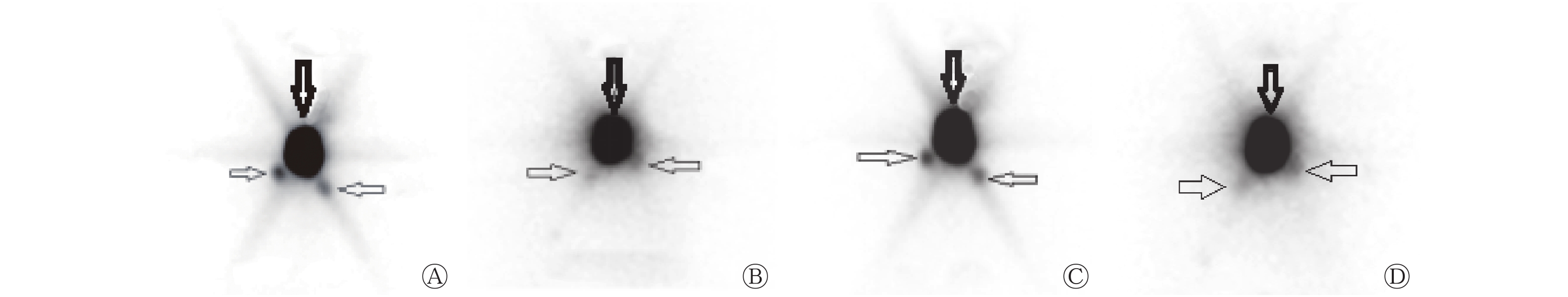
  • 将距离注射点最近、出现时间最早、浓聚程度最高的放射性热点确认为SLN(图1)。

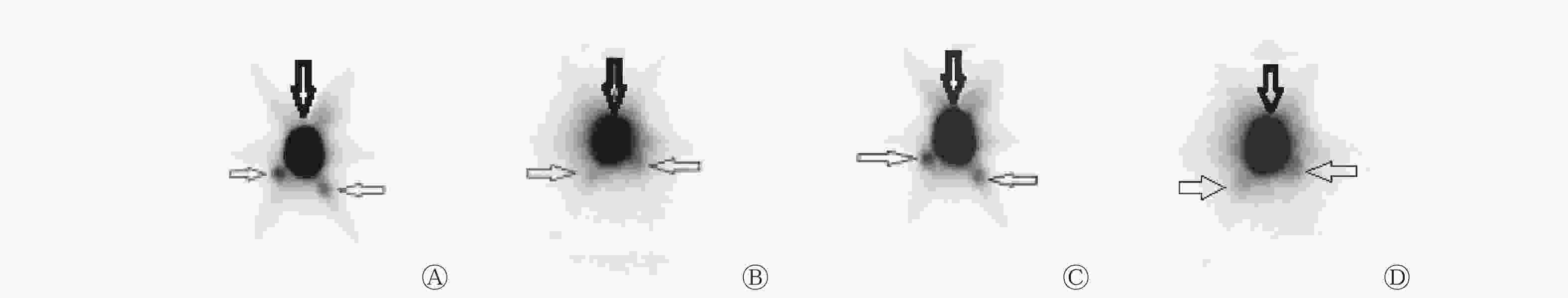
    Figure 1.  Preoperative sentinel lymph node planar images of a papillary thyroid carcinoma patient(female,54 years old)
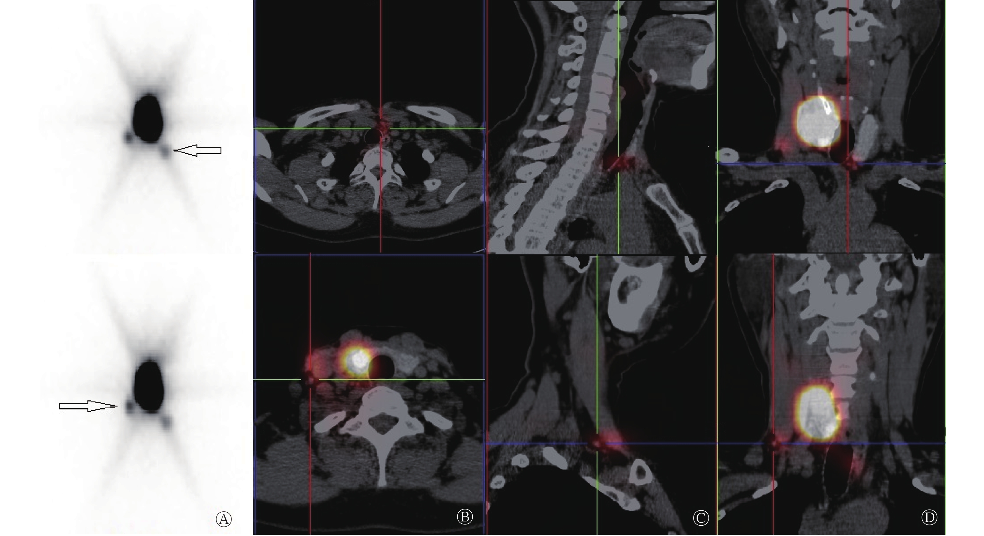
    SPECT/CT横断面、矢状面、冠状面断层融合显像见图2

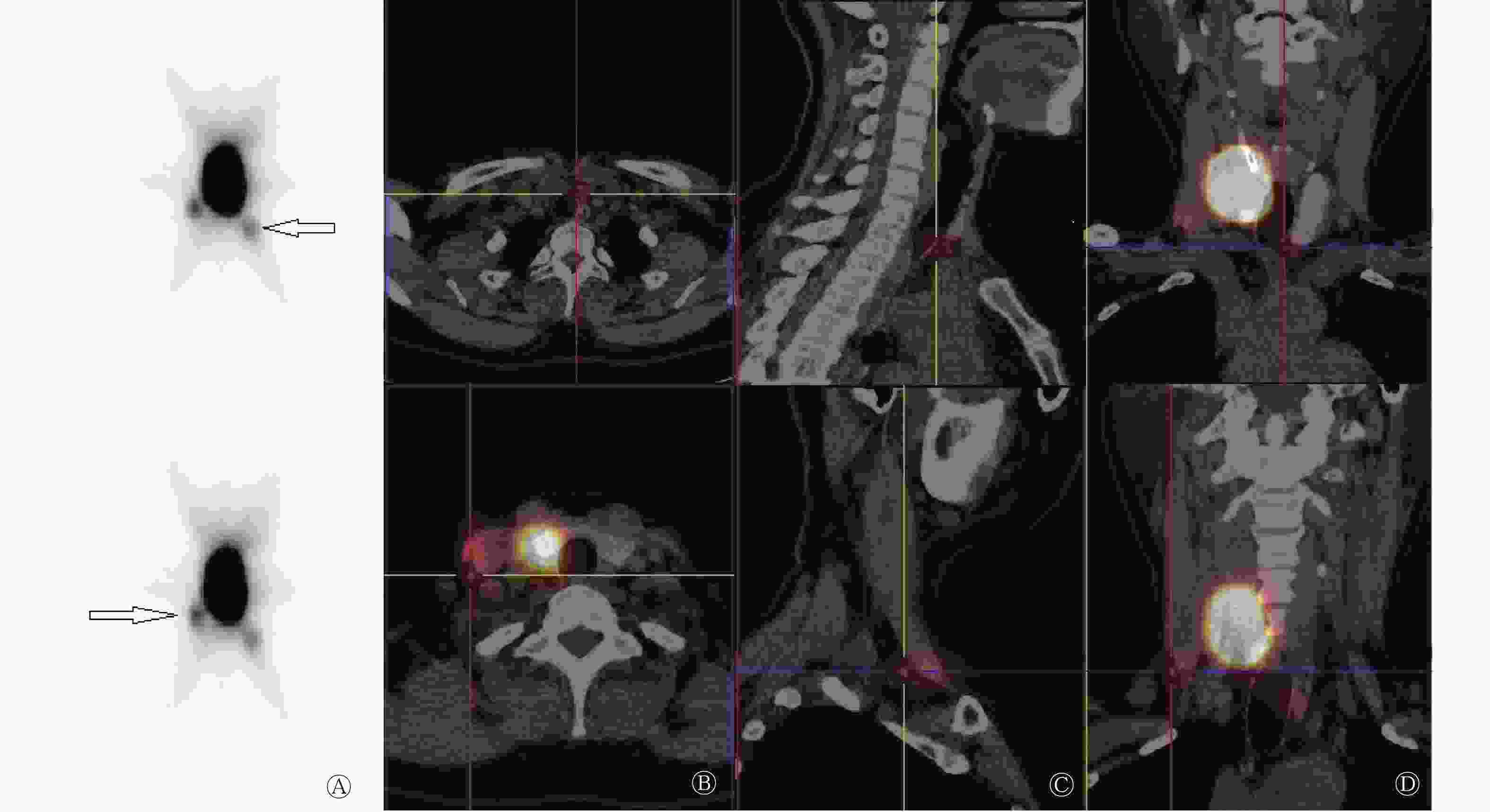
    Figure 2.  Preoperative sentinel lymph node SPECT/CT images of a papillary thyroid carcinoma patient(female,54 years old)

    术前99Tcm-SC平面显像共检出SLN患者14例,检出率为87.50%(14/16)。99Tcm-SC平面显像共检出31枚SLN,SPECT/CT共检出35枚SLN。SPECT/CT检出1枚SLN者5例(31.25%,5/16),检出2枚SLN者1例(6.25%,1/16),检出3枚SLN者5例(31.25%,5/16),检出4枚SLN者2例(12.50%,2/16),检出5枚SLN者1例(6.25%,1/16)。

  • 2.2.   手持式γ探测仪的检测

  • γ探测仪探测到13例患者的SLN,共计37枚,检出率为81.25%(13/16),有3例未探测到SLN(18.75%,3/16)。在探测到SLN的13例患者中,有3例探测到1枚(18.75%,3/16),1例探测到2枚(6.25%,1/16),5例探测到3枚(31.25%,5/16),3例探测到4枚(18.75%,3/16),1例探测到5枚(6.25%,1/16)。γ探测仪探测的SLN数目与SPECT/CT淋巴显像有4例不一致,12例一致,两种方法的一致率为75.00%(12/16)。

  • 2.3.   SLN的分布情况

  • 16例患者中,SPECT/CT检出14例有SLN。共检出SLN 35枚:其中位于Ⅵ区(中央区)的15枚;位于侧颈区共20枚,包括Ⅱ区的SLN 5枚、Ⅲ区的SLN 2枚、Ⅳ区的SLN 11枚、Ⅴ区的SLN 2枚。

    将肿瘤部位分为上极、中极、下极、腺体。肿瘤位于上极者4例,共检出SLN 8枚:中央区4枚,有3枚转移;侧颈区4枚(Ⅲ区1枚、Ⅳ区1枚、Ⅴ区2枚),有3枚转移(Ⅲ区1枚、Ⅳ区1枚、Ⅴ区1枚)。肿瘤位于中极者3例,共检出SLN 7枚:中央区1枚,无转移;侧颈区6枚(Ⅱ区4枚、Ⅳ区2枚),有4枚转移(Ⅱ区2枚、Ⅳ区2枚)。肿瘤位于下极者4例,共检出SLN 14枚,中央区7枚,有7枚转移;侧颈区7枚(Ⅱ区1枚、Ⅲ区1枚、Ⅳ区5枚),有3枚转移(Ⅲ区1枚、Ⅳ区2枚);肿瘤位于腺体者3例,共检出SLN 6枚,中央区3枚,有3枚转移;侧颈区3枚,均位于Ⅳ区,有2枚转移。肿瘤位于下极,SLN最容易发生转移,中央区比侧颈区更常见,占转移总数的28%(7/25),占SLN总数的20%(7/35)。肿瘤位于下极,侧颈部Ⅳ区SLN检出数目相对较多,占侧颈区SLN总数的25%(5/20)。

  • 2.4.   SPECT/CT淋巴显像与病理检查结果对照

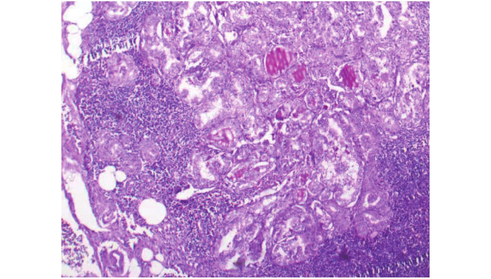
  • 16例患者中,15例(93.75%)常规病理学检查有淋巴结转移(图3),其中SLN阴性2例。中央区SLN有13枚转移,阳性率为86.67%(13/15),侧颈区SLN有12枚发生转移,阳性率为60.00% (12/20)。SPECT/CT检测SLN的灵敏度、准确率、假阴性率分别为86.67%(13/15)、81.25%(13/16)、13.33% (2/15)。

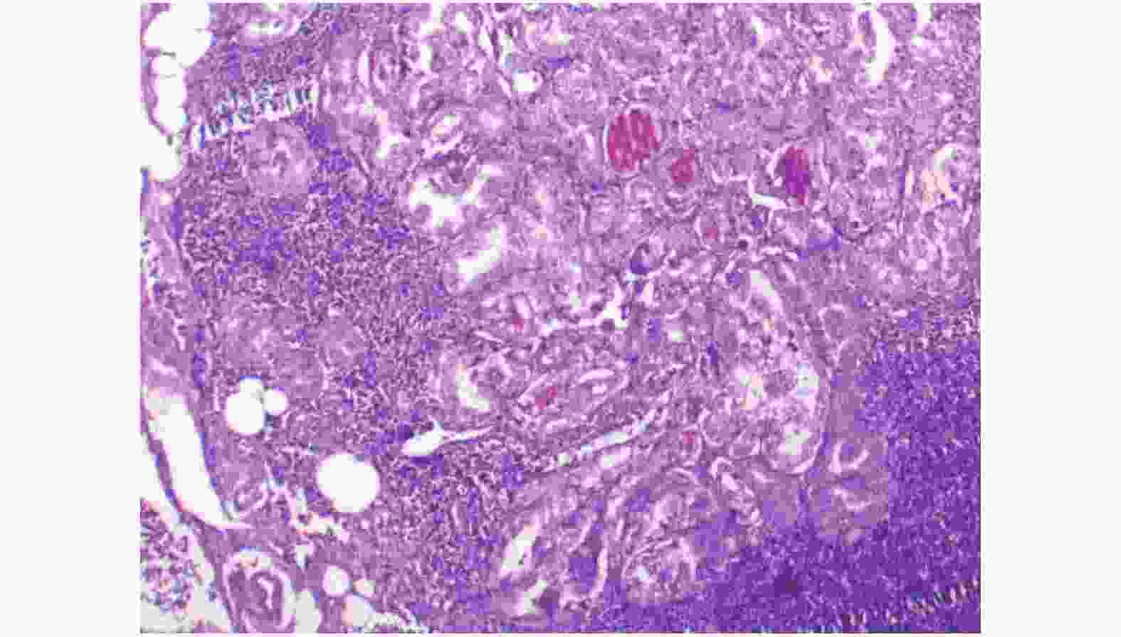
    Figure 3.  Sentinel lymph node pathology with positive metastasis in the right Ⅳ region of a papillary thyroid carcinoma patient (female, 54 years old)

3.   讨论
  • PTC是分化型甲状腺癌最常见的类型,其隐匿性淋巴结转移率在70%~80%[8]。区域淋巴结转移与PTC的局部复发有显著相关性,以往观点认为颈部淋巴结转移不会影响PTC患者的总体生存率,但近年来有学者认为PTC区域淋巴结转移会降低患者的生存率,是影响预后的重要因素[9]

    目前,cN0期PTC患者的分期主要依靠临床检查和颈部超声检查。临床检查无法对颈部淋巴结进行精确定位,导致假阴性率高达30%[10]。超声检测淋巴结转移有较高的准确率,但特异度范围较大(52%~84%),主要原因可能是颈部存在其他隐匿性疾病[11]。由此可见,这两种方法证实有无淋巴结隐匿性转移的可靠性有限。

    美国甲状腺协会指南建议,只有肿瘤细胞侵犯中央区淋巴结才能进行该区的淋巴结清扫,临床上未累及中央区淋巴结的原发肿瘤晚期(T3、T4期)或侧颈区淋巴结受累患者进行预防性中央区淋巴结清扫,对于T1期和T2期PTC患者,尚不推荐行预防性颈淋巴结清扫[12]。这可能会遗漏发生淋巴结转移的T1期、T2期患者,并扩大了未发生淋巴结转移的T3、T4期患者的手术范围。因此,临床上需要一种创伤小并能准确评估PTC患者颈部淋巴结状态、预测有无淋巴结隐匿性转移的技术。

    SLN活检在1998年首次被应用于检测甲状腺癌淋巴结的转移情况,现在已成为cN0期PTC患者选择性淋巴结清扫的一种可供选择的方法[13-14]。SLN活检的主要方法:①生物染料示踪法;②放射性核素淋巴显像+ γ探针法;③两者联合法。与生物染料示踪法相比,淋巴显像+ γ探针法有明显优势:术前注射放射性药物,避免了术中清除病灶时破坏淋巴管使淋巴引流中断;中央区以外的SLN清晰显影,并能进行精确定位;甲状旁腺不显影,可避免误切除甲状旁腺[15]

    SPECT/CT将功能代谢影像与解剖结构影像实时融合。本研究中,SPECT/CT显像较平面显像多检出4枚SLN,提高了SLN的检出率,可能原因是SPECT/CT能发现平面显像上被邻近病灶掩盖的SLN,同时其可以鉴别体外污染、皮肤皱壁摄取显像剂的情况,从而降低假阳性率。

    多个研究结果显示,术前SPECT/CT对SLN的检出率为79%~92%,术中γ探测仪对SLN的检出率为96%~100%[10, 16-18]。本研究中,术前SPECT/CT的SLN检出率为87.50%,而术中γ探测仪的SLN检出率为81.25%,稍低于96%。Portinari和Carcoforo[19]认为,影响术中γ探测仪SLN检出率的主要因素有显像剂注射的部位(瘤内或瘤外)和学习曲线效应等。由于本研究与多个研究[16, 20-21]均采用瘤内注射显像剂,所以受注射部位这一因素的影响较小。那么,在本研究中,学习曲线效应是否与术中γ探测仪的SLN检出率相关呢?结果显示,在第1例患者SPECT/CT显像中可见SLN显影,但γ探测仪未探及,并且前4例患者γ探测仪探测的SLN数目与SPECT/CT淋巴显像数目不一致,因此我们认为术中γ探测仪对SLN的检出率很可能与学习曲线效应有关。使用γ探测仪是逐步学习的过程,随着临床实践次数增多,熟练程度也随之增加,对SLN的检出也趋于准确。

    本研究中共检出35枚SLN,中央区SLN检出率为42.86%(15/35),转移阳性率为86.67%(13/15);侧颈区SLN检出率为57.14%(20/35),转移阳性率为60.00%(12/20);与鄢丹桂等[22]研究结果相似(中央区SLN检出率为37.1%)。本研究通过术前淋巴显像+γ探测仪探测,检出SLN的准确率高达81.25%(13/16)、灵敏度为86.67%(13/15)、假阴性率为13.33%(2/15)。多项研究结果显示,通过对PTC患者行SLN活检的诊断准确率为86%~100%、灵敏度为67.0%~100%、假阴性率为0%~33.3%[22-26]。与以上研究结果相比,本研究对SLN的诊断准确率略低,考虑可能与样本量少有关,而灵敏度、假阴性率均与活检相似。本研究16例PTC患者中有2例行SPECT/CT淋巴显像时未发现SLN,但经病理学检查提示有淋巴结转移,导致诊断假阴性率为13.33%,分析原因可能有:肿瘤组织体积过大,引流淋巴管被肿瘤细胞侵袭,导致癌栓堵塞淋巴管,从而使淋巴引流方向改变,使有转移的SLN不能显示;老年患者淋巴组织萎缩被脂肪组织替代,使SLN难以显示;操作失误,将放射性显像剂注入肿瘤周围,导致显像剂溢出到周围组织。

    Kaczka等[27]研究结果表明,绝大多数PTC患者的SLN分布于中央区,但也有部分出现在侧颈区[28-29]。本研究结果显示,肿瘤位于下极,侧颈部Ⅳ区检出SLN相对较多(25%,5/20)。4例患者有10枚SLN仅位于侧颈区,占28.57%(10/35),10枚SLN均发生转移。PTC患者颈部淋巴引流途径主要是以分区为导向,淋巴结转移一般首先出现在中央区。但临床上也发现部分PTC患者无中央区淋巴结转移而有侧颈区或上纵隔淋巴结转移,即淋巴结跳跃性转移,其发生率为2%~38%[30-31]。孙荣华等[32]研究认为,部分患者颈部淋巴结“跳跃转移”可能并非真正意义上的跳跃转移,而是PTC第一站引流淋巴结位于侧颈区。同时有研究证实肿瘤位置与PTC侧颈区淋巴结跳跃性转移有显著的相关性,如肿瘤位于甲状腺上极,其淋巴引流可能越过中央区直接流向侧颈区[29, 33]。本研究结果显示,当肿瘤位于上极,中央区与侧颈区发生转移的SLN数目一致,均占总转移数目12%(3/25)。

    我们认为,若SLN为转移性淋巴结,这说明该区域淋巴结很可能也已发生转移,则需行该区域淋巴结清扫;如果SLN为阴性(经病理学检查证实无肿瘤转移),则说明该区域淋巴结尚未发生转移,暂不需行颈部淋巴结清扫。本研究结果显示,利用术前SPECT/CT淋巴显像,结合术中γ探测仪检测SLN,可以进一步准确定位SLN,预测cN0期PTC患者的颈部隐匿性淋巴结转移,指导术中选择合理的术式,以减轻手术负担,减少术后并发症。

    本研究样本量有限,后续仍需扩大样本量进行前瞻性随机研究和长期随访,进一步证实术前SPECT/CT淋巴显像探测cN0期PTC患者SLN的临床价值。

    利益冲突 本研究由署名作者按以下贡献声明独立开展,不涉及任何利益冲突。

    作者贡献声明 胡珊负责病例的收集、辅助显像药物的准备及图像的采集分析、文献资料的查询及论文的撰写;杜芬负责图像的采集分析、文献资料的查询;白涛负责肿瘤穿刺及药物的注射;杨子瑶负责病例的收集;刘静负责手术及术中前哨淋巴结的定位;武晨负责病例收集及文献资料的查询;程艳、武志芳负责论文的指导和审校。

Reference (33)

Catalog

/

DownLoad:  Full-Size Img  PowerPoint
Return
Return