Volume 44 Issue 1
Mar.  2020
Article Contents

Citation:

Retrospective analysis of 18F-FDG PET/CT imaging in 22 cases of nodular type pulmonary cryptococcosis

  • Corresponding author: Duanyu Lin, lindy120@163.com
  • Received Date: 2019-03-19
  • Objective To investigate the 18F-fluorodeoxyglucose (FDG) PET/CT imaging characteristics of pulmonary cryptococcosis (PC). Methods A retrospective study was performed in 22 patients with PC (17 male and 5 female; aged 54.77±7.93 years), confirmed through etiology or pathological examination from January 2011 to January 2017 in Fujian Provincial Cancer Hospital. 18F-FDG PET/CT was performed. The number, size, distribution, maximum standardized uptake value (SUVmax), and nodule sign of the nodules were analyzed. T test or Pearson correlation analysis was used in the comparison in measurement data. The differences in nodule signs among the different groups were determined by χ2 test, continuous correction χ2 test, or Fisher exact probability method. Results ① A total of 235 nodules were found by 18F-FDG PET/CT imaging in the 22 cases of PC patients with the diameter of (0.7±0.5) cm. Of these nodules, 130 showed high 18F-FDG metabolism with an SUVmax of (3.5±2.9) and a diameter of (0.9±0.5) cm. A positive correlation was found between the two (r=0.702, P=0.000). Meanwhile, 105 nodules showed no 18F-FDG metabolism with a diameter of (0.3±0.1) cm. The diameter in the group with high 18F-FDG metabolism was greater than that in the group without 18F-FDG metabolism, and the difference was statistically significant (t=13.621, P=0.000). ② The nodules were mainly found in the right lung (57.4%, 135/235), lower lobe (64.3%, 151/235), and outer zone or subpleura (80.0%, 188/235). ③ The SUVmax in the nine cases of immunocompromised host with high 18F-FDG metabolism was 5.7±4.7 higher than that in the 13 cases of non-immunocompromised host (SUVmax was 3.0±2.0), and the difference was statistically significant (t=2.731, P=0.011). The ratio of wide base posted to the pleural sign was 17.5 (10/57) of the former and higher the 6.2% (11/178) of the latter. The ratio of halo sign was 25.3% (45/178) in the latter and higher than the former of 10.5% (6/57). All the differences were statistically significant (χ2=7.911, 4.628; P=0.005, 0.031). ④ The nodules of the group with high 18F-FDG metabolism accounted for 80% (16/20) and 53% (114/215), respectively, in the each of 11 cases of one lobe or multiple lobar involvement patients. The former SUVmax was 5.6±3.4 and higher than the latter of 3.2±2.7. The difference was statistically significant (t=2.652, P=0.016). The lobulation sign (35%, 7/20), spicule sign (30%, 6/20), and pleural indentation sign (15%, 3/20) in the former were higher than the 3.3% (7/215), 0.9% (2/215) and 0.9% (2/215) in the latter, respectively. The difference was statistically significant (χ2=32.911, 47.022, 17.395, all P<0.01). ⑤ The SUVmax of the 21 nodules with high 18F-FDG metabolism in the 44 nodules found in the 11 misdiagnosed cases was 5.0±4.6, which was higher than 3.2±2.4 in the 11 other correctly diagnosed cases where 191 nodules were found, including 109 nodules with high 18F-FDG metabolism. The difference was not statistically significant (t=16.825, P=0.106). The lobulation sign, spicule sign and pleural indentation sign in the former were 15.9% (7/44), 18.2% (8/44), 6.8% (3/44) respectively, which are higher than the 3.7% (7/191), 0.0% (0/191), and 1.0% (2/191) in the latter. The difference was statistically significant (χ2=9.570, 35.951, 5.720; all P<0.05). The halo sign was 26.2% (50/191) in the latter, and higher than the 2.3% (1/44) in the former. The difference was statistically significant (χ2=12.027, P=0.000). Conclusions The SUVmax of the nodules is positively correlated with diameter in cases of PC by 18F-FDG PET/CT imaging. Halo sign is a reliable sign of diagnosis. The resembling tumor signs and the high 18F-FDG metabolism, caused the 18F-FDG PET/CT imaging to misdiagnose easily in patients of PC with single lobe involvment.
  • 加载中
  • [1] Chang CC, Sorrell TC, Chen SC, et al. Pulmonary Cryptococcosis[J]. Semin Respir Crit Care Med, 2015, 36(5): 681−691. DOI: 10.1055/s−0035−1562895.
    [2] 中华医学会呼吸病学分会感染学组, 中华结核和呼吸杂志编辑委员会. 肺真菌病诊断和治疗专家共识[J]. 中华结核和呼吸杂志, 2007, 30(11): 821−834. DOI: 10.3760/j.issn:1001−0939.2007.11.008.Infectious Diseases Group of Chinese Thoracic Society, Editorial Board of Chinese Journal of Tuberculosis and Respiratory Diseases. Expert consensus on diagnosis and treatment of pulmonary mycosis[J]. Chin J Tube Resp Dis, 2007, 30(11): 821−834. DOI: 10.3760/j.issn:1001−0939.2007.11.008.
    [3] 刘又宁, 佘丹阳, 孙铁英, 等. 中国1998年至2007年临床确诊的肺真菌病患者的多中心回顾性调查[J]. 中华结核和呼吸杂志, 2011, 34(2): 86−90. DOI: 10.3760/cma.j.issn.1001−0939.2011.02.004.Liu YN, She DY, Sun TY, et al. A multicentre retrospective study of pulmonary mycosis clinically proven from 1998 to 2007[J]. Chin J Tuberc Respir Dis, 2011, 34(2): 86−90. DOI: 10.3760/cma.j.issn.1001−0939.2011.02.004.
    [4] 兰长青, 黄进宝, 黄梅萍, 等. 结节肿块型肺隐球菌病CT特征分析[J]. 中华放射学杂志, 2015, 49(10): 741−744. DOI: 10.3760/cma.j.issn.1005−1201.2015.10.005.Lan CQ, Huang JB, Huang MP, et al. Analysis of CT features of nodular or mass-like type of pulmonary cryptococcosis[J]. Chin J Radiol, 2015, 49(10): 741−744. DOI: 10.3760/cma.j.issn.1005−1201.2015.10.005.
    [5] Severo CB, Gazzoni AF, Severo LC. Chapter 3-Pulmonary cryptococcosis[J]. J Bras Pneumol, 2009, 35(11): 1136−1144. DOI: 10.1590/S1806−37132009001100012.
    [6] 王丽芳, 施毅, 丁媛, 等. 肺隐球菌病65例回顾分析[J]. 中华结核和呼吸杂志, 2014, 37(10): 764−768. DOI: 10.3760/cma.j.issn.1001−0939.2014.10.015.Wang LF, Shi Y, Ding Y, et al. Pulmonary cryptococcosis: a retrospective analysis of 65 cases[J]. Chin J Tuberc Respir Dis, 2014, 37(10): 764−768. DOI: 10.3760/cma.j.issn.1001−0939.2014.10.015.
    [7] 刘佳, 吴泰华, 翟文爽, 等. 22例误诊为周围型肺癌的肺隐球菌病患者临床分析[J]. 大连医科大学学报, 2017, 39(4): 349−353. DOI: 10.11724/jdmu.2017.04.08.Liu J, Wu TH, Zhai WS, et al. Analysis on 22 cases of pulmonary cryptococcosis misdiagnosed as peripheral lung cancer[J]. J Dalian Med Univ, 2017, 39(4): 349−353. DOI: 10.11724/jdmu.2017.04.08.
    [8] 谢丽璇, 李国雄, 刘志军, 等. 33例肺良性病变的18F-FDG PET/CT误诊原因分析[J]. 放射学实践, 2014, 29(5): 541−544.Xie LX, Li GX, Liu ZJ, et al. Analysis in 33 cases of misdiagnosed pulmonary benign lesions by 18F-FDG PET/CT imaging[J]. Radiol Practice, 2014, 29(5): 541−544.
    [9] 梅周芳, 秦欣宇, 潘星月, 等. 无免疫功能缺陷的肺隐球菌病19例误诊分析[J]. 临床误诊误治, 2016, 29(1): 50−52. DOI: 10.3969/j.issn.1002−3429.2016.01.019.Mei ZF, Qin XY, Pan XY, et al. Misdiagnosis analysis on 19 cases of non-immunodeficiency pulmonary cryptococcosis[J]. Clin Misdiag Misther, 2016, 29(1): 50−52. DOI: 10.3969/j.issn.1002−3429.2016.01.019.
    [10] Ye F, Xie JX, Zeng QS, et al. Retrospective analysis of 76 immunocompetent patients with primary pulmonary cryptococcosis[J]. Lung, 2012, 190(3): 339−346. DOI: 10.1007/s00408−011−9362−8.
    [11] Qu YJ, Liu GB, Ghimire P, et al. Primary pulmonary cryptococcosis: evaluation of CT characteristics in 26 immunocompetent Chinese patients[J]. Acta Radiol, 2012, 53(6): 668−674. DOI: 10.1258/ar.2012.110612.
    [12] 兰长青, 翁恒, 李红艳, 等. 肺隐球菌病117例回顾分析[J]. 中华结核和呼吸杂志, 2016, 39(11): 862−865. DOI: 10.3760/cma.j.issn.1001−0939.2016.11.008.Lan CQ, Weng H, Li HY, et al. Retrospective analysis of 117 cases of pulmonary cryptococcosis[J]. Chin J Tuberc Respir Dis, 2016, 39(11): 862−865. DOI: 10.3760/cma.j.issn.1001−0939.2016.11.008.
    [13] De Pauw B, Walsh TJ, Donnelly JP, et al. Revised Definitions of Invasive Fungal Disease from the European Organization for Research and Treatment of Cancer/Invasive Fungal Infections Cooperative Group and the National Institute of Allergy and Infectious Diseases Mycoses Study Group (EORTC/MSG) Consensus Group[J]. Clin Infect Dis, 2008, 46(12): 1813−1821. DOI: 10.1086/588660.
    [14] 杨海, 刘子姗, 陈盈, 等. 免疫正常患者肺隐球菌病的CT征象分析[J]. 中华全科医学, 2018, 16(2): 279−282, 290.Yang H, Liu ZS, Chen Y, et al. CT findings of pulmonary cryptococcosis in immunocompetent patients[J]. Chin J General Pract, 2018, 16(2): 279−282, 290.
    [15] 冯瑞枝, 林波淼, 梁文, 等. 肺隐球菌病的CT影像表现[J]. 临床肺科杂志, 2016, 21(11): 1986−1989. DOI: 10.3969/j.issn.1009−6663.2016.11.014.Feng RZ, Lin BM, Liang W, et al. CT appearance of pulmonary cryptococcosis[J]. J Clin Pulm Med, 2016, 21(11): 1986−1989. DOI: 10.3969/j.issn.1009−6663.2016.11.014.
    [16] Zinck SE, Leung AN, Frost M, et al. Pulmonary Cryptococcosis: CT and Pathologic Findings[J]. J Comput Assist Tomogr, 2002, 26(3): 330−334. DOI: 10.1097/00004728−200205000−00002.
    [17] Song KD, Lee KS, Chung MP, et al. Pulmonary Cryptococcosis: Imaging Findings in 23 Non-AIDS Patients[J]. Korean J Radiol, 2010, 11(4): 407−416. DOI: 10.3348/kjr.2010.11.4.407.
    [18] Sim YT, Goh YG, Dempsey MF, et al. PET-CT Evaluation of Solitary Pulmonary Nodules: Correlation with Maximum Standardized Uptake Value and Pathology[J]. Lung, 2013, 191(6): 625−632. DOI: 10.1007/s00408−013−9500−6.
    [19] Chang CY, Tzao C, Lee SC, et al. Incremental value of integrated FDG-PET/CT in evaluating indeterminate solitary pulmonary nodule for malignancy[J]. Mol Imaging Biol, 2010, 12(2): 204−209. DOI: 10.1007/s11307−009−0241−0.
    [20] Chang JM, Lee HJ, Goo JM, et al. False positive and false negative FDG-PET scans in various thoracic diseases[J]. Korean J Radiol, 2006, 7(1): 57−69. DOI: 10.3348/kjr.2006.7.1.57.
    [21] Ghimire P, Sah AK. Pulmonary cryptococcosis and tuberculoma mimicking primary and metastatic lung cancer in 18F-FDG PET/CT[J]. Nepal Med Coll J, 2011, 13(2): 142−143.
    [22] Wang J, Ju HZ, Yang MF. Pulmonary cryptococcosis and cryptococcal osteomyelitis mimicking primary and metastatic lung cancer in 18F-FDG PET/CT[J]. Int J Infect Dis, 2014, 18: 101−103. DOI: 10.1016/j.ijid.2013.08.009.
    [23] Lee CH, Tzao C, Chang TH, et al. Case of pulmonary cryptococcosis mimicking hematogeneous metastases in an immuocompetent patient: value of absent 18F-fluorodeoxylucose uptake on positron emission tomography/CT scan[J]. Korean J Radiol, 2013, 14(3): 540−543. DOI: 10.3348/kjr.2013.14.3.540.
    [24] 陈涛, 朱晔涵, 蒋军红, 等. 肺隐球菌病PET/CT误诊肺癌一例并文献复习[J]. 中国血液流变学杂志, 2013, 23(1): 76−77. DOI: 10.3969/j.issn.1009−881X.2013.01.025.Chen T, Zhu YH, Jiang JH, et al. A case of pulmonary cryptococcosis misdiagnosed by PET/CT and literature review[J]. Chin J Hemorheol, 2013, 23(1): 76−77. DOI: 10.3969/j.issn.1009−881X.2013.01.025.
    [25] 李建南, 冯洪波, 蔡博文, 等. 非小细胞肺癌病灶大小及密度与PET/CT标准摄取值相关性研究[J]. 大连医科大学学报, 2012, 34(1): 56−59. DOI: 10.11724/jdmu.2012.01.12.Li JN, Feng HB, Cai BW, et al. Covariance analysis between SUVmax of NSCLC and influencing clinical factors[J]. J Dalian Med Univ, 2012, 34(1): 56−59. DOI: 10.11724/jdmu.2012.01.12.
    [26] 周锦, 姜东朗, 谢芳, 等. 肺隐球菌病18F-FDG PET/CT显像特征分析[J]. 国际放射医学核医学杂志, 2018, 42(6): 481−485. DOI: 10.3760/cma.j.issn.1673−4114.2018.06.001.Zhou J, Jiang DL, Xie F, et al. The characteristics of 18F-FDG PET/CT in pulmonary cryptococcosis[J]. Int J Radiat Med Nucl Med, 2018, 42(6): 481−485. DOI: 10.3760/cma.j.issn.1673−4114.2018.06.001.
  • 加载中
通讯作者: 陈斌, bchen63@163.com
  • 1. 

    沈阳化工大学材料科学与工程学院 沈阳 110142

  1. 本站搜索
  2. 百度学术搜索
  3. 万方数据库搜索
  4. CNKI搜索

Figures(1) / Tables(3)

Article Metrics

Article views(12398) PDF downloads(31) Cited by()

Related
Proportional views

Retrospective analysis of 18F-FDG PET/CT imaging in 22 cases of nodular type pulmonary cryptococcosis

    Corresponding author: Duanyu Lin, lindy120@163.com
  • Department of Nuclear Medicine, Fujian Provincial Cancer Hospital, Fujian Medical University Cancer Hospital, Fuzhou 350014, China

Abstract:  Objective To investigate the 18F-fluorodeoxyglucose (FDG) PET/CT imaging characteristics of pulmonary cryptococcosis (PC). Methods A retrospective study was performed in 22 patients with PC (17 male and 5 female; aged 54.77±7.93 years), confirmed through etiology or pathological examination from January 2011 to January 2017 in Fujian Provincial Cancer Hospital. 18F-FDG PET/CT was performed. The number, size, distribution, maximum standardized uptake value (SUVmax), and nodule sign of the nodules were analyzed. T test or Pearson correlation analysis was used in the comparison in measurement data. The differences in nodule signs among the different groups were determined by χ2 test, continuous correction χ2 test, or Fisher exact probability method. Results ① A total of 235 nodules were found by 18F-FDG PET/CT imaging in the 22 cases of PC patients with the diameter of (0.7±0.5) cm. Of these nodules, 130 showed high 18F-FDG metabolism with an SUVmax of (3.5±2.9) and a diameter of (0.9±0.5) cm. A positive correlation was found between the two (r=0.702, P=0.000). Meanwhile, 105 nodules showed no 18F-FDG metabolism with a diameter of (0.3±0.1) cm. The diameter in the group with high 18F-FDG metabolism was greater than that in the group without 18F-FDG metabolism, and the difference was statistically significant (t=13.621, P=0.000). ② The nodules were mainly found in the right lung (57.4%, 135/235), lower lobe (64.3%, 151/235), and outer zone or subpleura (80.0%, 188/235). ③ The SUVmax in the nine cases of immunocompromised host with high 18F-FDG metabolism was 5.7±4.7 higher than that in the 13 cases of non-immunocompromised host (SUVmax was 3.0±2.0), and the difference was statistically significant (t=2.731, P=0.011). The ratio of wide base posted to the pleural sign was 17.5 (10/57) of the former and higher the 6.2% (11/178) of the latter. The ratio of halo sign was 25.3% (45/178) in the latter and higher than the former of 10.5% (6/57). All the differences were statistically significant (χ2=7.911, 4.628; P=0.005, 0.031). ④ The nodules of the group with high 18F-FDG metabolism accounted for 80% (16/20) and 53% (114/215), respectively, in the each of 11 cases of one lobe or multiple lobar involvement patients. The former SUVmax was 5.6±3.4 and higher than the latter of 3.2±2.7. The difference was statistically significant (t=2.652, P=0.016). The lobulation sign (35%, 7/20), spicule sign (30%, 6/20), and pleural indentation sign (15%, 3/20) in the former were higher than the 3.3% (7/215), 0.9% (2/215) and 0.9% (2/215) in the latter, respectively. The difference was statistically significant (χ2=32.911, 47.022, 17.395, all P<0.01). ⑤ The SUVmax of the 21 nodules with high 18F-FDG metabolism in the 44 nodules found in the 11 misdiagnosed cases was 5.0±4.6, which was higher than 3.2±2.4 in the 11 other correctly diagnosed cases where 191 nodules were found, including 109 nodules with high 18F-FDG metabolism. The difference was not statistically significant (t=16.825, P=0.106). The lobulation sign, spicule sign and pleural indentation sign in the former were 15.9% (7/44), 18.2% (8/44), 6.8% (3/44) respectively, which are higher than the 3.7% (7/191), 0.0% (0/191), and 1.0% (2/191) in the latter. The difference was statistically significant (χ2=9.570, 35.951, 5.720; all P<0.05). The halo sign was 26.2% (50/191) in the latter, and higher than the 2.3% (1/44) in the former. The difference was statistically significant (χ2=12.027, P=0.000). Conclusions The SUVmax of the nodules is positively correlated with diameter in cases of PC by 18F-FDG PET/CT imaging. Halo sign is a reliable sign of diagnosis. The resembling tumor signs and the high 18F-FDG metabolism, caused the 18F-FDG PET/CT imaging to misdiagnose easily in patients of PC with single lobe involvment.

    HTML

  • 肺隐球菌病(pulmonary cryptococcosis,PC)是一种侵袭性肺部真菌病,主要致病菌为新生隐球菌或格特隐球菌,呼吸道吸入是常见的致病途经[1-2]。近年来,PC的发病率呈逐年增加的趋势,据多中心回顾性调查显示,隐球菌已位居侵袭性肺真菌感染常见病原体的第3位[3],而且PC临床症状不典型,影像改变形态各异,可表现为结节肿块型、浸润实变型、弥漫性粟粒型和弥漫混合病变型,其中结节肿块型最多见且具有类似肿瘤的征象,常与肺癌、肺转移癌混淆[4]。本研究回顾性分析22例结节型PC患者的临床及18F-FDG PET/CT显像资料,现将结果报道如下。

  • 1.   资料与方法

      1.1.   临床资料

    • 收集2011年1月至2017年1月于我院手术病理确诊的PC患者22例,其中男性17例、女性5例,年龄(54.77±7.93)岁。就诊原因包括咳嗽、咳痰(伴或不伴胸闷、气促、胸痛)12例,癌胚抗原增高1例,体检发现肺部结节9例。免疫功能受损宿主(immunocompromised host,ICH)9例,免疫受损疾病包括糖尿病3例、乙型肝炎3例(其中1例伴肝癌、肝硬化)、类风湿性关节炎2例、肾病综合征1例,均无人类免疫缺陷病毒感染。非免疫功能受损宿主(non-immunocompromised host,NICH)13例。所有患者均在术前1周内行18F-FDG PET/CT显像,术后病理证实均为PC。所有患者或家属均签署了知情同意书。

    • 1.2.   显像方法

    • 采用荷兰Philips公司生产的Gemini TF 64 PET/CT仪,18F-FDG由日本住友公司HM-10回旋加速器生产,放射化学纯度>95%。患者检查前禁食6 h以上,控制血糖范围在3.9~7.5 mmol/L,静脉注射185~370 MBq的18F-FDG后,安静休息约60 min,排尿后仰卧位上机扫描。CT扫描采集条件:电压120 kV、电流200 mA,矩阵512×512,层厚5 mm。以1.0 min/床位的速度采集从颅底至股骨上段的PET图像,采用三维模式采集,应用CT数据进行衰减校正。在EBW2.0后处理工作站上,将PET图像和CT图像进行融合,分别得到横断面、矢状面及冠状面的PET、CT和PET/CT融合图像。

    • 1.3.   图像分析

    • 由两位有5年以上PET/CT诊断经验的核医学科医师共同对PET图像、CT图像及PET/CT融合图像分别判读,包括以下6个方面。(1)病灶数目;(2)病灶大小:手动测量病灶的直径;(3)病灶分布:分为左肺上叶、下叶,右肺上叶、中叶、下叶,肺野内带、中带、外带及胸膜下;(4)SUVmax:选择病灶横断面放射性摄取程度最高层面,以病灶大小的90%为ROI,计算机自动计算SUVmax,并结合目测法,以正常肺野为本底,SUVmax≥2倍肺本底视为18F-FDG代谢增高;(5)结节征象及伴随征象:分叶征、毛刺征、支气管充气征、晕征、胸膜凹陷征、宽基底贴胸膜征、肺门或纵隔淋巴结肿大、胸腔积液等;(6)出现意见分歧以主任医师意见为准。

    • 1.4.   统计学分析

    • 采用SPSS 22.0软件进行统计学分析。符合正态分布的计量资料以${\bar{x}} \pm s$表示。在方差齐的条件下,组内两两比较采用配对资料的t检验,结节SUVmax与直径间的关系采用Pearson相关性分析。计数资料以构成比(%)表示。不同组别结节征象间差异的比较采用χ2检验、连续校正χ2检验(当n≥40但有1≤T<5时)或Fisher确切概率法(当n<40或T<1时)。P<0.05表示差异有统计学意义。

    2.   结果

      2.1.   结节数目及直径

    • 22例PC患者共发现235个结节,直径(0.7±0.5) cm,范围0.2~2.8 cm。其中,18F-FDG代谢增高结节130个,SUVmax=3.5±2.9(0.9~20.1),直径(0.9±0.5) cm,范围0.3~2.8 cm,二者存在正相关(r=0.702,P=0.000);无18F-FDG代谢结节105个,直径(0.3±0.1) cm,范围0.2~0.6 cm;代谢增高结节的直径大于无代谢者,差异有统计学意义(t=13.621,P=0.000)。

    • 2.2.   结节分布

    • 以右肺(57.4%,135/235)、下叶(64.3%,151/235)多见结节,多数结节(80.0%,188/235)位于肺野外带或胸膜下,仅少数(11.5%,27/235)位于中带和内带(8.5%,20/235),其余数据见表1

      病灶位置上叶中叶下叶合计
      左肺35(14.9)0(0.0)65(27.7)100(42.6)
      右肺43(18.3)6(2.6)86(36.6)135(57.4)
      合计78(33.2)6(2.6)151(64.3) 235(100.0)

      Table 1.  The distribution of 235 nodules in 22 patients with pulmonary ryptococcosis

    • 2.3.   PC患者18F-FDG PET/CT显像结节特征

    • 单个肺叶受累者(单发结节8例、单叶散在结节3例)与多个肺叶受累者(多叶散在结节9例、多叶聚集分布结节2例)各11例;PET/CT正确诊断与误诊的患者各11例,其中误诊组中单个肺叶受累者8例。235个结节中出现分叶征、毛刺征、支气管充气征、晕征、空泡征、胸膜凹陷征和宽基底贴胸膜征的个数分别为14、8、28、51、2、5和21,具体数据见表2。典型病例的PET/CT显像见图1,此病例误诊为左侧肺癌右肺转移。

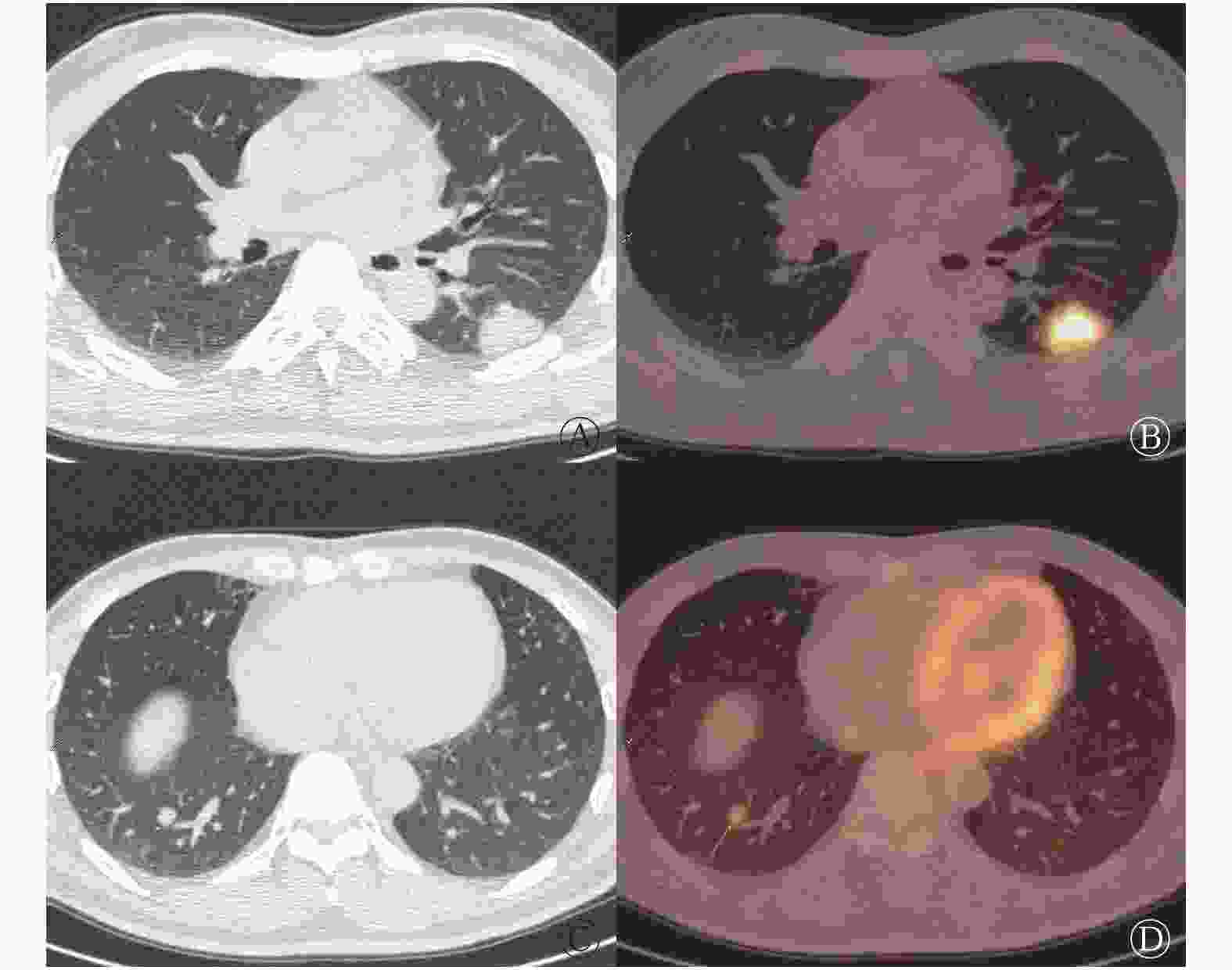
      Figure 1.  18F-FDG PET/CT imaging of pulmonary cryptococcosis (male, 35 years old)

      序号性别年龄免疫功能受损影像诊断结节分布及数目[a+b(个)]结节直径(cm)结节SUVmax结节征象及个数(个)
      右肺上叶右肺中叶右肺下叶左肺上叶左肺下叶分叶征毛刺征支气管充气征晕征空泡征胸膜凹陷征宽基底贴胸膜
      1 56 LC 1+0 0.6 1.1 1
      2 54 LC 1+0 2.8 9.6 1 1 1 1
      3 53 LC 1+0 1.4 7.9 1 1 1
      4 49 LC 1+0 1.5 6.2 1 1 1
      5 43 MC 1+0 1.6 3.1 1 1 1
      6 61 MC 1+0 1.0 4.3 1 1 1
      7 53 LC 1+0 1.3 5.7 1 1 1
      8 55 LC 1+0 1.0 7.9 1 1 1
      9 54 LM 2+0 0.3/0.5 1.4/3.5 1
      10 69 MC 4+3 0.2~1.6 2.7~13.6 1 1
      11 57 LWM 2+1 0.2~1.0 2.5~6.8 1
      12 63 LWM 4+3 0+1 0.3~1.1 2.1~9.3 1 1 1
      13 41 MC 12+22 20+7 0.3~2.0 1.0~3.8 5 17 18
      14 35 LWM 1+0 1+0 0.7/2.3 2.3/20.1 1 2
      15 60 MC 2+1 9+1 5+1 0.2~1.4 1.8~6.5 6
      16 67 LWM 2+10 2+5 1+3 0.2~1.2 1.1~1.7 1 1 1 1 1
      17 50 MC 0+2 4+0 10+2 1+0 6+1 0.3~1.3 0.9~6.8 1 7 1
      18 57 MC 0+6 0+1 4+4 3+10 1+2 0.2~1.2 1.0~6.0 1 5
      19 58 MC 0+2 4+2 3+4 0.2~1.5 1.3~6.4 1 3
      20 58 MC 1+1 2+1 3+2 0.2~2.3 2.2~10.5 2 5 5
      21 53 MC 2+2 1+3 4+1 0.2~1.5 1.1~7.6 6 1 1
      22 59 MC 1+0 1+1 3+0 1+0 0.4~1.7 1.5~12.3 4 2
      注:表中,PC:肺隐球菌病;18F-FDG:氟脱氧葡萄糖F18;PET/CT:正电子发射断层显像计算机体层摄影术;LC:肺癌;MC:真菌感染;LM:肺转移癌;LWM:肺癌伴肺内转移;a:18F-FDG代谢增高结节个数;b:无18F-FDG代谢结节个数;SUVmax:最大标准化摄取值;/:结节数为2枚;~:结节数≥3枚;−:无此项数据

      Table 2.  The 18F-FDG PET/CT imaging characteristics of 22 patienst with pulmonary cryptococcosis

      ICH患者共发现57个结节,18F-FDG代谢增高者25个,SUVmax=5.7±4.7(1.1~20.1);NICH患者共发现178个结节,18F-FDG代谢增高者105个,SUVmax=3.0±2.0(0.9~12.3),二者SUVmax的比较差异有统计学意义(t=2.731,P=0.011)。前者出现宽基底贴胸膜征象的比例高于后者,后者晕征的比例高于前者,差异均有统计学意义。具体数据见表3

      征象NICH
      n/178(%)
      ICH
      n/57(%)
      χ2P单叶受累
      n/20(%)
      多叶受累
      n/215(%)
      χ2P正确诊断
      n/191(%)
      误诊
      n/44(%)
      χ2P
      LS 9(5.0) 5(8.8) 1.364 0.243 7(35.0) 7(3.3) 32.911 0.000 7(3.7) 7(15.9) 9.570 0.006
      SS 4(2.2) 4(7.0) 3.417 0.065 6(30.0) 2(0.9) 47.022 0.000 0(0.0) 8(18.2) 35.951 0.000
      PS 23(12.9) 5(8.8) 0.471 0.492 4(20.0) 24(11.2) 1.362 0.243 23(12.0) 5(11.4) 0.016 0.900
      PIS 3(1.7) 2(3.5) 0.836 0.360 3(15.0) 2(0.9) 17.395 0.005 2(1.0) 3(6.8) 5.720 0.047
      HS 45(25.3) 6(10.5) 4.628 0.031 3(15.0) 48(22.3) 0.578 0.447 50(26.2) 1(2.3) 12.027 0.000
      VS 1(0.6) 1(1.8) 0.832 0.362 1(5.0) 1(0.5) 4.459 0.163 1(0.5) 1(2.3) 1.297 0.340
      BBPS 11(6.2) 10(17.5) 7.911 0.005 3(15.0) 18(8.4) 0.988 0.320 16(8.4) 5(11.4) 3.920 0.531
      注:表中,18F-FDG:氟脱氧葡萄糖F18;PET/CT:正电子发射断层显像计算机体层摄影术;LS:分叶征;SS:毛刺征;PS:支气管充气征;PIS:胸膜凹陷征;HS:晕征;VS:空泡征;BBPS:宽基底贴胸膜征;NICH:非免疫功能受损宿主;ICH:免疫功能受损宿主

      Table 3.  The comparison of nodular signs on 18F-FDG PET/CT imaging between different groups

      单个肺叶受累者11例共发现20个结节,其中16个18F-FDG代谢增高,SUVmax=5.6±3.4(1.1~13.6);多个肺叶(多叶散在结节)受累者11例共发现215个结节,其中114个18F-FDG代谢增高,SUVmax=3.2±2.7(0.9~20.1),SUVmax差异有统计学意义(t=2.652,P=0.016)。前者出现分叶征、毛刺征、胸膜凹陷征的比例高于后者,差异有统计学意义。具体数据见表3

      18F-FDG PET/CT显像误诊的11例患者共发现44个结节,其中21个18F-FDG代谢增高,SUVmax=5.0±4.6;正确诊断真菌感染的11例患者共发现191个结节,其中109个18F-FDG代谢增高,SUVmax=3.2±2.4,二者SUVmax差异无统计学意义(t=16.825,P=0.106)。前者出现分叶征、毛刺征、胸膜凹陷征的比例高于后者,差异有统计学意义;后者出现晕征的比例高于前者,差异有统计学意义。具体数据见表3

    3.   讨论
    • PC是由隐球菌感染引起的一种急性、亚急性或慢性真菌病,呼吸系统是感染的主要门户,好发于中青年男性,本研究中男性患者占多数(17 /22,77.3%),与Severo等[5]报道相符。本研究中54.5%(12/22)的患者主要临床症状为咳嗽、咳痰,而体检发现肺部结节占40.9%(9/22),这说明PC发病隐匿,临床症状轻或无临床症状,这与王丽芳等[6]报道相符。Chang等[1]指出PC好发于免疫功能受损的患者,尤其是HIV感染、使用免疫抑制剂或皮质类固醇治疗的患者,以及有肝炎、糖尿病、结核等基础疾病的患者。但是,近年来免疫功能正常的患者出现隐球菌感染的报道[7-8]并不少见。本研究22例患者均无HIV感染,ICH患者占40.9%(9/22),而NICH的患者占59.1%(13/22),这说明PC在非免疫功能受损人群中也常见,这也与刘又宁等[3]报道一致。

      PC的影像学表现各异,临床误诊率高,需与机化性肺炎、肺癌、肺结核等相鉴别[7-9]。目前CT对于PC诊断的报道较多,研究结果显示PC以结节或肿块型多见,分布以肺野外带为主,晕征与支气管充气征对诊断有一定帮助[4, 10-12]。本研究22例PC患者均为结节型,发现的235个结节以右肺、下叶、肺野外带或胸膜下多见,符合PC的分布特征。本研究还显示结节中出现分叶征、毛刺征、支气管充气征、晕征、空泡征、胸膜凹陷征和宽基底贴胸膜征的个数分别为14、8、28、51、2、5和21个,出现晕征的结节个数最多,占21.7%(51/235)。在CT图像上,结节、肿块、实变及空洞等病灶周围环绕密度较淡、均匀的磨玻璃样淡片影,即晕征,病理证实晕征为水肿和(或)出血,欧洲癌症治疗组织及真菌病研究组制定的诊断标准中认为,晕征是肺部真菌感染的特征性表现[13]。杨海等[14]报道,免疫正常患者PC好发于下叶胸膜下,以单发结节和(或)肿块型为主。 本研究正确诊断组中,晕征的比例要高于误诊组,差异有统计学意义,这说明晕征在PC的诊断中是比较可靠的征象;同时在NICH患者中晕征出现的比例略高于ICH,差异有统计学意义,这与兰长青等[12]报道一致。本研究结果显示,单个肺叶受累组以及误诊组中结节出现分叶征、毛刺征及胸膜凹陷征比例高于多个肺叶受累组及正确诊断组,差异有统计学意义,由于误诊组中单个肺叶受累者占72.7%(8/11),所以两组比较结果类似,这也说明单个肺叶受累PC患者结节易出现类肿瘤征象,给诊断带来困扰。冯瑞枝等[15]报道,孤立结节或肿块型PC主要的CT征象为病灶呈宽基底分布并向外紧贴胸膜。本研究中21个结节(21/235,8.9%)出现此征象,仅在ICH组出现的比例高于NICH组,差异有统计学意义,而在其他组别中差异无统计学意义,有待进一步研究。支气管充气征指在病变的肺组织内见透亮的细支气管影,在普通肺炎及肺癌均常见,本研究中该征象在各个组别中的差异均无统计学意义,且本组病例均为结节型,没有出现大片实变或肿块征象。同样,本研究中空泡征在各个组别中的差异均无统计学意义,文献报道PC空洞型少见,多见于免疫缺陷的患者[16],本组病例中仅出现2例,需更大样本量进行研究。本研究并无纵隔或肺门淋巴结肿大以及胸腔积液的病例,说明这两种伴随征象在结节型PC中少见,这与Song等[17]报道类似。

      18F-FDG PET/CT作为功能显像与解剖显像融合为一体的影像学检查工具,其在肺结节良恶性鉴别中的应用广泛[18-19],因此18F-FDG PET/CT会在细菌感染(如结核分枝杆菌、真菌等)、放射性炎症、结节病或者手术后局部出现18F-FDG高摄取,出现假阳性[20]。PC作为肺内活动性炎性反应的良性病变也会出现18F-FDG摄取增高,文献多为个案报道[21-24]。本研究中22例PC患者共发现235个结节,18F-FDG代谢增高130个,SUVmax为0.9~20.1,直径(0.9±0.5) cm,明显大于18F-FDG代谢不增高者(0.3±0.1) cm,且SUVmax与直径存在正相关,这与李建南等[25]报道类似。ICH组的SUVmax高于NICH组,差异有统计学意义,这可能与免疫功能受损PC患者炎性细胞活动更活跃有关。PC以单发或多发结节为主,葡萄糖代谢SUV差异大,误诊率较高[26]。单个肺叶受累组中SUVmax高于多个肺叶受累组,这对单个肺叶受累病灶良恶性的判断造成困扰,而且本研究显示误诊组与正确诊断组之间SUVmax的差异无统计学意义,这也说明了18F-FDG代谢增高不能有效提高单个肺叶受累PC患者术前诊断的准确率。

      综上所述,PC多见于中青年男性,好发于右肺、下叶、肺野外带或胸膜下。结节SUVmax与直径呈正相关。免疫功能正常的PC患者,结节易出现晕征,在多个肺叶受累者中,晕征是可靠的征象,但对于单个肺叶受累者,PC的类肿瘤征象及增高的SUVmax使得18F-FDG PET/CT显像易误诊,需引起临床重视。

      利益冲突 本研究由署名作者按以下贡献声明独立开展,不涉及任何利益冲突。

      作者贡献声明 李生栩负责命题的提出与设计、论文的撰写;唐明灯负责论文的修订与审校、图像的分析;林端瑜负责图像的分析、英文的校对;刘道佳、张杰平、吕清湖负责图像的采集、数据的统计分析。

Reference (26)

Catalog

/

DownLoad:  Full-Size Img  PowerPoint
Return
Return