Volume 38 Issue 5
Oct.  2014
Article Contents

Citation:

18F-FDG PET/CT: the prevalence of activated brown adipose tissue and outdoor temperature

  • Objective To investigate the association between the prevalence of activated brown adipose tissue(BAT)and the average outdoor temperature in 18F-FDG PET/CT. Methods 18F-FDG PET/CT scans were performed in 20 949 cases in nuclear medicine department of Changhai hospital and BAT studies from PubMed(2002-2013) were reviewed, and the average outdoor temperatures during the study periods were obtained.A simple linear regression was used to analyze the association between the prevalence of activated BAT(P) and the average outdoor temperature(T). Results The review analysis for 11 BAT studies(n=44 581) (including 10 BAT studies from PubMed that meet the criteria for this study) showed a significant negative correlation(r=-0.718, P=0.013) between the prevalence of activated BAT and the average outdoor temperature.The equation of the regression line is P(%)=6.87-0.22×T(℃).The prevalence of activated BAT decreased by about 1% for each 5 ℃ increase in average outdoor temperature. Conclusion The prevalence of activated BAT is low in subtropics, there is a significant linear negative correlation between the prevalence of activated BAT and the average outdoor temperature.
  • 加载中
  • [1] Cohade C, Osman M, Pannu HK, et al.Uptake in supraclavicular area fat("USA"-fat): description on 18F-FDG PET/CT[J].J Nucl Med, 2003, 44(2): 170-176.
    [2] Morrison SF.Central pathways controlling brown adipose tissue thermogenesis[J].News Physiol Sci, 2004, 19(2): 67-74.
    [3] Cohade C, Mourtzikos KA, Wahl RL."USA-Fat": prevalence is related to ambient outdoor temperature-evaluation with 18F-FDG PET/CT[J].J Nucl Med, 2003, 44(8): 1267-1270.
    [4] Au-Yong IT, Thorn N, Ganatra R, et al.Brown adipose tissue and seasonal variation in humans[J].Diabetes, 2009, 58(11): 2583-2587.
    [5] Ouellet V, Routhier-Labadie A, Bellemare W, et al.Outdoor temperature, age, sex, body mass index, and diabetic status determine the prevalence, mass, and glucose-uptake activity of 18F-FDG-detected BAT in humans[J].J Clin Endocrin Metabol, 2011, 96(1): 192-199.
    [6] Hany TF, Gharehpapagh E, Kamel EM, et al.Brown adipose tissue: a factor to consider in symmetrical tracer uptake in the neck and upper chest region[J].Eur J Nucl Med Mol Imaging, 2002, 29(10): 1393-1398.
    [7] Yeung HW, Grewal RK, Gonen M, et al.Patterns of 18F-FDG uptake in adipose tissue and muscle: a potential source of false-positives for PET[J].J Nucl Med, 2003, 44(11): 1789-1796.
    [8] Kim S, Krynykyi BR, Machac J, et al.Temporal relation between temperature change and FDG uptake in brown adipose tissue[J].Eur J Nucl Med Mol Imaging, 2008, 35(5): 984-989.
    [9] Cypess AM, Lehman S, Williams G, et al.Identification and importance of brown adipose tissue in adult humans[J].N Engl J Med, 2009, 360(15): 1509-1517.
    [10] Esen Akkas B, Gokaslan D, Guner L, et al.FDG uptake in brown adipose tissue-a brief report on brown fat with FDG uptake mechanisms and quantitative analysis using dual-time-point FDG PET/CT[J].Rev Esp Med Nucl, 2011, 30(1): 14-18.
    [11] Huang YC, Hsu CC, Wang PW, et al.Review analysis of the association between the prevalence of activated brown adipose tissue and outdoor temperature[J/OL].ScientificWorld Jouranl, 2012: 2012: 793039[2013-08-19].http://www.hindawi.com/journals/tswj/2012/793039.
    [12] Cronin CG, Prakash P, Daniels GH, et al.Brown fat at PET/CT: correlation with patient characteristics[J].Radiology, 2012, 263(3): 836-842.
    [13] Fruhbeck G, Becerrll S, Sainz N, et al. BAT: A new target for human obesity?[J].Trends Pharmaeol Sci, 2009, 30(8): 387-396.
    [14] Saito M, Okamatsu-Ogura Y, Matsushita M, et al.High incidence of metabolically active brown adipose tissue in healthy adult humans: effects of cold exposure and adiposity[J].Diabetes, 2009, 58(7): 1526-1531.
    [15] Zukotynski KA, Fahey FH, Lassin S, et al.Seasonal variation in the effect of constant ambient temperature of 24 degrees in reducing FDG uptake by brown adipose tissue in Children[J].Eur J Nucl Med Mol Imaging, 2010, 37(10): 1854-1860.
    [16] Arch JR, Ainsworth AT, Cawthorne MA, et al. Atypical beta-adrenoceptor on brown adipocytes as target for anti-obesity drugs[J].Nature, 1984, 309(5964): 163-165.
    [17] Kosari S, Rathner JA, Chen F, et al.Centrally administered resistin enhances sympathetic nerve activity to the hindlimb but attenuates the activity to brown adipose tissue[J].Endocrinology, 2011, 152(7): 2626-2633.
    [18] Cypess AM, Chen YC, Sze C, et al.Cold but not sympathomimetics activates human brown adipose tissue in vivo[J].Proc Natl Acad Sci USA, 2012, 109(25): 10001-10005.
  • 加载中
通讯作者: 陈斌, bchen63@163.com
  • 1. 

    沈阳化工大学材料科学与工程学院 沈阳 110142

  1. 本站搜索
  2. 百度学术搜索
  3. 万方数据库搜索
  4. CNKI搜索

Figures(1) / Tables(1)

Article Metrics

Article views(2316) PDF downloads(6) Cited by()

Related
Proportional views

18F-FDG PET/CT: the prevalence of activated brown adipose tissue and outdoor temperature

    Corresponding author: Changjing Zuo, changjing.zuo@qq.com
  • Department of Nuclear Medicine, Changhai Hospital, Shanghai 200433, China

Abstract:  Objective To investigate the association between the prevalence of activated brown adipose tissue(BAT)and the average outdoor temperature in 18F-FDG PET/CT. Methods 18F-FDG PET/CT scans were performed in 20 949 cases in nuclear medicine department of Changhai hospital and BAT studies from PubMed(2002-2013) were reviewed, and the average outdoor temperatures during the study periods were obtained.A simple linear regression was used to analyze the association between the prevalence of activated BAT(P) and the average outdoor temperature(T). Results The review analysis for 11 BAT studies(n=44 581) (including 10 BAT studies from PubMed that meet the criteria for this study) showed a significant negative correlation(r=-0.718, P=0.013) between the prevalence of activated BAT and the average outdoor temperature.The equation of the regression line is P(%)=6.87-0.22×T(℃).The prevalence of activated BAT decreased by about 1% for each 5 ℃ increase in average outdoor temperature. Conclusion The prevalence of activated BAT is low in subtropics, there is a significant linear negative correlation between the prevalence of activated BAT and the average outdoor temperature.

    HTML

  • 棕色脂肪组织(brown adipose tissue,BAT)是哺乳动物代谢产热的重要组织,以往人们认为BAT在新生儿时期帮助人体维持正常的体温,后来随着年龄的增长BAT会不断退化,成年时则完全消失,但现在已经证明,BAT仍存在于成人体内,并且在寒冷环境下易被激活,激活的BAT能够被18F-FDG PET/CT检查清晰地显示出来[1-2]。以往对于BAT激活与室外温度关系的研究多局限在温带或热带气候带(例如北美、欧洲和中国台湾),且研究样本量不大(均小于5000例)[3-12],为了研究亚热带地区BAT激活与室外温度之间的相关性,以及从更广的范围来研究室外温度与BAT激活发生率之间的关系,本研究对我院核医学科的检查数据和Pubmed数据库的相关研究进行了回顾性分析。

1.   资料与方法

    1.1.   一般资料

  • 2010年8月至2013年6月在我院行18F-FDG PET/CT全身检查的20 949例受检者,其中,男性13 038例、女性7911例,所有受检者检查前均未采取防止BAT激活的相关措施、未服用β受体阻滞剂(如倍他乐克)或镇静类药物(如地西泮)。登陆Pubmed数据库在医学类别下使用下列主题词进行搜索,检索式为“BAT-18F-FDG或PET-文献语言为英文”,论文发表时间为2002年1月至2013年6月。排除标准包括:研究对象为非人类、病例报道、无法获取全文、特定的研究对象(如儿童、某种疾病患者、单为男性或者女性)、采取了预防BAT激活的措施或者仅对某个特定部位的BAT进行研究。此外,入组的研究必须包含BAT摄取发生率的确切数据、研究地点以及具体时间跨度。

  • 1.2.   18F-FDG PET/CT显像方法

  • 所有受检者均使用德国SIMENS Biograph TruPoint 64层52环高分辨率PET/CT仪,18F-FDG由上海科兴公司和复旦大学附属华山医院提供,放化纯度>95%。

    扫描前确保受检者禁食至少6 h,血糖<150 mg/dl,常规问诊病史,按4.4~6.7 MBq/kg的标准静脉注射18F-FDG,安静平卧休息45~60 min后进行显像。受检者平卧于检查床上,首先行体部Topogram定位扫描,电流35 mA,电压120 kV,扫描时间10.5~15.6 s,扫描层厚0.6 mm; 接着行体部CT扫描,电流170 mA,电压120 kV,扫描时间18.67~21.93 s,扫描层厚3 mm; 然后行体部PET扫描,采集5~6个床位,2.5 min/床位。头部扫描顺序同体部扫描,Topogram参数:电流35 mA,电压120 kV,扫描时问2.8 s,扫描层厚0.6 mm; CT扫描参数:电流190 mA,电压120 kV,扫描时间11.2 s,扫描层厚3.0 mm; PET采集1个床位。注射室、休息室及检查室温度为22 ℃~25 ℃。采用后处理工作站TureD系统进行图像重建,形成横断面、冠状面、矢状面断层图像及三维投影图像。

  • 1.3.   PET/CT图像分析

  • 对PET/CT图像采用视觉和半定量方法即测定最大标准化摄取值(maximum standardized uptake value,SUVmax)进行分析。由3名有经验的核医学医师共同对每例受检者的PET/CT图像进行分析和讨论,取18F-FDG高摄取部位在相应融合图像上的CT值为脂肪密度,根据CT图像上相应部位有无占位进行判别。

  • 1.4.   室外温度数据获取及统计学分析

  • 通过登录天气网(www.tianqi.com)获取与本研究相关的气温信息,对于经Pubmed查询纳入的研究,通过登录对应城市或国家的官方气象网站查询月平均气温信息。

    采用SPSS 16.0统计学软件进行数据处理和分析。采用简单线性回归来分析BAT激活发生率(P)与室外平均温度(T)之间的相关性。P<0.05为差异有统计学意义。

2.   结果
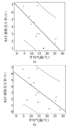
  • 在20 949例受检者中,共发现215例受检者出现BAT摄取,PubMed数据库查询共得到141项关于使用18F-FDG PET/CT评估BAT摄取的相关研究,其中有10项研究符合所有入组标准[3-12],其中包括北美和欧洲8项、土耳其1项、中国台湾地区1项。在包括本研究在内的11项研究中(表 1),研究人数范围为638~20949例,总人数为44 581例,简单线性回归分析结果表明,BAT摄取发生率与室外温度呈显著负相关(r=-0.718,P=0.013)(图 1a),回归方程为P(%)=6.87-0.22×T(℃)。对本研究中发生BAT激活的215例受检者进行季节分组,然后再进行简单线性回归分析(图 1b),其结果与上述结果基本相符(r=-0.663,slope=-0.19,P=0.010)。简单线性回归分析结果表明,室外平均温度每上升5 ℃,BAT摄取发生率下降约1%。

    BAT相关研究 研究地点 BAT摄取发生率(%) 研究时间(年/月) 平均室外温度(℃)
    Cohade等[3] 美国,巴尔的摩 6.85(62/905) 2001.7~2002.6 13.7
    Au-Yong等[4] 英国,诺丁汉 4.62(167/3614) 2006.3~2008.10 11.5
    Ouellet等[5] 加拿大,魁北克 6.77(328/4842) 2007.1~2008.12 4.6
    Hany等[6] 瑞士,苏黎世 2.66(17/638) 2001.1~2001.11 12.7
    Yeung等[7] 美国,纽约 2.32(20/863) 2002.7~2002.8 25.7
    Kim等[8] 美国,纽约 3.02(35/1159) 2000.3~2003.11 13.4
    Cypess等[9] 美国,波士顿 5.38(106/1972) 2003.8~2006.5 8.4
    Esen Akkas等[10] 土耳其,安卡拉 3.00(31/1032) 2008.1~2008.10 14.7
    Huang等[11] 中国,台湾 1.72(30/1740) 2005.6~2009.5 25.4
    Cronin等[12] 美国,波士顿 4.33(298/6867) 2004.1~2008.11 9.6
    本研究 中国,上海 1.03(215/20949) 2010.8~2013.6 17.7
        春季 1.86(92/4959) 7.2
        夏季 0.47(30/6336) 21.3
        秋季 0.30(13/4275) 28.1
        冬季 1.49(80/5379) 14.0
    注:表中,BAT:棕色脂肪细胞。

    Table 1.  Prevalence of activated brown adipose tissue(detected by 18F-FDG uptake) and average outdoor temperature during the study period of enrolled studies

    Figure 1.  Simple linear regression diagram of the association between the prevalence of activated brown adipose tissue and average outdoor temperature

3.   讨论
  • 脂肪组织分为BAT和白色脂肪组织,两者在形态、作用和分子水平上存在显著差异,与白色脂肪组织细胞相比,几乎每个BAT细胞都直接与毛细血管接触并受到交感神经末梢的支配,细胞内线粒体大而丰富、嵴窄而密集,线粒体内膜有大量的嘌呤核苷结合位点及酰基肉毒碱转移酶[13],这是BAT作为成人冷适应和非战栗产热重要来源的结构基础。

    本研究中,18F-FDG PET/CT探测到的BAT摄取发生率非常低(1.03%~6.85%),简单线性回归分析表明,BAT摄取发生率与室外温度之间呈显著负相关,每当室外温度上升5 ℃,BAT摄取发生率会下降约1%,表明室外温度是导致BAT激活的重要因素。对处于本研究温度范围内的18F-FDG PET/CT显像,P(%)=6.87-0.22×T(℃)这个公式可以作为基线参考来估计BAT摄取发生率。

    在一项研究中,把56名健康成人短时冷暴露于19 ℃环境下并赤脚站立于冰块上,然后进行18F-FDG PET/CT显像,发现BAT摄取发生率高达33%[14]。而在另一项针对儿童的研究中则发现当环境温度从21 ℃上升至24 ℃时,BAT摄取发生率下降了约2/3[15]。在本研究回顾性分析中,也发现BAT摄取发生率是与研究所在地环境温度的高低呈负相关的,而且本研究中发现BAT摄取发生率有明显的四季分别。

    虽然冷刺激会激活BAT产热,但其中的机制仍然存在一些疑惑。目前已发现BAT细胞表面有着丰富的β3受体[16],冷刺激能够兴奋交感神经释放大量的去甲肾上腺素并与β3受体结合,通过环腺苷酸-蛋白激酶A途径最终引起产热增加。抑制交感神经兴奋可以降低BAT的温度,表明交感神经兴奋可以激活或者提高BAT的产热代谢[17],但在Cypess等[18]针对10名健康志愿者的研究中,分别给予冷刺激和拟交感神经药物麻黄素后行18F-FDG PET/CT显像,结果表明麻黄素虽然能够引起受试者的血压、心率、能量消耗增高,但不能激活BAT; 相比之下冷刺激也能导致血压增高和能量消耗水平增加,心率反而下降,此时18F-FDG PET/CT能够探测到激活的BAT,因此作者推断冷刺激可通过特殊的交感神经通路激活BAT。

    当然本研究也存在一定的局限性,首先本研究是回顾性研究,其次PET/CT机房平均温度存在一些波动,受试者检查前所处环境温度及逗留时间长短难以精确考量并加以分析。BAT的激活是由多个因素导致的,但本研究只对其中最重要的环境温度因素进行了大样本研究,结果表明BAT摄取发生率与环境温度间存在显著的负相关关系。

Reference (18)

Catalog

/

DownLoad:  Full-Size Img  PowerPoint
Return
Return