Volume 48 Issue 2
Feb.  2024
Article Contents

Citation:

Diagnostic value of ultrasonography, MRI and mammography features of breast cancer with different molecular types

  • Corresponding author: Haiyang Dai, d.ocean@163.com
  • Received Date: 2022-12-26
  • Objective To explore the diagnostic value of ultrasound, MRI, and mammography in different molecular subtypes of breast cancer. Methods A cohort of 212 female patients, aged (47.6±9.3) years old and with confirmed breast cancer, from Huizhou Municipal Central Hospital from January 2018, to April 2022, was retrospectively analyzed. In accordance with the results of pathology and immunohistochemistry, the patients were grouped into four groups: 31 cases of Luminal A, aged (49.2±10.8) years; 104 cases of Luminal B, aged (46.1±8.9) years; 45 cases of human epidermal growth factor receptor-2 (HER-2) overexpression, aged (49.6±10.1) years; and 32 cases of triple-negative breast cancer (TNBC), aged (48.5±8.5) years. The clinical characteristics of the patients and the imaging features of ultrasound, MRI, and mammography were analyzed. Intergroup comparisons of metric data conforming to normal distribution were performed using independent sample t-test (equal variance). Intergroup comparisons of count data were performed using chi-square test or Fisher’s exact probability method. Statistically significant features were evaluated for accuracy of the diagnostic efficacy by using ordered logistic regression analysis and area under the curve (AUC). Results Among the clinical features, the differences in menopausal status and lymph node metastasis among patients with breast cancer of different molecular subtypes were statistically significant (χ2=10.345, 10.026; P=0.016, 0.018). The results of ultrasonography showed that the margins of Luminal A breast cancer tumor were mostly blurred, the margins of Luminal B and HER-2 overexpression breast cancer tumors were mostly angular or hairy, and the margins of TNBC tumor were mostly lobulated. Moreover, the internal echoes in the tumors of the four subtypes were mostly heterogeneous echogenicity, which was more commonly seen in Luminal B. The blood flow Alder grade was mostly grade 0 or Ⅰ within Luminal A and TNBC, whereas the blood flow Alder grade within Luminal B and HER-2 overexpression breast cancer was mostly grade Ⅱ or Ⅲ. The differences were statistically significant (χ2=33.542, 12.283, 10.699; all P<0.05). The results of MRI showed that the tumor enhancement characteristics of Luminal A, Luminal B, and HER-2 overexpression breast cancer were mainly inhomogeneous enhancement, and the proportion of ring enhancement in TNBC tumors was higher than those in the other three, with a statistically significant difference (χ2=30.142, P<0.001). The results of mammography showed that calcification was mostly present in Luminal B and HER-2 overexpression breast cancer tumors, whereas no calcification was found in Luminal A and TNBC tumors. The calcification pattern in Luminal A and Luminal B breast cancer tumors was mainly punctate calcification, whereas that in HER-2 overexpression and TNBC tumors was mainly polypoidal or linear. The differences were all statistically significant (χ2=9.627, 11.792; both P<0.05). The patients' menopausal status and the tumor margins of breast cancer could be used as independent predictors of the molecular subtypes of breast cancer (B=0.729, 0.775; OR=7.053, 23.696; 95%CI: 0.193~1.267, 0.463~1.087; both P<0.05), with an AUC of 0.635 (95%CI: 0.528~0.742). Conclusion Differences were observed in the characteristics of ultrasound, MRI, and mammography in different molecular subtypes of breast cancer, indicating their certain diagnostic value.
  • 加载中
  • [1] Sung H, Ferlay J, Siegel RL, et al. Global cancer statistics 2020: GLOBOCAN estimates of incidence and mortality worldwide for 36 cancers in 185 countries[J]. CA Cancer J Clin, 2021, 71(3): 209−249. DOI: 10.3322/caac.21660.
    [2] Singer CF, Balmaña J, Bürki N, et al. Genetic counselling and testing of susceptibility genes for therapeutic decision-making in breast cancer—an European consensus statement and expert recommendations[J]. Eur J Cancer, 2019, 106: 54−60. DOI: 10.1016/j.ejca.2018.10.007.
    [3] Goldhirsch A, Winer EP, Coates AS, et al. Personalizing the treatment of women with early breast cancer: highlights of the St Gallen International Expert Consensus on the Primary Therapy of Early Breast Cancer 2013[J]. Ann Oncol, 2013, 24(9): 2206−2223. DOI: 10.1093/annonc/mdt303.
    [4] 陈进, 王振平. 多模态MR成像技术在不同病理分型乳腺癌中的诊断价值对比[J]. 临床放射学杂志, 2019, 38(5): 821−824. DOI: 10.13437/j.cnki.jcr.2019.05.016.Chen J, Wang ZP. Comparison of the diagnostic values of multimodal MR imaging in different pathological types of breast cancer[J]. J Clin Radiol, 2019, 38(5): 821−824. DOI: 10.13437/j.cnki.jcr.2019.05.016.
    [5] 王琰娟, 米成嵘, 王文. 超声造影诊断乳腺癌不同分子分型腋窝淋巴结转移的价值[J]. 中国医学影像技术, 2013, 29(7): 1122−1125. DOI: 10.13929/j.1003-3289.2013.07.046.Wang YJ, Mi CR, Wang W. CEUS in diagnosis of axillary lymph node metastasis in different molecular types of breast cancer[J]. Chin J Med Imaging Technol, 2013, 29(7): 1122−1125. DOI: 10.13929/j.1003-3289.2013.07.046.
    [6] 黄秒, 苏丹柯, 金观桥, 等. 乳腺癌X线摄影表现与ER、PR和HER-2表达的相关性研究[J]. 实用放射学杂志, 2020, 36(2): 214−218, 222. DOI: 10.3969/j.issn.1002-1671.2020.02.011.Huang M, Su DK, Jin GQ, et al. Correlation study of mammography findings and expression of ER, PR and HER-2 in breast cancer[J]. J Pract Radiol, 2020, 36(2): 214−218, 222. DOI: 10.3969/j.issn.1002-1671.2020.02.011.
    [7] 方琦, 王旭芬. 非激素依赖性乳腺癌CerbB-2表达与P53、Ki-67的关系研究[J]. 南方医科大学学报, 2011, 31(2): 380−382. DOI: 10.12122/j.issn.1673-4254.2011.02.046.Fang Q, Wang XF. Correlation of CerbB-2 to Ki-67 and P53 expressions in hormone-independent breast cancer[J]. J South Med Univ, 2011, 31(2): 380−382. DOI: 10.12122/j.issn.1673-4254.2011.02.046.
    [8] 杨欢. 乳腺癌超声、MRI、X线征象与分子分型的相关性[J]. 中国医学影像技术, 2017, 33(5): 675−678. DOI: 10.13929/j.1003-3289.201610069.Yang H. Correlation of molecular subtypes of breast cancer with ultrasound, MRI and mammography features[J]. Chin J Med Imaging Technol, 2017, 33(5): 675−678. DOI: 10.13929/j.1003-3289.201610069.
    [9] 康佳, 吴桐, 张蕾, 等. 不同分子分型乳腺癌的多模态超声特征和临床病理对照研究[J]. 中华超声影像学杂志, 2020, 29(4): 330−336. DOI: 10.3760/cma.j.cn131148-20190926-00591.Kang J, Wu T, Zhang L, et al. Comparative study of multimodal ultrasonographic features and clinicopathological appearances of breast cancer with different molecular subtypes[J]. Chin J Ultrasonogr, 2020, 29(4): 330−336. DOI: 10.3760/cma.j.cn131148-20190926-00591.
    [10] 孙斌, 曾林胜, 李江, 等. 乳腺癌边缘超声特征与相应病灶边缘免疫组化结果相关性分析[J]. 现代医院, 2018, 18(11): 1700−1702, 1705. DOI: 10.3969/j.issn.1671-332X.2018.11.041.Sun B, Zeng LS, Li J, et al. Correlation between ultrasound characteristics of breast cancer margins and immunohistochemical results of corresponding lesion margins[J]. Modern hospitals, 2018, 18(11): 1700−1702, 1705. DOI: 10.3969/j.issn.1671-332X. 2018.11.041.
    [11] Wang YB, Ikeda DM, Narasimhan B, et al. Estrogen receptor-negative invasive breast cancer: imaging features of tumors with and without human epidermal growth factor receptor type 2 overexpression[J]. Radiology, 2008, 246(2): 367−375. DOI: 10.1148/radiol.2462070169.
    [12] Yamashita H. Tumor biology in estrogen receptor-positive, human epidermal growth factor receptor type 2-negative breast cancer: mind the menopausal status[J/OL]. World J Clin Oncol, 2015, 6(6): 220−224[2022-12-25]. https://www.wjgnet.com/2218-4333/full/v6/i6/220.htm. DOI: 10.5306/wjco.v6.i6.220.
    [13] Wang DM, Zhu K, Tian JW, et al. Clinicopathological and ultrasonic features of triple-negative breast cancers: a comparison with hormone receptor-positive/human epidermal growth factor receptor-2-negative breast cancers[J]. Ultrasound Med Biol, 2018, 44(5): 1124−1132. DOI: 10.1016/j.ultrasmedbio. 2018.01.013.
    [14] Hasan R, Bhatt D, Khan S, et al. Association of Her-2 expression and clinicopathological parameters in colorectal carcinoma in Indian population[J/OL]. Open Access Maced J Med Sci, 2019, 7(1): 6−11[2022-12-25]. https://oamjms.eu/index.php/mjms/article/view/oamjms.2019.008. DOI: 10.3889/oamjms.2019.008.
    [15] 李宏, 李德华, 胡波. 乳腺癌超声征象与Ki67、p53表达的相关性及临床意义[J]. 中国临床医学影像杂志, 2015, 26(5): 321−324.Li H, Li DH, Hu B. Correlation between ultrasonography features and expression of Ki67 and p53 in breast cancer and its clinical significance[J]. J China Clin Med Imaging, 2015, 26(5): 321−324.
    [16] Yerushalmi R, Woods R, Ravdin PM, et al. Ki67 in breast cancer: prognostic and predictive potential[J]. Lancet Oncol, 2010, 11(2): 174−183. DOI: 10.1016/S1470-2045(09)70262-1.
    [17] 毛锡金, 冯艳, 曹新山, 等. 三阴性乳腺癌临床病理及影像学征象分析[J]. 临床放射学杂志, 2014, 33(4): 503−508. DOI: 10.13437/j.cnki.jcr.2014.04.007.Mao XJ, Feng Y, Cao XS, et al. Clinical analysis of the pathological and imaging features of triple negative breast cancer[J]. J Clin Radiol, 2014, 33(4): 503−508. DOI: 10.13437/j.cnki.jcr.2014.04.007.
    [18] 胡仰玲, 曾辉, 何子龙, 等. 钙化型乳腺癌的分子分型特点及其预后分析[J]. 实用医学杂志, 2020, 36(10): 1354−1359. DOI: 10.3969/j.issn.1006-5725.2020.10.017.Hu YL, Zeng H, He ZL, et al. Analysis of molecular typing and prognosis of breast cancer with calcification[J]. J Pract Med, 2020, 36(10): 1354−1359. DOI: 10.3969/j.issn.1006-5725.2020.10.017.
    [19] Cai SQ, Yao MM, Cai DL, et al. Association between digital breast tomosynthesis and molecular subtypes of breast cancer[J]. Oncol Lett, 2019, 17(3): 2669−2676. DOI: 10.3892/ol.2019.9918.
  • 加载中
通讯作者: 陈斌, bchen63@163.com
  • 1. 

    沈阳化工大学材料科学与工程学院 沈阳 110142

  1. 本站搜索
  2. 百度学术搜索
  3. 万方数据库搜索
  4. CNKI搜索

Figures(2) / Tables(4)

Article Metrics

Article views(8566) PDF downloads(8) Cited by()

Related
Proportional views

Diagnostic value of ultrasonography, MRI and mammography features of breast cancer with different molecular types

    Corresponding author: Haiyang Dai, d.ocean@163.com
  • 1. Department of Ultrasound, Huizhou Municipal Central Hospital, Huizhou 516001, China
  • 2. Department of Radiology, Huizhou Municipal Central Hospital, Huizhou 516001, China
  • 3. Department of Ultrasound , Huizhou First Hospital, Huizhou 516001, China

Abstract:  Objective To explore the diagnostic value of ultrasound, MRI, and mammography in different molecular subtypes of breast cancer. Methods A cohort of 212 female patients, aged (47.6±9.3) years old and with confirmed breast cancer, from Huizhou Municipal Central Hospital from January 2018, to April 2022, was retrospectively analyzed. In accordance with the results of pathology and immunohistochemistry, the patients were grouped into four groups: 31 cases of Luminal A, aged (49.2±10.8) years; 104 cases of Luminal B, aged (46.1±8.9) years; 45 cases of human epidermal growth factor receptor-2 (HER-2) overexpression, aged (49.6±10.1) years; and 32 cases of triple-negative breast cancer (TNBC), aged (48.5±8.5) years. The clinical characteristics of the patients and the imaging features of ultrasound, MRI, and mammography were analyzed. Intergroup comparisons of metric data conforming to normal distribution were performed using independent sample t-test (equal variance). Intergroup comparisons of count data were performed using chi-square test or Fisher’s exact probability method. Statistically significant features were evaluated for accuracy of the diagnostic efficacy by using ordered logistic regression analysis and area under the curve (AUC). Results Among the clinical features, the differences in menopausal status and lymph node metastasis among patients with breast cancer of different molecular subtypes were statistically significant (χ2=10.345, 10.026; P=0.016, 0.018). The results of ultrasonography showed that the margins of Luminal A breast cancer tumor were mostly blurred, the margins of Luminal B and HER-2 overexpression breast cancer tumors were mostly angular or hairy, and the margins of TNBC tumor were mostly lobulated. Moreover, the internal echoes in the tumors of the four subtypes were mostly heterogeneous echogenicity, which was more commonly seen in Luminal B. The blood flow Alder grade was mostly grade 0 or Ⅰ within Luminal A and TNBC, whereas the blood flow Alder grade within Luminal B and HER-2 overexpression breast cancer was mostly grade Ⅱ or Ⅲ. The differences were statistically significant (χ2=33.542, 12.283, 10.699; all P<0.05). The results of MRI showed that the tumor enhancement characteristics of Luminal A, Luminal B, and HER-2 overexpression breast cancer were mainly inhomogeneous enhancement, and the proportion of ring enhancement in TNBC tumors was higher than those in the other three, with a statistically significant difference (χ2=30.142, P<0.001). The results of mammography showed that calcification was mostly present in Luminal B and HER-2 overexpression breast cancer tumors, whereas no calcification was found in Luminal A and TNBC tumors. The calcification pattern in Luminal A and Luminal B breast cancer tumors was mainly punctate calcification, whereas that in HER-2 overexpression and TNBC tumors was mainly polypoidal or linear. The differences were all statistically significant (χ2=9.627, 11.792; both P<0.05). The patients' menopausal status and the tumor margins of breast cancer could be used as independent predictors of the molecular subtypes of breast cancer (B=0.729, 0.775; OR=7.053, 23.696; 95%CI: 0.193~1.267, 0.463~1.087; both P<0.05), with an AUC of 0.635 (95%CI: 0.528~0.742). Conclusion Differences were observed in the characteristics of ultrasound, MRI, and mammography in different molecular subtypes of breast cancer, indicating their certain diagnostic value.

    HTML

  • 乳腺癌已经成为全球女性癌症病死最主要的原因,其发病率居于女性恶性肿瘤首位[1]。乳腺癌是一种高度异质性的恶性肿瘤,在病理特征、临床表现和治疗疗效等方面均有明显的个体差异[2]。根据其免疫组织化学检查结果[如雌激素受体(ER)、孕激素受体(PR)、人表皮生长因子受体2(human epidermal growth factor receptor-2,HER-2)、细胞增殖核抗原(Ki67)的水平],可将乳腺癌分子分型分为Luminal A型、Luminal B型、HER-2过表达型、三阴性乳腺癌(triple negative breast cancer,TNBC)及其他特殊类型[3]。不同分子分型的乳腺癌具有不同的生物学特点,不同的生物学特点决定了其组织病理学改变不同,这也是影像表现的基础;同时乳腺癌不同的生物学特点也影响临床治疗方案的制定及疗效的评估[4]。本研究旨在探讨不同分子分型的乳腺癌在超声、MRI及乳腺X射线摄影检查中影像特征的差异。

  • 1.   资料与方法

      1.1.   一般资料

    • 回顾性分析2018年1月至2022年4月在惠州市中心人民医院就诊的212例女性乳腺癌患者的临床资料和影像资料,年龄(47.6±9.3)岁。根据其组织病理学和免疫组织化学检查结果分为4组:Luminal A型31例,年龄(49.2±10.8)岁;Luminal B型104例,年龄(46.1±8.9)岁;HER-2过表达型45例,年龄(49.6±10.1)岁;TNBC 32例,年龄(48.5±8.5)岁。纳入标准:(1)乳腺癌术后病理分子分型为Luminal A型、Luminal B型、HER-2过表达型及TNBC;(2)乳腺癌术前均行超声、MRI及乳腺X射线摄影等影像检查;(3)临床资料完整。排除标准:(1)乳腺癌术后病理分子分型不明确或为良性病灶;(2)患者术前已接受其他全身性治疗;(3)正值哺乳期或妊娠期的患者。所有患者均于检查前签署了知情同意书。本研究经惠州市中心人民医院伦理委员会批准(批准号:kyll2024008)。

    • 1.2.   显像方法

    • 所有患者在术前一周内完成超声、MRI及乳腺X射线摄影检查。应用荷兰Philips公司的Affiniti 50型和Affiniti 70型彩色多普勒超声诊断仪,探头频率为7.5~12 MHz。患者取仰卧位,上臂外展,以乳头为中心呈放射状扫查,发现病灶时进行十字形交叉纵横扫查,同时扫查双侧腋窝。

      应用德国Siemens公司的Prisma 3.0T型MRI扫描仪,对患者行双乳及双侧腋窝MRI平扫及动态增强扫描。患者取俯卧位,双乳下垂。扫描序列及参数:采用Dixon fast序列进行横断面T2WI,重复时间4360 ms、回波时间81 ms、层厚4 mm、层间距0.8 mm、矩阵384×326;采用resolve序列进行横断面DWI,重复时间5510 ms、回波时间49 ms、层厚4 mm、层间距0.8 mm、矩阵204×101,b值为800 s/mm2。动态增强扫描对比剂采用钆特酸葡胺注射液(购自恒瑞医药),以高压注射器经手背静脉注射,剂量0.1 mmol/kg,流率2.0 ml/s,注射结束后用生理盐水冲管。采用T1高分辨各向同性容积激发序列进行动态增强扫描:重复时间4.21 ms、回波时间1.87 ms、层厚1 mm、层间距0 mm、矩阵352×352;先获取平扫图像,注入对比剂后连续采集5个时相,每个时相扫描117 s。

      应用荷兰Philips公司的MicroDose SI型乳腺X射线机,双侧乳腺均行轴位(CC)及斜位(MLO)摄影,全自动曝光,必要时局部加压放大摄影。摄影条件为电压30~35 kV、电流15~20 mA。

    • 1.3.   图像分析

    • 分别由2位副高及以上职称的诊断医师进行独立阅片,观察并记录病灶的影像特征,意见不一致时经协商达成一致。在超声图像上提取病灶位置(左侧、右侧)、长径(≤20 mm、>20 mm~≤40 mm、>40 mm)、形态(不规则形、规则形)、边缘(光滑、模糊、分叶、毛刺或成角)、纵横比值(≥1、<1)、内部回声(均匀回声、不均匀回声)、后方回声(衰减或增强、无改变)、肿瘤内血流Alder分级(0或Ⅰ级、Ⅱ或Ⅲ级)。在MRI图像上提取乳腺实质背景强化(无或轻度、中度或重度)、强化形式(肿块型、非肿块型或点状)、强化特征(均匀强化、不均匀强化、环形强化)、动态增强曲线(流入型、平台型、流出型)、DWI弥散情况(高信号、等信号)。在乳腺X射线图像上提取乳腺腺体量(致密型、非致密型)、肿瘤内钙化情况(有钙化、无钙化)、钙化形态(点状、多形性或线样)等特征征象。分析并记录不同分子分型乳腺癌的影像特征以及患者的临床特征。

    • 1.4.   统计学方法

    • 应用SPSS 23.0软件对数据进行统计学分析。符合正态分布的计量资料以$\bar x\pm s $表示,组间比较采用两独立样本t检验(方差齐)。计数资料采用频数和百分比表示,组间比较采用χ2检验或Fisher确切概率法。具有统计学意义的特征进一步采用有序Logistic回归分析及AUC对回归模型的诊断效能进行评估。P<0.05为差异有统计学意义。

    2.   结果

      2.1.   不同分子分型乳腺癌患者的临床特征

    • 表1可知,不同分子分型乳腺癌患者的绝经情况、淋巴结转移情况差异均有统计学意义(均P<0.05),其中Luminal A型、Luminal B型及TNBC癌患者确诊时多处于未绝经时期;Luminal B型乳腺癌患者确诊时淋巴结转移阳性较高。

      分型 年龄(岁,$\bar x\pm s $ 绝经情况[例(%)] 淋巴结转移情况[例(%)]
      已绝经 未绝经 阳性 阴性
      Luminal A型(n=31) 49.2±10.8 9(29.0) 22(71.0) 12(38.7) 19(61.3)
      Luminal B型(n=104) 46.1±8.9 29(27.9) 75(72.1) 64(61.5) 40(38.5)
      HER-2过表达型(n=45) 49.6±10.1 24(53.3) 21(46.7) 18(40.0) 27(60.0)
      三阴性乳腺癌(n=32) 48.5±8.5 14(43.8) 18(56.2) 13(40.6) 19(59.4)
      检验值 t=0.113~1.037 χ2=10.345 χ2=10.026
      P 均>0.05 0.016 0.018
       注:HER-2为人表皮生长因子受体2

      Table 1.  Comparison of clinical characteristics of female breast cancer patients with different molecular types (n=212)

    • 2.2.   超声检查结果

    • 表2可知,不同分子分型的乳腺癌边缘、内部回声、肿瘤内血流Alder分级的差异均有统计学意义(均P<0.05)。Luminal A型乳腺癌肿瘤边缘多表现为模糊(图1A);Luminal B型和HER-2过表达型乳腺癌肿瘤边缘多表现为成角或毛刺(图1B1C);TNBC肿瘤边缘多为分叶状(图1D)。Luminal A型、Luminal B型、HER-2过表达型及TNBC肿瘤内部回声多为不均匀回声,其中更多见于Luminal B型。Luminal A型和TNBC肿瘤内血流信号不丰富,Alder分级主要为0或Ⅰ级血流信号(图2A2B);Luminal B型和HER-2过表达型乳腺癌肿瘤内多表现为丰富血流信号,即Alder分级主要为Ⅱ或Ⅲ级(图2C2D)。乳腺癌肿瘤发生位置、长径、形态、纵横比值、后方回声差异均无统计学意义(均P>0.05,表2)。

      分型 长径(mm) 位置[例(%)] 形态[例(%)] 边缘[例(%)]
      ≤20 >20~≤40 >40 左侧 右侧 不规则形 规则形 光滑 模糊 分叶 成角或毛刺
      Luminal A型(n=31) 11(35.5) 11(35.5) 9(29.0) 18(58.0) 13(42.0) 31(100.0) 0(0.0) 5(16.1) 15(48.4) 6(19.4) 5(16.1)
      Luminal B型(n=104) 25(24.0) 58(55.8) 21(20.2) 51(49.0) 53(51.0) 95(91.3) 9(8.7) 8(7.7) 22(21.2) 30(28.8) 44(42.3)
      HER-2过表达型(n=45) 12(26.7) 21(46.7) 12(26.6) 23(51.1) 22(48.9) 44(97.8) 1(2.2) 1(2.2) 3(6.7) 18(40.0) 23(51.1)
      三阴性乳腺癌(n=32) 11(34.4) 18(56.2) 3(9.4) 16(50.0) 16(50.0) 31(96.9) 1(3.1) 1(3.1) 4(12.5) 15(46.9) 12(37.5)
      χ2 8.183 0.792 3.900 33.542
      P 0.225 0.851 0.272 <0.001
      分型 纵横比值[例(%)] 内部回声[例(%)] 后方回声[例(%)] 肿瘤内血流Alder分级[例(%)]
      <1 ≥1 不均匀 均匀 衰减或增强 无改变 0或Ⅰ级 Ⅱ或Ⅲ级
      Luminal A型(n=31) 20(64.5) 11(35.5) 26(83.9) 5(16.1) 18(58.1) 13(41.9) 19(61.3) 12(38.7)
      Luminal B型(n=104) 83(79.8) 21(20.2) 98(94.2) 6(5.8) 44(42.3) 60(57.7) 43(41.3) 61(58.7)
      HER-2过表达型(n=45) 37(82.2) 8(17.8) 37(82.2) 8(17.8) 16(35.6) 29(64.4) 16(35.6) 29(64.4)
      三阴性乳腺癌(n=32) 27(84.4) 5(15.6) 23(71.9) 9(28.1) 13(40.6) 19(59.4) 21(65.6) 11(34.4)
      χ2 4.756 12.283 3.983 10.699
      P 0.191 0.006 0.263 0.013
       注:HER-2为人表皮生长因子受体2

      Table 2.  Comparison of ultrasonographic results of female breast cancer patients with different molecular types (n=212)

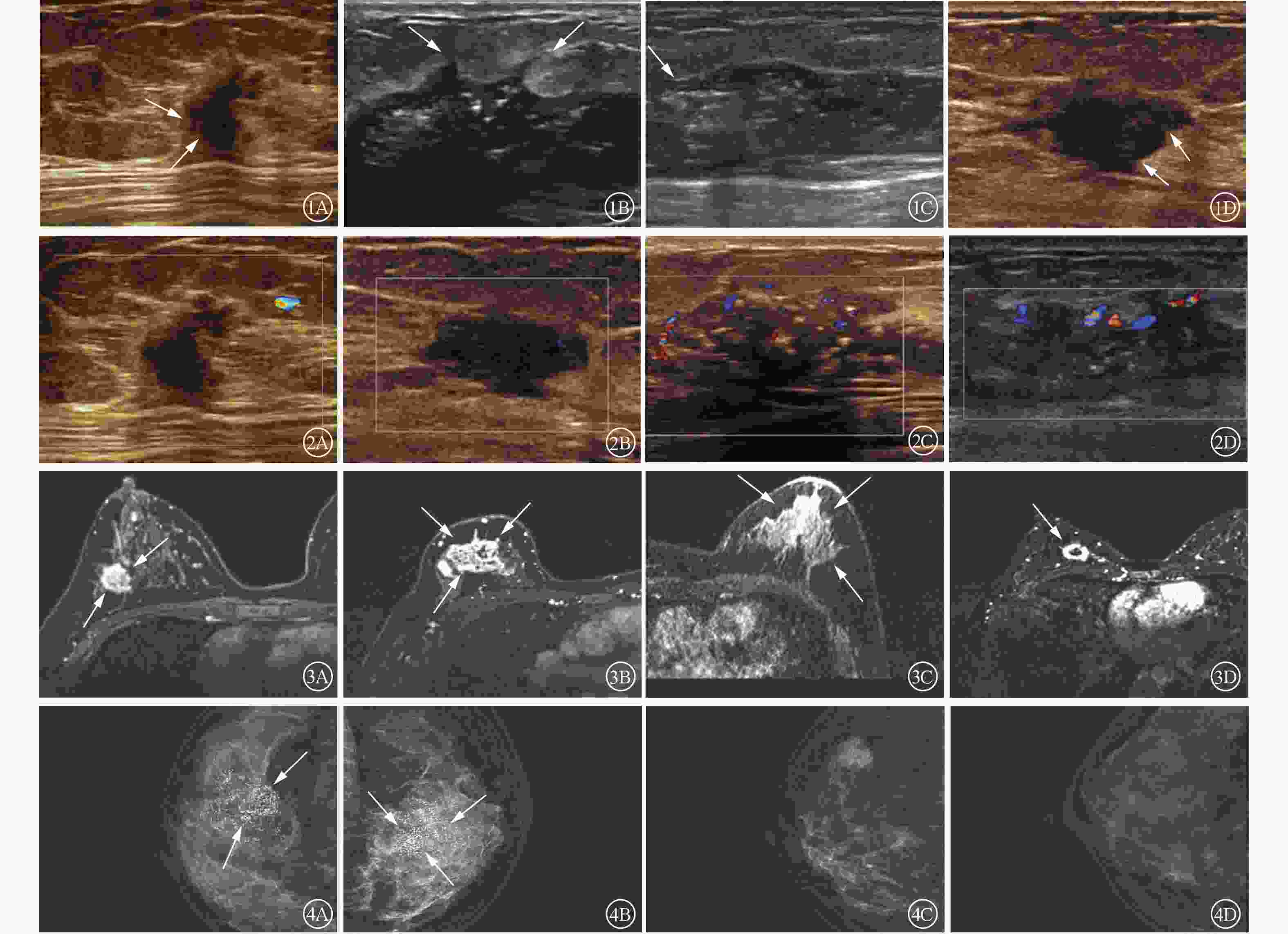
      Figure 1.  2-Dimensional ultrasound imagings of breast cancer with different molecular types Figure 2 Color doppler ultrasound imagings of breast cancer with different molecular types Figure 3 MRI enhanced images of breast cancer with different molecular typesFigure 4 Mammography images of breast cancer with different molecular types

    • 2.3.   MRI检查结果

    • 表3可知,不同分子分型乳腺癌的肿瘤强化特征存在差异,且差异有统计学意义(P<0.001)。其中Luminal A型、Luminal B型及HER-2过表达型乳腺癌肿瘤强化特征以不均匀强化为主(图3A~C),而TNBC肿瘤环形强化的占比高于其他三者(图3D)。MRI乳腺实质背景强化、肿瘤强化形式、动态增强曲线、DWI弥散情况在不同分子分型乳腺癌中的差异均无统计学意义(均P>0.05,表3)。

      分型 乳腺实质
      背景强化
      强化形式 强化特征 动态增
      强曲线
      DWI弥散
      情况
      无或
      轻度
      中度或
      重度
      肿块型 非肿块型
      或点状
      均匀
      强化
      不均匀
      强化
      环形
      强化
      流入型 平台型 流出型 高信号 等信号
      Luminal A型(n=31) 23(74.2) 8(25.8) 27(87.1) 4(12.9) 1(3.2) 27(87.1) 3(9.7) 1(3.2) 8(25.8) 22(71.0) 29(93.5) 2(6.5)
      Luminal B型(n=104) 69(66.3) 35(33.7) 80(76.9) 24(23.1) 19(18.3) 72(69.2) 13(12.5) 11(10.6) 18(17.3) 75(72.1) 99(95.2) 5(4.8)
      HER-2过表达型(n=45) 30(66.7) 15(33.3) 35(77.8) 10(22.2) 13(28.9) 27(60.0) 5(11.1) 0(0.0) 14(31.1) 31(68.9) 45(100.0) 0(0.0)
      三阴性乳腺癌(n=32) 23(71.9) 9(28.1) 28(87.5) 4(12.5) 6(18.8) 12(37.5) 14(43.7) 0(0.0) 8(25.0) 24(75.0) 32(100.0) 0(0.0)
      χ2 0.926 2.871 30.142 12.176 2.709
      P 0.819 0.412 <0.001 0.058 0.439
       注:MRI为磁共振成像;DWI为弥散加权成像;HER-2为人表皮生长因子受体2

      Table 3.  Comparison of MRI results of female breast cancer patients with different molecular types (n=212, cases (%))

    • 2.4.   乳腺X射线摄影检查结果

    • 表4可知,不同分子分型的乳腺癌肿瘤内钙化情况的表现差异有统计学意义(P=0.022),其中Luminal B型及HER-2过表达型乳腺癌肿瘤内多伴有钙化(图4A4B),而Luminal A型及TNBC肿瘤内多无钙化(图4C4D)。Luminal A型与Luminal B型乳腺癌肿瘤内钙化形态以点状钙化为主,HER-2过表达型及TNBC肿瘤内钙化形态以多形性或线样为主,差异有统计学意义(P=0.008);乳腺腺体量在不同分子分型的乳腺癌中差异无统计学意义(P=0.096,表4)。

      分型 腺体量 肿瘤内钙化情况 钙化形态
      致密型 非致密型 有钙化 无钙化 点状 多形性或线样
      Luminal A型(n=31) 25(80.6) 6(19.4) 12(38.7) 19(61.3) 10(83.3) 2(16.7)
      Luminal B型(n=104) 88(84.6) 16(15.4) 63(60.6) 41(39.4) 44(69.8) 19(30.2)
      HER-2过表达型(n=45) 30(66.7) 15(33.3) 31(68.9) 14(31.1) 14(45.2) 17(54.8)
      三阴性乳腺癌(n=32) 26(81.3) 6(18.7) 14(43.8) 18(56.2) 5(35.7) 9(64.3)
      χ2值  6.346 9.627 11.792
      P值  0.096 0.022 0.008
       注:HER-2为人表皮生长因子受体2

      Table 4.  Comparison of mammography results of female breast cancer patients with different molecular types (n=212, cases (%))

    • 2.5.   乳腺癌分子分型多分类有序Logistic回归分析及ROC曲线结果

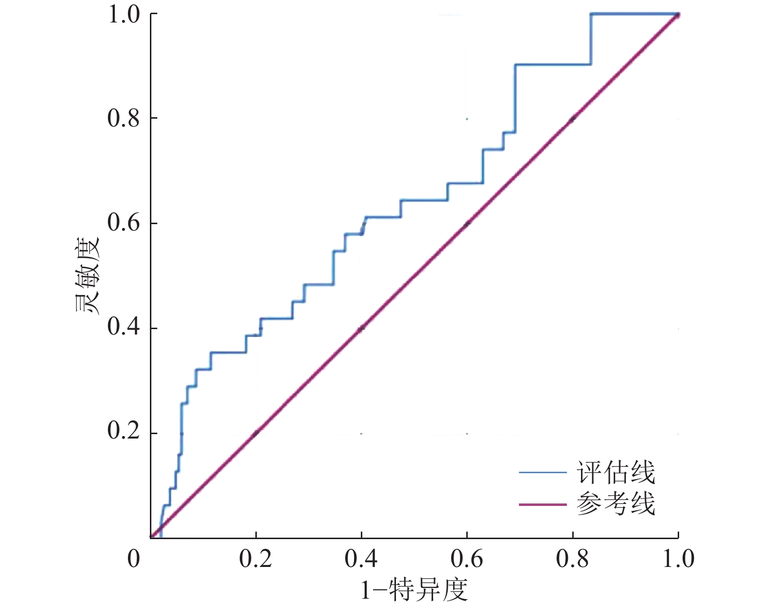
    • 将单因素Logistic回归分析中差异有统计学意义的变量进一步纳入多分类有序Logistic回归分析,结果显示,患者绝经情况与乳腺癌肿瘤边缘可作为乳腺癌分子分型的独立预测因子(B=0.729、0.775,OR=7.053、23.696,95%CI:0.193~1.267、0.463~1.087,P=0.008、<0.001),AUC为0.635(95%CI:0.528~0.742),这表明患者绝经情况与乳腺癌肿瘤边缘情况对乳腺癌分子分型具有一定的诊断效能(图5)。

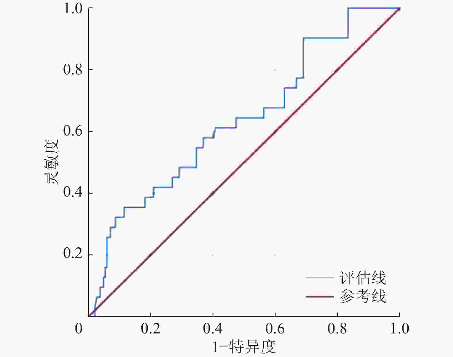
      Figure 5.  Receiver operating characteristic curves of the ordered Logistic regression model for predicting the breast cancer with different molecular typing

    3.   讨论
    • 不同分子分型乳腺癌的治疗方式不同。本研究探讨了超声、MRI和乳腺X射线摄影特征对不同分子分型乳腺癌的诊断价值,以期为乳腺癌的治疗提供一定的临床支持。

      本研究中,患者的发病年龄对乳腺癌分子分型的鉴别无统计学意义,患者的绝经情况对乳腺癌分子分型的鉴别有统计学意义。Luminal B型和TNBC患者发病时尚未绝经的可能性高于Luminal A型和HER-2过表达型乳腺癌,患者绝经情况可作为乳腺癌的风险因子,但对乳腺癌分子分型的影响目前尚无明确定论。本研究结果显示,Luminal B型乳腺癌患者的腋窝淋巴结转移率高于其他3种乳腺癌,但王琰娟等[5]认为TNBC和HER-2过表达型乳腺癌淋巴结转移率高于Luminal A型与Luminal B型乳腺癌,这与本研究结果不一致,这种差异可能与研究对象、研究环境的不同有关。黄秒等[6]的研究结果显示,腋窝淋巴结转移阳性提示HER-2表达阳性可能,这在一定程度上提示Luminal B型、HER-2过表达型乳腺癌的可能。方琦和王旭芬[7]的研究结果显示,HER-2可通过促进酶类分泌,从而提高细胞的运动能力,加快肿瘤侵袭及转移的能力。

      超声检查是目前重要的乳腺癌检查方法之一[8]。康佳等[9]的研究结果显示,不同分子分型的乳腺癌形态多表现为不规则形,这可能与研究的病理类型多数为浸润性乳腺癌有关,肿瘤向周围组织浸润生长,引起周围纤维结缔组织反应。Luminal A型乳腺癌肿瘤边缘多表现为模糊、成角或毛刺征,以Luminal B型和HER-2过表达型乳腺癌多见。孙斌等[10]的研究结果显示,雌激素受体(ER)表达与病灶边缘模糊及病灶边缘成角呈正相关。而Wang等[11]认为HER-2基因的表达与边缘毛刺征具有正相关性。TNBC的生长模式倾向于快速增殖,肿瘤细胞常呈推挤式增生,因此边缘易形成分叶状[12]。Luminal B型和HER-2过表达型乳腺癌肿瘤内血流信号较丰富,Alder分级主要为Ⅱ或Ⅲ级。Wang等[13]的研究结果显示,HER-2基因与血管内皮生长因子的表达相关,HER-2基因使血管内皮细胞增殖,促进血管生成,新生血管数目多,缺乏肌层,故血流信号丰富,进而使肿瘤生长、侵袭、转移。Hasan等[14]的研究结果显示,Luminal B型乳腺癌血流信号丰富可能与Ki-67高表达或HER-2表达阳性有关。Ki-67是一种增殖细胞抗原,与细胞有丝分裂密切相关,是乳腺癌预后的重要标志物,其高表达促进细胞活跃增殖,故而需要丰富的血流,使肿瘤快速生长[15]

      MRI检查具有较高的软组织分辨力,可以提供病灶形态学特征、血流动力学及血管生成等多参数信息,是检测乳腺癌的灵敏方法[16]。我们发现Luminal A型、Luminal B型和HER-2过表达型乳腺癌多表现为不均匀强化,TNBC在MRI强化特征中多表现为环形强化。MRI强化程度主要由肿瘤组织的血管密度及对比剂进入肿瘤组织的数量决定。毛锡金等[17]的研究结果显示,TNBC边缘区域微血管密度高,肿瘤生长较快,中心区域血流较少,中心易坏死。而HER-2和Ki-67的高表达促进大量的新生血管生长,进而使肿瘤生长加快,血流分布不规则,使Luminal B型和HER-2过表达型乳腺癌肿瘤的强化特征表现为不均匀强化。

      乳腺X射线摄影检查作为诊断乳腺癌常用的检查方法之一,可提供乳腺癌形态、密度、边界、钙化等重要信息。HER-2过表达型和Luminal B型乳腺癌的钙化率高于Luminal A型和TNBC,HER-2过表达型乳腺癌肿瘤钙化形态多以细小多形性及线样为主,Luminal A型多为点状钙化,这与胡仰玲等[18]的研究结果一致。肿瘤钙化与肿瘤坏死产物、肿瘤自主分泌的代谢产物或钙盐沉积有关,Cai等[19]的研究结果显示,HER-2基因具有促进肿瘤钙化的作用。雌激素受体(ER)与HER-2基因可能参与钙离子的转运过程,但目前尚无明确定论[6]

      综上所述,不同分子分型乳腺癌的临床特征及治疗方案存在较大差异,乳腺癌肿瘤边缘和患者绝经情况可作为乳腺癌分子分型的独立预测因子。超声、MRI及乳腺X射线摄影检查作为筛查乳腺癌无创且易行的影像检查方法各有优势,不同分子分型的乳腺癌在不同影像特征上具有一定的差异性。不同检查技术的联合将有助于提高乳腺癌分子分型诊断的准确性。

      利益冲突 所有作者声明无利益冲突

      作者贡献声明 刘晓瑜负责研究命题的设计与提出、数据的校对、论文的撰写与修改、研究过程的实施、数据的收集与统计分析;林翠君负责研究过程的实施、图像的校对与审阅;黄洁负责研究过程的实施、数据的收集与统计分析;代海洋负责研究命题的设计与提出、数据的校对、论文的审阅与修订

Reference (19)

Catalog

/

DownLoad:  Full-Size Img  PowerPoint
Return
Return