Volume 48 Issue 2
Feb.  2024
Article Contents

Citation:

Diagnosis consistency and influencing factors of DWI and MRA in patients with acute cerebral infarction

  • Corresponding author: Sangang Wang, 67410196@qq.com
  • Received Date: 2022-12-21
  • Objective To explore the consistency and influencing factors between diffusion-weighted imaging (DWI) and magnetic resonance angiography (MRA) in diagnosing acute cerebral infarct (ACI). Methods Ninety-eight suspected ACI patients who received treatment at Lai'an Jianing Hospital from January 2020 to February 2022 were selected as the study subjects and included in the training set. Among them, 58 were males and 40 were females, aged 45–80 (60.5±3.3) years old. Suspected ACI patients (33 cases) diagnosed and treated at Lai'an Jianing Hospital from March to October 2022 were selected for retrospective analysis according to the same criteria and included in the validation set. Among them were 18 males and 15 females, aged 42–79 (61.1±3.6) years old. Using clinical comprehensive diagnosis as the "gold standard", we analyzed the diagnostic efficacy, imaging manifestations, and consistency of examination results of DWI and MRA. Two independent sample t-tests were used for intergroup comparison of econometric data. The intergroup comparison of counting data was conducted using χ2 test. Kappa test with multiple classification data was performed to analyze the consistency between DWI and MRA in diagnosing ACI. Multiple Logistic regression analysis was conducted to screen for independent risk factors with inconsistent results between DWI and MRA examinations. Empower Stats and statistical software package "R" were used to draw a forest map, construct a risk column-chart prediction model, and evaluate the model. The discriminability and calibration of the risk-prediction model were determined using the receiver operating characteristic (ROC) curve and the Hosmer–Lemeshow goodness-of-fit test. Risk nomogram prediction model accuracy was evaluated using decision curve analysis. Results Among the 74 patients diagnosed with ACI clinically, 73 (98.65%) were positive for DWI and 71 (95.95%) were positive for MRA. The difference in apparent diffusion coefficient (ADC) values between the healthy (≤6 h: (1.06±0.24)×10–4 cm2/s; 6–24 h: (1.13±0.26)×10–4 cm2/s; 24–72 h: (1.05±0.17)×10–4 cm2/s) and affected (≤6 h: (0.59±0.11)×10–4 cm2/s; 6–24 h: (0.44±0.10)×10–4 cm2/s; 24–72 h: (0.53±0.09)×10–4 cm2/s) brain tissues of patients were statistically significant (t=10.227, 12.630, 7.646; all P<0.05). Within 24 h after the onset of the disease, the ADC value and rADC (≤6 h: (0.53±0.08); 6–24 h: (0.43±0.05)) in the affected side of the brain initially decreased significantly (t=5.410, 5.569; both P<0.05) and then increased significantly (24–72 h ADC: (0.53±0.09)×10–4 cm2/s, 24–72 h rADC: (0.49±0.06)) (t=2.274, 2.835; both P<0.05). A total of 68 patients had consistent results between DWI and MRA (Group A), whereas 30 had inconsistent ones (Group B). The consistency between DWI and MRA was good (Kappa=0.654, P<0.05). Results of multivariate Logistic regression analysis showed that onset time ≤24 h, posterior circulation, length of infarct lesion <2 cm were independent risk factors for inconsistent results between the DWI and MRA diagnosis of ACI patients (OR=1.119, 1.169, 1.567; all P<0.05). Evaluation results of the risk nomogram prediction model showed that its discrimination, accuracy, and effectiveness were all high, and the area under curve of the training and validation sets were 0.930 (95%CI: 0.899–0.961, P<0.001) and 0.855 (95% CI: 0.812–0.898, P<0.001). Conclusions DWI can clearly display the location and degree of ischemia of the lesion, whereas MRA can accurately locate the infarcted blood vessels and their stenosis. The consistency between the two examinations is good, and both can help diagnose and evaluate ACI. The onset time, posterior circulation, and length of infarct lesion are risk factors that affect the consistency of diagnosis between the two.
  • 加载中
  • [1] Hijazi Z, Yassi N, O'Brien JT, et al. The influence of cerebrovascular disease in dementia with Lewy bodies and Parkinson's disease dementia[J]. Eur J Neurol, 2021, 29(4): 1254−1265. DOI: 10.1111/ene.15211.
    [2] Xu XL, Hua Y, Wang LL, et al. Correlation between risk factors of cerebrovascular disease and calcified plaque characteristics in patients with atherosclerotic severe carotid stenosis[J]. Neurol Res, 2020, 42(1): 83−89. DOI: 10.1080/01616412.2019.1710403.
    [3] 张家莉. 脑血管意外患者精神焦虑抑郁的治疗[J]. 家庭生活指南, 2019, (9): 64.Zhang JL. Treatment of mental anxiety and depression in patients with cerebrovascular accident[J]. Family Life Guide, 2019, (9): 64.
    [4] Ablefoni M, Ullrich S, Surov A, et al. Diagnostic benefit of high b-value computed diffusion-weighted imaging in acute brainstem infarction[J]. J Neuroradiol, 2022, 49(1): 47−52. DOI: 10.1016/j.neurad.2020.09.004.
    [5] Wang ML, Yu MM, Li WB, et al. Application of neutrophil to lymphocyte ratio to identify CT-negative cerebral infarction with nonfocal symptoms[J]. Ann Transl Med, 2020, 8(21): 1359. DOI: 10.21037/atm-20-1640.
    [6] Lee SH, Jung JM, Kim KY, et al. Intramural hematoma shape and acute cerebral infarction in intracranial artery dissection: a high-resolution magnetic resonance imaging study[J]. Cerebrovasc Dis, 2020, 49(3): 269−276. DOI: 10.1159/00050 8027.
    [7] 傅小燕, 王小丽. 血清VCAM-1、PECAM-1水平与急性脑梗死患者并发卒中后认知功能障碍的相关性研究[J]. 中国实验诊断学, 2022, 26(6): 834−838. DOI: 10.3969/j.issn.1007-4287.2022.06.012.Fu XY, Wang XL. Correlation between serum levels of VCAM-1, PECAM-1 and post-stroke cognitive impairment in patients with acute cerebral infarction[J]. Chin J Lab Diagn, 2022, 26(6): 834−838. DOI: 10.3969/j.issn.1007-4287.2022.06.012.
    [8] 中华医学会神经病学分会, 中华医学会神经病学分会脑血管病学组. 中国急性缺血性脑卒中诊治指南2014[J]. 中华神经科杂志, 2015, 48(4): 246−257. DOI: 10.3760/cma.j.issn.1006-7876.2015.04.002.Neurology Branch of the Chinese Medical Association, Cerebrovascular Disease Group, Neurology Branch, Chinese Medical Association. Guidelines for the diagnosis and treatment of acute ischemic stroke in China 2014[J]. Chin J Neurol, 2015, 48(4): 246−257. DOI: 10.3760/cma.j.issn.1006-7876.2015.04.002.
    [9] Xu M, He XY, Huang P. The relationship between the mean platelet volume and carotid atherosclerosis and prognosis in patients with acute cerebral infarction[J]. Biomed Res Int, 2020, 2020: 6685740. DOI: 10.1155/2020/6685740.
    [10] Liu YP, Ju ML, Yu FQ. Clinical significance of FSTL 1, Bax, Bcl-2 in acute cerebral infarction and its relationship with hemorrhagic transformation[J]. Eur Rev Med Pharmacol Sci, 2020, 24(16): 8447−8457. DOI: 10.26355/eurrev_202008_22642.
    [11] 杨桂林, 张效杰, 余光权, 等. 磁共振弥散加权成像联合磁共振血管成像在急性脑梗死患者诊断中应用研究[J]. 现代医用影像学, 2022, 31(3): 479−481. DOI: 10.3969/j.issn.1006-7035.2022.3.xdyyyxx202203022.Yang GL, Zhang XJ, Yu GQ, et al. Application of diffusion weighted magnetic resonance imaging combined with magnetic resonance angiography in the diagnosis of acute cerebral infarction patients[J]. Mod Med Imagel, 2022, 31(3): 479−481. DOI: 10.3969/j.issn.1006-7035.2022.3.xdyyyxx202203022.
    [12] Razek AAKA, Gadelhak BN, El Zahabey IA, et al. Diffusion-weighted imaging with histogram analysis of the apparent diffusion coefficient maps in the diagnosis of parotid tumours[J]. Int J Oral Maxillofac Surg, 2022, 51(2): 166−174. DOI: 10.1016/j.ijom.2021.03.019.
    [13] Zhu N, Shu H, Jiang WB, et al. Mean platelet volume and mean platelet volume/platelet count ratio in nonvalvular atrial fibrillation stroke and large artery atherosclerosis stroke[J]. Medicine, 2020, 99(28): e21044. DOI: 10.1097/MD.00000000 00021044.
    [14] 刘巍. 急性期脑梗死中DWI和MRA的临床应用价值[J]. 影像研究与医学应用, 2019, 3(1): 133−134. DOI: 10.3969/j.issn.2096-3807.2019.01.086.Liu W. The clinical application value of DWI and MRA in acute cerebral infarction[J]. J Imaging Res Med Appl, 2019, 3(1): 133−134. DOI: 10.3969/j.issn.2096-3807.2019.01.086.
    [15] 王继蕊, 孙丽红, 张元超, 等. 磁共振DWI早期呈阴性的急性缺血性脑卒中的临床特点及原因分析[J]. 医学临床研究, 2017, 34(2): 264−267, 271. DOI: 10.3969/j.issn.1671-7171.2017. 02.019.Wang JR, Sun LH, Zhang YC, et al. Clinical characteristics analysis of acute ischemic stroke with early stage negative magnetic resonance diffusion-weighted imaging[J]. J Clin Res, 2017, 34(2): 264−267, 271. DOI: 10.3969/j.issn.1671-7171.2017. 02.019.
    [16] 白向东, 马艳, 魏欣, 等. 早期急性脑梗死患者颅脑核磁DWI/FLAIR不匹配的临床特点[J]. 宁夏医学杂志, 2020, 42(8): 703−705. DOI: 10.13621/j.1001-5949.2020.08.0703.Bai XD, Ma Y, Wei X, et al. Clinical research of DWI/FLAIR mismatch in patients with early acute cerebral infarction[J]. Ningxia Med J, 2020, 42(8): 703−705. DOI: 10.13621/j.1001-5949.2020.08.0703.
  • 加载中
通讯作者: 陈斌, bchen63@163.com
  • 1. 

    沈阳化工大学材料科学与工程学院 沈阳 110142

  1. 本站搜索
  2. 百度学术搜索
  3. 万方数据库搜索
  4. CNKI搜索

Figures(6) / Tables(5)

Article Metrics

Article views(11075) PDF downloads(8) Cited by()

Related
Proportional views

Diagnosis consistency and influencing factors of DWI and MRA in patients with acute cerebral infarction

    Corresponding author: Sangang Wang, 67410196@qq.com
  • 1. Department of General Medicine, Lai'an Jianing Hospital, Chuzhou 239200, China
  • 2. Department of General Medicine, Chuzhou First People's Hospital, Chuzhou 239200, China

Abstract:  Objective To explore the consistency and influencing factors between diffusion-weighted imaging (DWI) and magnetic resonance angiography (MRA) in diagnosing acute cerebral infarct (ACI). Methods Ninety-eight suspected ACI patients who received treatment at Lai'an Jianing Hospital from January 2020 to February 2022 were selected as the study subjects and included in the training set. Among them, 58 were males and 40 were females, aged 45–80 (60.5±3.3) years old. Suspected ACI patients (33 cases) diagnosed and treated at Lai'an Jianing Hospital from March to October 2022 were selected for retrospective analysis according to the same criteria and included in the validation set. Among them were 18 males and 15 females, aged 42–79 (61.1±3.6) years old. Using clinical comprehensive diagnosis as the "gold standard", we analyzed the diagnostic efficacy, imaging manifestations, and consistency of examination results of DWI and MRA. Two independent sample t-tests were used for intergroup comparison of econometric data. The intergroup comparison of counting data was conducted using χ2 test. Kappa test with multiple classification data was performed to analyze the consistency between DWI and MRA in diagnosing ACI. Multiple Logistic regression analysis was conducted to screen for independent risk factors with inconsistent results between DWI and MRA examinations. Empower Stats and statistical software package "R" were used to draw a forest map, construct a risk column-chart prediction model, and evaluate the model. The discriminability and calibration of the risk-prediction model were determined using the receiver operating characteristic (ROC) curve and the Hosmer–Lemeshow goodness-of-fit test. Risk nomogram prediction model accuracy was evaluated using decision curve analysis. Results Among the 74 patients diagnosed with ACI clinically, 73 (98.65%) were positive for DWI and 71 (95.95%) were positive for MRA. The difference in apparent diffusion coefficient (ADC) values between the healthy (≤6 h: (1.06±0.24)×10–4 cm2/s; 6–24 h: (1.13±0.26)×10–4 cm2/s; 24–72 h: (1.05±0.17)×10–4 cm2/s) and affected (≤6 h: (0.59±0.11)×10–4 cm2/s; 6–24 h: (0.44±0.10)×10–4 cm2/s; 24–72 h: (0.53±0.09)×10–4 cm2/s) brain tissues of patients were statistically significant (t=10.227, 12.630, 7.646; all P<0.05). Within 24 h after the onset of the disease, the ADC value and rADC (≤6 h: (0.53±0.08); 6–24 h: (0.43±0.05)) in the affected side of the brain initially decreased significantly (t=5.410, 5.569; both P<0.05) and then increased significantly (24–72 h ADC: (0.53±0.09)×10–4 cm2/s, 24–72 h rADC: (0.49±0.06)) (t=2.274, 2.835; both P<0.05). A total of 68 patients had consistent results between DWI and MRA (Group A), whereas 30 had inconsistent ones (Group B). The consistency between DWI and MRA was good (Kappa=0.654, P<0.05). Results of multivariate Logistic regression analysis showed that onset time ≤24 h, posterior circulation, length of infarct lesion <2 cm were independent risk factors for inconsistent results between the DWI and MRA diagnosis of ACI patients (OR=1.119, 1.169, 1.567; all P<0.05). Evaluation results of the risk nomogram prediction model showed that its discrimination, accuracy, and effectiveness were all high, and the area under curve of the training and validation sets were 0.930 (95%CI: 0.899–0.961, P<0.001) and 0.855 (95% CI: 0.812–0.898, P<0.001). Conclusions DWI can clearly display the location and degree of ischemia of the lesion, whereas MRA can accurately locate the infarcted blood vessels and their stenosis. The consistency between the two examinations is good, and both can help diagnose and evaluate ACI. The onset time, posterior circulation, and length of infarct lesion are risk factors that affect the consistency of diagnosis between the two.

    HTML

  • 脑血管疾病是指脑部血管的各种疾病,包括脑动脉粥样硬化、脑梗死等[1-2],其可引起脑组织缺血或出血性意外的发生,导致患者认知障碍、残疾甚至死亡。目前,神经系统性疾病住院患者中脑血管疾病的发病率为25%~50%[3],且仍有继续上升的趋势,其中缺血性脑血管病占所有脑血管疾病的70%~80%,动脉粥样硬化是脑血管疾病发病的最主要病因。快速发现、准确定位病灶并确定责任血管对于脑血管病的诊断和治疗十分重要。

    目前,磁共振DWI被认为是诊断急性期脑梗死(acute cerebral infarct,ACI)灵敏度最高的影像检查方法,其可反映脑内病灶局部区域水分子的扩散状态,并可通过表观扩散系数(apparent diffusion coefficient,ADC)直观地反映水分子的状态,ADC值越大,组织内的自由水分子越多。与常规MRI相比,DWI对新旧脑梗死的区分以及对腔隙脑梗死的诊断具有明显优势[4]。Wang等[5]的研究结果表明,DWI可以显示脑梗死病灶的部位、分布及其形态。磁共振血管成像(magnetic resonance angiography,MRA)具有操作简单、扫描时间短、灵敏度高、无创、无辐射、无需注射对比剂的优点[6],在临床上的应用较为广泛,其图像可辅助医师明确患者责任血管的位置和范围。DWI和MRA对于ACI患者病灶部位的诊断有时会存在差异,目前尚缺乏关于DWI与MRA对ACI患者诊断一致性的研究。本文对ACI患者的DWI和MRA影像资料进行综合分析,探究DWI与MRA对ACI患者的诊断一致性及其影响因素,从而为ACI患者的诊断及预后提供参考依据。

  • 1.   资料与方法

      1.1.   一般资料

    • 回顾性分析2020年1月至2022年2月于来安家宁医院接受诊治的98例疑似ACI患者的临床资料并纳入训练集,其中男性58例、女性40例,年龄45~80(60.5±3.3)岁,身体质量指数(body mass index,BMI)为(23.30±1.46) kg/m2。按照相同标准选取2022年3至10月于来安家宁医院接受诊治的33例疑似ACI患者纳入验证集,其中男性18例、女性15例,年龄42~79(61.1±3.6)岁,BMI为(23.21±1.23) kg/m2。纳入标准:(1)符合ACI的诊断标准[7],经MRI及CT检查确诊为ACI;(2)临床资料完整。排除标准:(1)合并严重心脏、肝脏、肾脏等器官功能障碍;(2)合并恶性肿瘤;(3)患有其他非血管性脑病;(4)有精神类疾病病史。所有患者均行DWI和MRA检查并在检查前签署了知情同意书。本研究经来安家宁医院医学伦理委员会的批准(批准号:20210817)。

    • 1.2.   方法

      1.2.1.   一般资料的收集
    • 收集所有患者的临床资料,包括年龄、性别、BMI、发病时间、吸烟史、饮酒史、高血压病史、糖尿病病史、高血脂病病史、临床表现、临床综合诊断[8]、美国国立卫生研究院卒中量表(national institute of health stroke scale, NIHSS)评分。

    • 1.2.2.   影像检查方法
    • 使用美国GE公司1.5T HDxt MRI,8通道头部线圈对所有患者进行检查。DWI:横断面平扫自旋回波(SE)序列为T1WI、液体衰减反转回复(fluid attenuated inversion recovery,FLAIR)序列和T2WI,b值分别为1 000 s/mm和0 s/mm。扫描参数:回波时间104 ms、重复时间 2 701 ms,层数18,矩阵152×113,层间距0 mm,激励1次,层厚5.5 mm,扫描时间34 s,视野射野23 cm。计算ADC和相对表观扩散系数(relative apparent diffusion coefficient,rADC),rADC=患侧ADC/健侧ADC。

      MRA:以三维立体飞行时间法进行扫描。扫描参数:回波时间 6.9 ms、重复时间 25 ms,层数140,矩阵396×226,层间距10 mm,激励4次,层厚20 mm,扫描时间285 s,视野射野20 cm。

      DWI和MRA的扫描范围均包括大脑前动脉,大脑中动脉,大脑后动脉主干、分支和Willis动脉环,观察病灶的位置、大小、狭窄程度(0级为闭塞,远端血管无显影;1级为缺失,远端血管部分显影;2级为狭窄>50%,远端血管完全显影;3级为狭窄≤50%,远端血管完全显影)等。

    • 1.3.   统计学方法

    • 应用SPSS 23.0软件对数据进行统计学分析。符合正态分布的计量资料以$\bar x \pm s $表示,计数资料以频数(%)表示。符合正态分布的计量资料的组间比较采用两独立样本t检验(方差齐);计数资料的组间比较采用χ2检验;以临床综合诊断为“金标准”,采用多分类资料的Kappa检验分析DWI与MRA诊断ACI的一致性,Kappa值<0.4表示一致性较差,0.4<Kappa值<0.6表示一致性中等,0.6<Kappa值<0.8表示一致性较高;采用多因素Logistic回归分析筛选DWI与MRA检查结果不一致的独立危险因素;采用Empower Stats和统计软件包“R”绘制森林图,构建风险列线图预测模型,并对模型进行评价;采用ROC曲线和霍斯默莱梅肖拟合优度(Hosmer-Lemeshow goodness-of-fit)检验风险列线图预测模型的区分度和精准度;采用决策曲线(decision curve analysis,DCA)评价风险列线图预测模型的精准度。检验水准α=0.05。P<0.05为差异有统计学意义。

    2.   结果

      2.1.   诊断效能

    • 98例疑似ACI的患者中,74例经临床综合诊断为ACI患者,其中DWI检出73例(假阴性7例,假阳性6例)(98.65%);MRA检出71例(假阴性9例,假阳性6例)(95.95%)。DWI与MRA诊断ACI的灵敏度、特异度、准确率、阳性预测值和阴性预测值的差异均无统计学意义(均P>0.05,表1)。

      影像检查方法 灵敏度 特异度 准确率 阳性预测值 阴性预测值
      弥散加权成像  90.54(67/74) 75.00(18/24) 86.73(85/98) 91.78(67/73) 72.00(18/25)
      磁共振血管成像 87.84(65/74) 75.00(18/24) 84.69(83/98) 91.55(65/71) 66.67(18/27)
      χ2 0.280 0.000 0.167 0.003 0.173
      P 0.597 1.000 0.683 0.960 0.677

      Table 1.  Comparison of diagnostic efficacy between diffusion weighted magnetic resonance imaging and magnetic resonance angiography in 98 suspected acute cerebral infarction patients (%)

    • 2.2.   DWI与MRA的影像表现

    • 74例确诊ACI的患者中,DWI检查结果为阳性的67例(90.54%),患者病灶表现为高信号,病灶定位与临床组织病理学检查结果一致。其中发病时间≤6 h的患者33例,DWI检出33例(100.00%),病灶表现为异常高信号;发病时间为6~24 h的患者30例,DWI检出26例(86.67%);发病时间为24~72 h的患者11例,DWI检出8例(72.73%)。67例ACI患者健侧与患侧脑组织ADC值的差异均有统计学意义(均P<0.05);与发病≤6 h相比,发病6~24 h的ACI患者患侧ADC值和rADC均明显降低,且差异均有统计学意义(均P<0.05);发病24~72 h的ACI患者患侧ADC值、rADC较发病6~24 h明显升高,且差异均有统计学意义(均P<0.05,表2)。74例ACI患者中,MRA检查结果为阳性的65例(87.84%)患者病变脑血管的分布情况及其狭窄程度见表3,病变血管多为大血管狭窄,其中以大脑中动脉狭窄最多,其次为大脑前动脉、大脑后动脉和颈内动脉、椎-基动脉和左椎动脉、右椎动脉。典型病例的DWI与MRA图像见图1

      发病时间(h) ADC(×10−4 cm2/s) rADC
      患侧 健侧
      ≤6(n=33) 0.59±0.11a 1.06±0.24 0.53±0.08
      6~24(n=26) 0.44±0.10a,b 1.13±0.26 0.43±0.05b
      24~72(n=8) 0.53±0.09a,c 1.05±0.17 0.49±0.06c
      注:a表示与健侧相比,差异均有统计学意义(t=10.227、12.630、7.646,均P<0.05);b表示与≤6 h相比,差异均有统计学意义(t=5.410、5.569,均P<0.05);c表示与6~24 h相比,差异均有统计学意义(t=2.274、2.835,均P<0.05)。ADC为表观扩散系数;rADC为相对表观扩散系数

      Table 2.  Comparison of apparent diffusion coefficient and relative apparent diffusion coefficient values in diffusion weighted magnetic resonance imaging of 67 patients with acute cerebral infarction at different onset times

      病变血管 狭窄程度 合计
      0级 1级 2级 3级
      颈内动脉 0 2 3 4 9
      大脑前动脉 1 1 2 6 10
      大脑中动脉 1 1 3 6 11
      大脑后动脉 0 1 3 5 9
      椎-基动脉 0 2 4 3 9
      左椎动脉 1 1 2 5 9
      右椎动脉 1 1 2 4 8
      合计 4 9 19 33 65

      Table 3.  Distribution and stenosis degree of cerebrovascular lesions on magnetic resonance angiography in 65 patients with acute cerebral infarction (case)

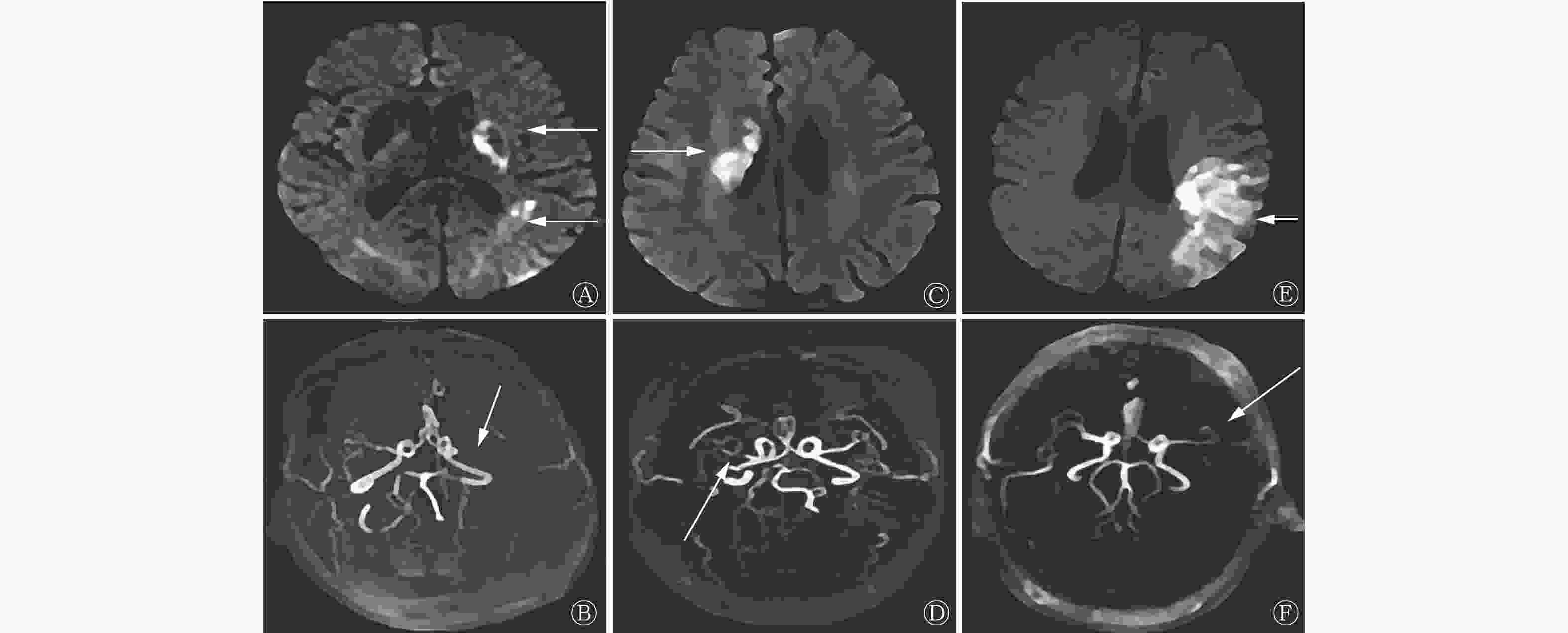
      Figure 1.  Diffusion weighted imaging and magnetic resonance angiography images of 3 patients with acute cerebral infarction

    • 2.3.   DWI与MRA诊断结果的一致性

    • 98例疑似ACI的患者中,DWI显示的脑梗死病灶位置与MRA病变血管一致的患者68例(69.39%),不一致的患者30例(30.61%),DWI与MRA检查结果的一致性良好(Kappa值=0.654,P<0.05)。DWI检查结果为阳性的73例患者中有16例MRA检查结果为阴性,DWI检查结果为阴性的25例患者中有14例MRA检查结果为阳性。DWI与MRA的检查结果均为阳性的患者的例数、病变血管和脑梗死病灶位置的分布情况:MRA表现为颈内动脉狭窄的患者6例,DWI显示为丘脑梗死灶;MRA表现为大脑前动脉主干狭窄的患者9例,DWI显示为额叶梗死灶;MRA表现为大脑中动脉主干狭窄的患者10例,其中DWI显示为颞叶梗死灶3例、基底节区梗死灶4例、半卵圆中心梗死灶3例;MRA表现为大脑后动脉主干狭窄的患者7例,DWI显示为枕叶梗死灶;MRA表现为椎-基动脉狭窄的患者8例,其中DWI显示为小脑梗死灶4例、脑干梗死灶4例;MRA表现为左椎动脉狭窄的患者9例,DWI显示为枕叶梗死灶;MRA表现为右椎动脉狭窄的患者8例,DWI显示为枕叶梗死灶。

    • 2.4.   两组患者临床资料的比较

    • DWI与MRA检查结果一致的患者为A组(68例),不一致的患者为B组(30例),比较两组患者的临床资料。与A组相比,B组中发病时间≤24 h、多发病灶、后循环、梗死病灶长径<2 cm患者的比例明显增加,NIHSS评分明显降低,且差异均有统计学意义(均P<0.05)。两组患者年龄、性别、BMI、吸烟史、饮酒史、糖尿病病史、高血压病史、高血脂病史以及临床表现的差异均无统计学意义(均P>0.05,表4)。

      组别 年龄(岁) 性别[例(%)] BMI(kg/m2 发病时间[例(%)] 吸烟史
      [例(%)]
      饮酒史
      [例(%)]
      ≤24 h >24 h
      A组(n=68) 60.11±3.17 38(55.88) 30(44.12) 23.14±1.05 45(66.18) 23(33.82) 22(32.35) 22(32.35)
      B组(n=30) 60.97±3.39 19(63.33) 11(36.67) 23.55±1.67 27(90.00) 3(10.00) 10(33.33) 9(30.00)
      检验值 t=1.212 χ2=0.475 t=1.473 χ2=6.061 χ2=0.009 χ2=0.053
      P 0.229 0.491 0.144 0.014 0.924 0.817
      组别 高血压病史
      [例(%)]
      糖尿病病史
      [例(%)]
      高血脂病史
      [例(%)]
      临床表现[例(%)]
      头痛、头晕 偏瘫 感觉功能障碍 失语 其他
      A组(n=68) 51(75.00) 24(35.29) 21(30.88) 27(39.71) 35(51.47) 12(17.65) 2(2.94) 4(5.88)
      B组(n=30) 20(66.67) 12(40.00) 9(30.00) 9(30.00) 15(50.00) 7(23.33) 1(3.33) 2(6.67)
      检验值 χ2=0.724 χ2=0.198 χ2=0.008 χ2=0.844 χ2=0.018 χ2=0.431 χ2=0.011 χ2=0.022
      P 0.395 0.656 0.930 0.358 0.893 0.512 0.917 0.881
      组别 病灶情况[例(%)] 病灶位置[例(%)] 梗死病灶长径[例(%)] NIHSS评分(分)
      单发病灶 多发病灶 前循环 后循环 <2 cm ≥2 cm
      A组(n=68) 53(77.94) 15(22.06) 55(80.88) 13(19.12) 5(7.35) 63(92.65) 7.36±2.50
      B组(n=30) 17(56.67) 13(43.33) 15(50.00) 15(50.00) 21(70.00) 9(30.00) 6.31±1.93
      检验值 χ2=4.617 χ2=9.728 χ2=41.913 t=2.045
      P 0.032 0.002 <0.001 0.044
      注:BMI为身体质量指数;NIHSS为美国国立卫生研究院卒中量表

      Table 4.  Comparison of clinical data between diffusion weighted magnetic resonance imaging and magnetic resonance angiography in patients with acute cerebral infarction in the consistent group (Group A) and inconsistent group (Group B)

    • 2.5.   多因素Logistic回归分析

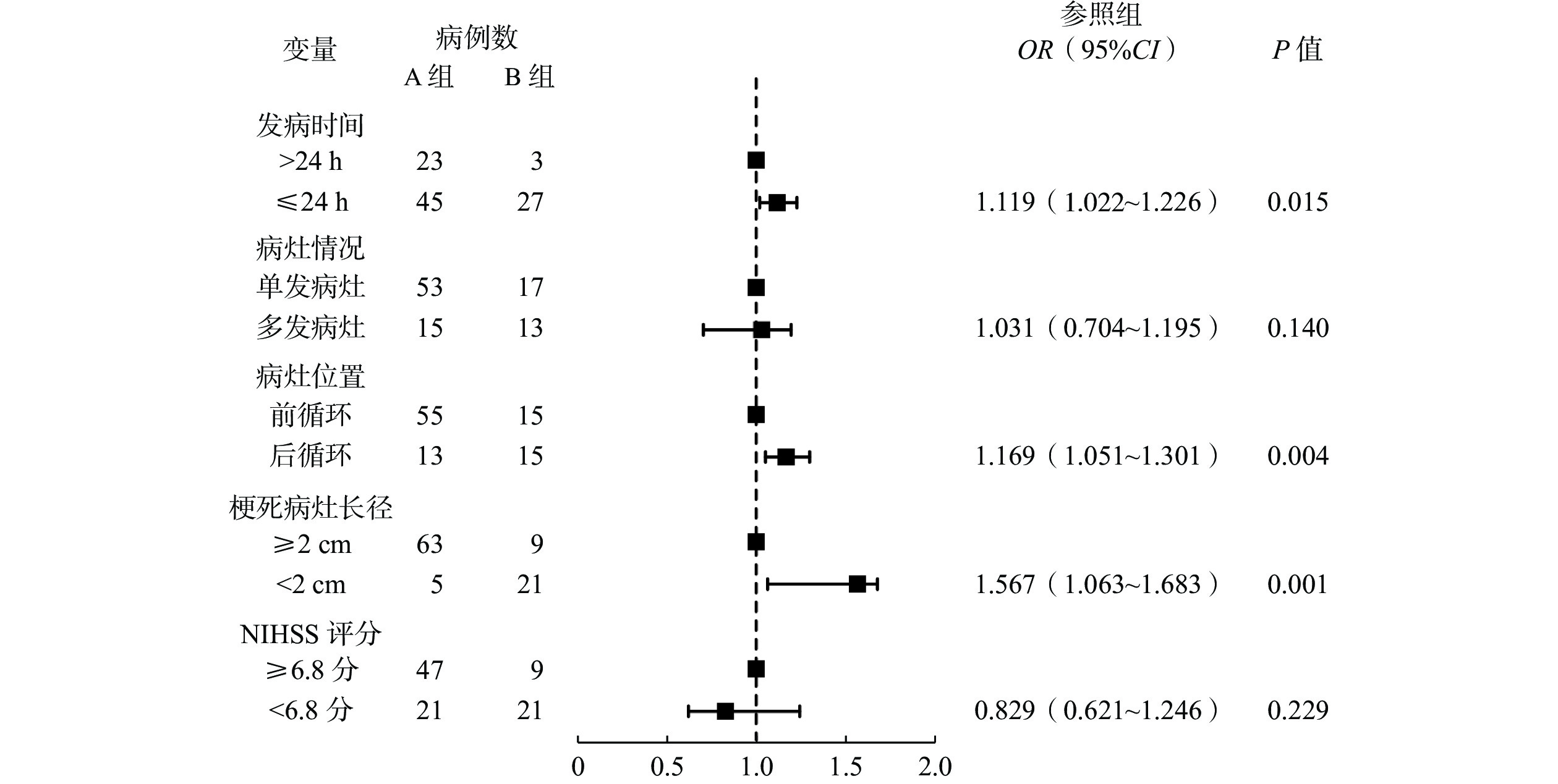
    • 以DWI与MRA检查结果是否一致为因变量,以发病时间、病灶情况、病灶位置、梗死病灶长径、NIHSS评分为自变量,进行多因素Logistic回归分析,结果表明,发病时间≤24 h、后循环、梗死病灶长径<2 cm均是DWI与MRA检查结果不一致的独立危险因素(均P<0.05,图2表5)。

      Figure 2.  Forest map of risk factors for inconsistent results between diffusion weighted magnetic resonance imaging and magnetic resonance angiography in patients with acute cerebral infarction

      指标 量化赋值
      发病时间 >24 h=0,≤24 h=1
      病灶情况 单发病灶=0,多发病灶=1
      病灶位置 前循环=0,后循环=1
      梗死病灶长径 ≥2 cm=0,<2 cm=1
      NIHSS评分 ≥6.8分=0,<6.8分=1
      DWI与MRA检查结果是否一致 否=0,是=1
      注:DWI为弥散加权成像;MRA为磁共振血管成像;NIHSS为美国国立卫生研究院卒中量表

      Table 5.  Multivariate Logistic regression analysis quantitative assignment table for inconsistent results between diffusion weighted magnetic resonance imaging and magnetic resonance angiography in patients with acute cerebral infarction

    • 2.6.   风险列线图预测模型

    • 根据ACI患者DWI与MRA检查结果不一致的独立危险因素构建风险列线图预测模型,其中发病时间≤24 h赋20分、后循环赋28分,梗死病灶长径<2 cm赋80分,总分为128分(图3)。

      Figure 3.  Risk column chart prediction model for inconsistent results between diffusion weighted magnetic resonance imaging and magnetic resonance angiography in patients with acute cerebral infarction

    • 2.7.   风险列线图预测模型的评价

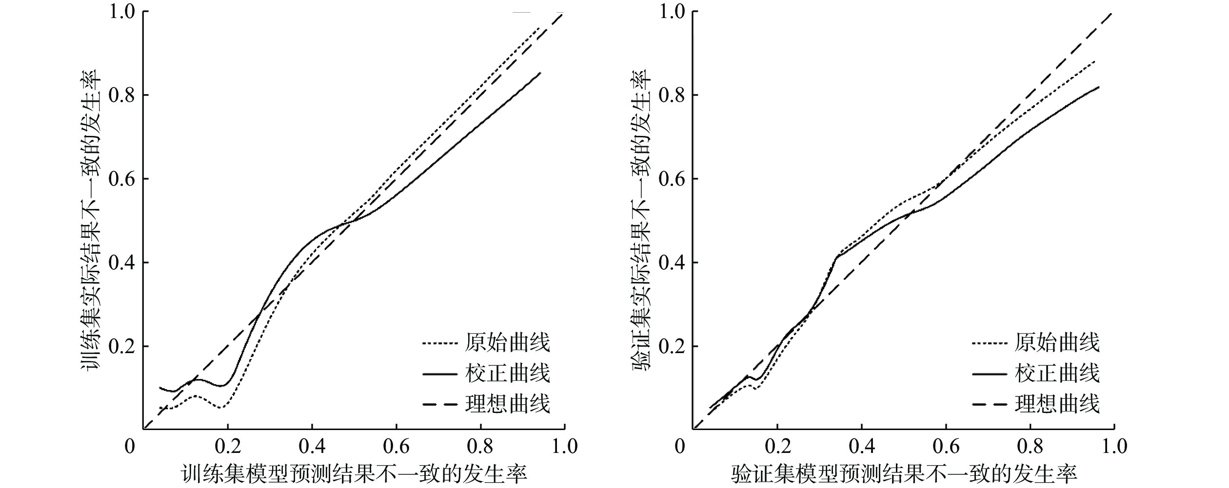
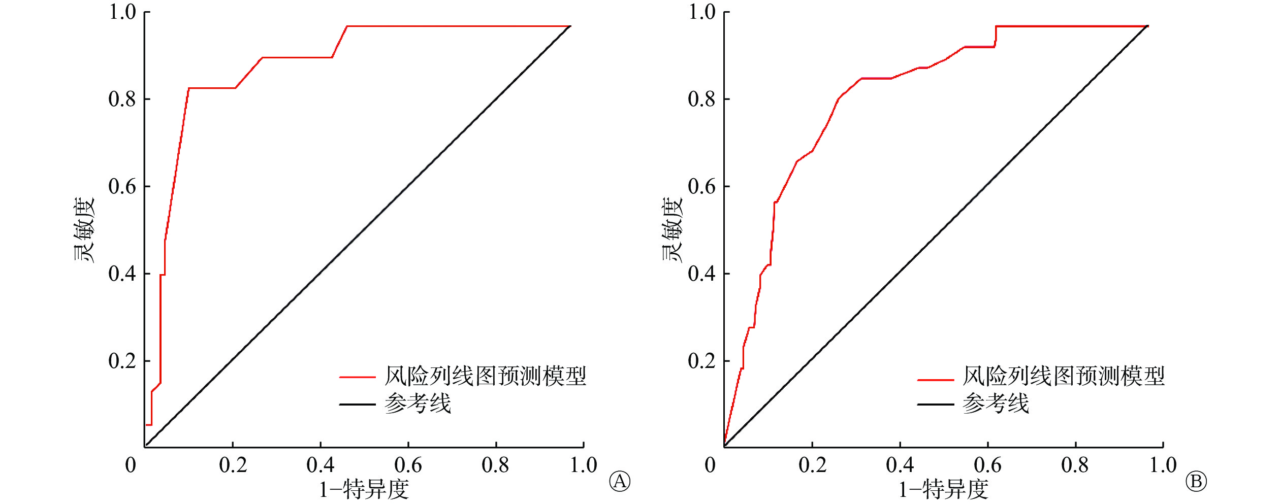
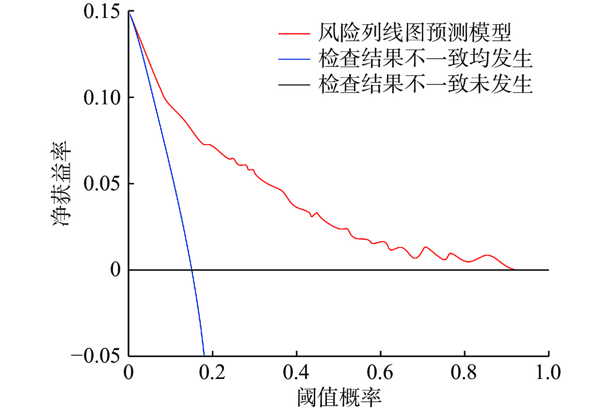
    • 采用Bootstrap法(原始数据重复抽样1 000次后)对风险列线图预测模型进行内部验证,外部验证通过验证集进行验证。训练集和验证集的一致性指数(concordance index,C-index)分别为0.880(95%CI:0.844~0.916,P<0.001)和0.812(95%CI:0.785~0.839,P<0.001)。训练集和验证集的校正曲线与理想曲线均拟合良好(图4)。训练集和验证集ROC的AUC分别为0.930(95%CI:0.899~0.961,P<0.001)、0.855(95%CI:0.812~0.898,P<0.001)(图5)。风险列线图预测模型的DCA曲线显示阈值概率在1%~92%时,净获益率>0(图6)。以上结果均显示该风险列线图预测模型具有较高的精准度,且有效性较好,安全可靠,实用性强。

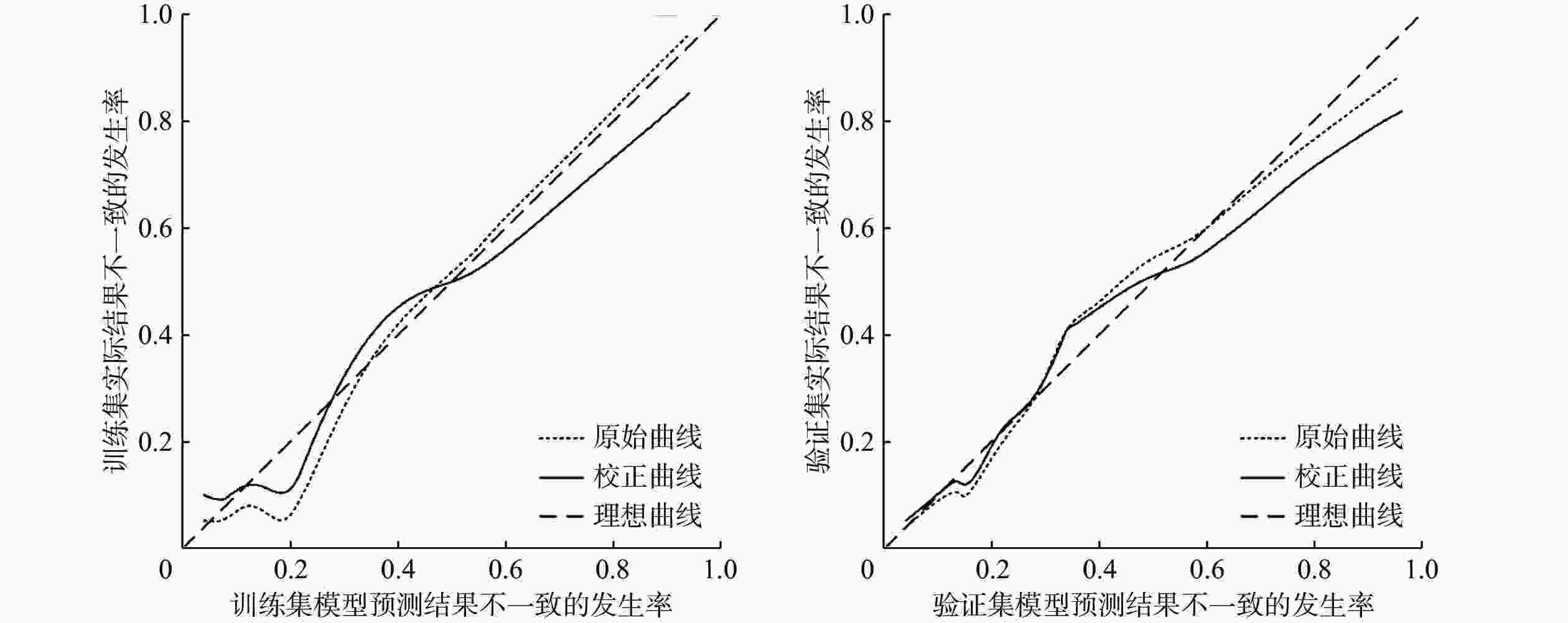
      Figure 4.  Correction curves of the training and validation sets of the risk column chart prediction model for inconsistent results between diffusion weighted magnetic resonance imaging and magnetic resonance angiography in patients with acute cerebral infarction

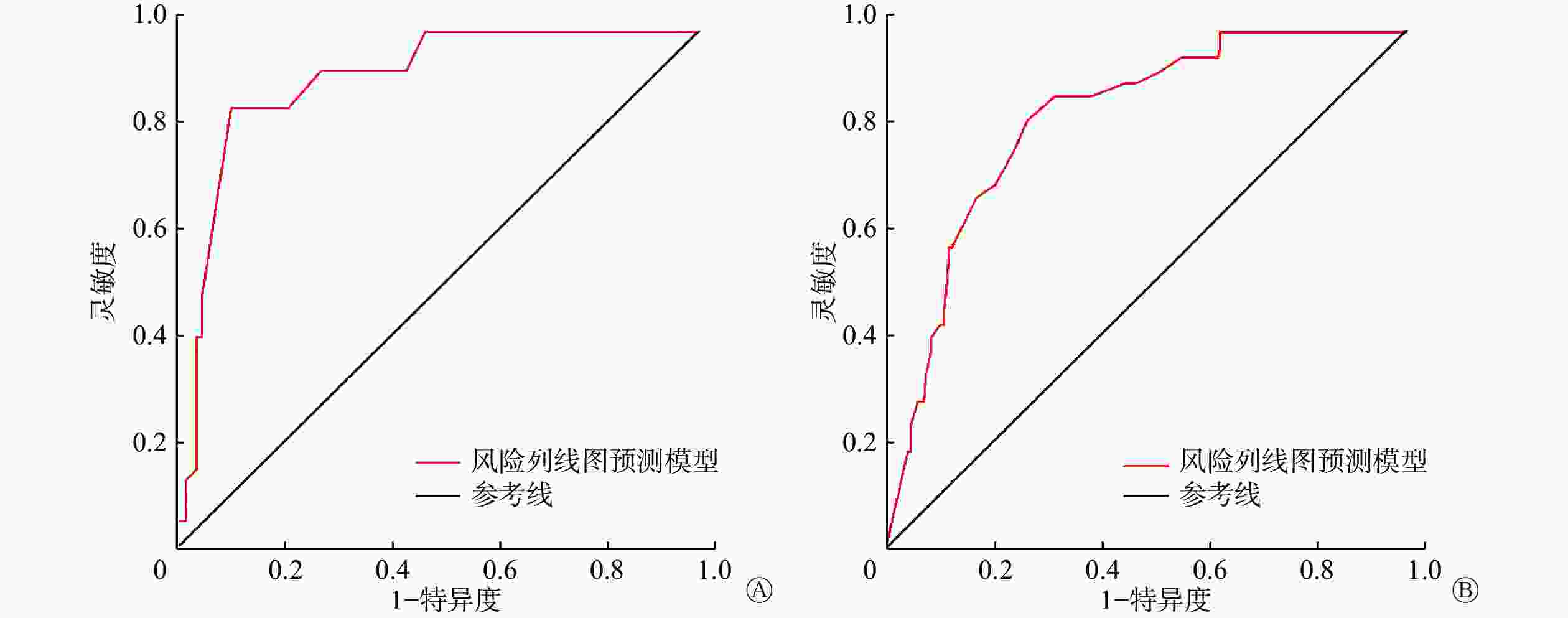
      Figure 5.  The receiver operating characteristic curve of the risk column chart prediction model training set (A) and validation set (B) for acute cerebral infarction patients with inconsistent results between diffusion weighted magnetic resonance imaging and magnetic resonance angiography

      Figure 6.  Decision curve analysis of risk column chart prediction model for inconsistent results between diffusion weighted magnetic resonance imaging and magnetic resonance angiography in patients with acute cerebral infarction

    3.   讨论
    • ACI的发病机制较复杂且危险因素较多,其中以颈动脉粥样硬化最常见[9]。随着人口老龄化的加剧,ACI的发病率不断上升,且其致残、致死率均较高[10]。因此,早期诊断,及时预防、干预,改善ACI患者的预后是临床工作、研究的重点。

      近年来,影像检查作为ACI诊断和辅助治疗的主要方法迅速发展。传统CT和常规MRI的诊断灵敏度较低,需要在ACI发病较长时间后才能定位病灶,且难以界定病灶的范围,因此临床应用的价值较低[11]。DWI可早期发现ACI病灶,并准确判断病灶的部位和体积,对ACI患者神经功能的受损情况进行定量评估,为临床医师的诊断及治疗提供依据[12]。本研究结果显示,ACI患者患侧和健侧脑组织ADC值的差异均有统计学意义。患侧ADC值和rADC在患者发病后先明显降低,发病24 h后明显升高,其原因可能是ACI患者在发病24 h内病情逐渐加重,脑细胞的代谢紊乱,导致细胞膜Na+/K+-ATP酶失活,造成水钠潴留,限制了水分子的弥散,使rADC降低。ACI患者发病24 h后rADC升高可能与病灶部位血液灌注的恢复有关。因此,ACI患者发病后72 h内,临床医师可通过rADC了解患者的病理、生理改变和病情变化。

      脑部主要供血动脉狭窄或闭塞是ACI发病的主要机制,虽然DWI可定位病灶并对病情进行评估,但无法显示主要的责任血管[13],因此本研究采用MRA检查主要责任血管的位置。MRA可显示三级以上分支动脉血管的狭窄或闭塞情况,且MRA检测出的病变血管与DWI图像显示的病灶位置具有解剖关系,但血液涡流会使MRA图像出现伪影进而造成误诊[14]。MRA可根据血管的狭窄程度和远端血管的显影情况对患者的严重程度进行分级,评估病情,用以指导临床治疗。

      本研究中,DWI与MRA检查结果一致的患者68例(69.39%),不一致患者30例(30.61%),二者的一致性较好。多因素Logistic回归分析结果显示,发病时间≤24 h、后循环、梗死病灶长径<2 cm均是DWI与MRA检查结果不一致的独立危险因素。ACI发病早期,患者脑组织的缺血程度或血管狭窄程度较轻,或疾病处于逐渐进展的状态,可能出现漏诊,导致DWI与MRA检查结果不一致。王继蕊等[15]的研究结果表明,在ACI患者发病早期,较多患者的DWI结果为阴性,起病后120 h DWI结果才为阳性。ACI患者脑部梗死病灶过小可能造成漏诊,因此DWI结果为阴性的病灶一般较小,且发生在后循环的概率大于前循环。白向东等[16]的研究结果亦表明,ACI患者DWI-FLAIR不匹配组发生后循环梗死的比例较DWI-FLAIR匹配组高,证实了后循环对脑缺血的耐受性更强、溶栓时间可以适当延长的结论。本研究中,风险列线图预测模型结果显示各危险因素总计128分。ROC曲线、校正曲线和DCA曲线的评价结果显示,该风险列线图预测模型的区分度、准确率和有效性均较高。

      本研究具有一定的局限性,如样本量相对不足,且研究周期较短等,因此难免存在一些偏倚。

      综上所述,DWI与MRA检查对于ACI患者诊断的一致性较好。发病时间≤24 h、后循环、梗死病灶长径<2 cm均是DWI与MRA检查结果不一致的独立危险因素。DWI可显示早期ACI病灶的位置和缺血程度,MRA则可准确定位责任血管及其狭窄程度,DWI和MRA的广泛应用,使ACI的早期诊断成为可能,从而使医师及时干预,缓解病情,改善患者预后。

      利益冲突 所有作者声明无利益冲突

      作者贡献声明 高爽负责命题的提出与设计、方法的建立、数据的分析、论文的撰写;王三刚负责论文的修订、数据的收集

Reference (16)

Catalog

/

DownLoad:  Full-Size Img  PowerPoint
Return
Return