


-
上皮样血管内皮瘤(epithelioid angiosarcoma,EA)是一种来源于血管内皮细胞的恶性肿瘤,该病具有高度侵袭性和破坏性[1],在深部软组织较常见[2],原发于骨组织较为罕见[3] 。99Tcm-MDP全身骨显像可观察全身骨质情况,并且SPECT/CT融合显像作为全身骨显像的有效补充,将组织代谢情况与形态学表现相结合能更好地了解病灶的改变[4]。本文报道了1例发生于股骨的EA病例,查阅文献并分析其99Tcm-MDP SPECT/CT 显像特点,旨在为临床鉴别诊断提供参考。
-
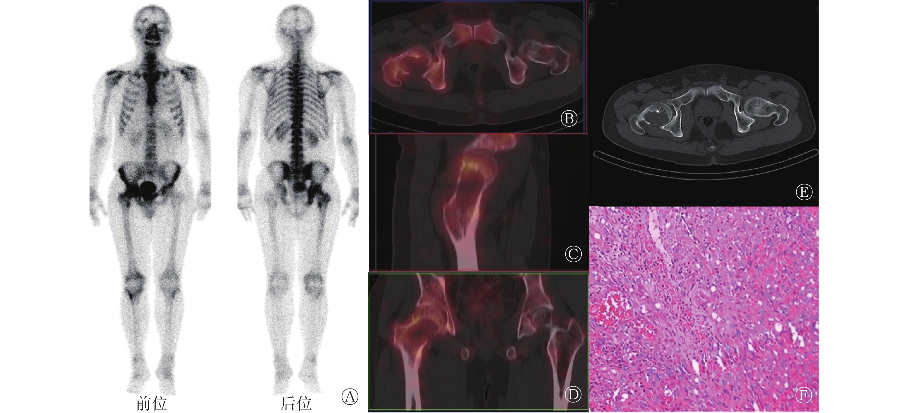
患者男性,38岁,因“右侧髋部疼痛4个月,伴行走障碍,疼痛放射至右侧膝关节”入院就诊。既往无骨关节手术史及外伤骨折史。体格检查结果:右侧髋部软组织无肿胀,未触及包块,压痛不明显,右侧股四头肌未见萎缩,患肢末梢血液循环及感觉正常。实验室检查结果(括号内为正常参考值):血常规、肝、肾功能正常;碱性磷酸酶(ALP) 92(25~90) U/L、血钙2.30(2.30~2.55) mmol/L、血磷1.44(0.87~1.60) mmol/L、超敏C反应蛋白24.49(0~8) mg/L;癌胚抗原(CEA)、甲胎蛋白(AFP)、糖类抗原199(CA199)均为阴性。为明确诊断,患者行99Tcm-MDP全身骨平面显像,示右侧股骨近端及股骨颈放射性浓聚影(图1A),其余骨骼未见异常;SPECT/CT融合显像示右侧股骨溶骨性改变,病灶内部放射性分布减低,病灶边缘见不均匀放射性浓聚(图1B~D),考虑原发性骨肿瘤,恶性病变可能性大。同机CT检查示:右侧股骨头至股骨颈局限性溶骨性改变,边界清楚,无硬化边,周围见残存骨皮质(图1E)。患者行右侧股骨肿瘤病灶清除术,免疫组化检测结果:CD31(+)、E26转录因子相关基因(ERG,+)、波形蛋白(vimentin,+)、CD68(部分+)、转录因子E3(TFE3,弱+)、细胞角蛋白(CK,散灶+)、 CK19(散灶+)、抗胰糜蛋白酶(AACT,+)、传导样增强因子1(TLE1,部分+)、细胞增殖核抗原Ki-67(30%+)、抗凋亡蛋白63(P63,−)、CD34(−)、上皮膜抗原(EMA,−)、酸性钙结合蛋白(S-100,−)、CD56(−)、抗黑色素瘤特异性单抗(HMB45,−)。组织病理学检查诊断结果:(右股骨近端及股骨颈)符合EA(图1F)。

Figure 1. 99Tcm-MDP SPECT/CT fusion imaging and histopathological examination images of a patient with the epithelioid angiosarcoma of the femur (male, 38 years old)
-
骨EA是上皮样血管源性的高度恶性骨肿瘤,原发于骨的EA极为罕见,占骨原发恶性肿瘤的0.5%~1%[5]。骨EA以老年人多见,男性多于女性。EA好发于长骨,偶见于颅骨、脊柱及骨盆[6]。骨EA临床表现无特异性,以病变部位持续性疼痛常见,实验室检查也无特殊表现。
骨EA的影像学表现无特异性。CT平扫主要表现为溶骨性骨质破坏,低度恶性EA可呈局限性骨质破坏、边界清晰、无硬化边、瘤内一般无钙化、骨膜反应少见,高度恶性EA呈大片状溶骨性骨质破坏[7];CT增强扫描表现为肿瘤实质部分可见强化,但强化程度差异较大,可表现为轻度至明显强化[8]。CT可显示发病部位、大小、形态、病变与周边组织的关系,为制定手术治疗方案及预后观察提供重要依据。MRI软组织分辨能力极佳,对骨肿瘤的软组织显示清楚。MRI平扫在T1加权成像上呈等信号或低信号,在T2加权成像上呈混杂稍高信号;增强扫描表现为明显不均匀性强化[3]。本例骨EA患者右侧股骨头至股骨颈的CT骨窗可见溶骨性骨质破坏,骨皮质变薄,软组织窗病灶内见分叶状软组织肿块,该结果与李翠翠等[9]文献报道的CT特征相似。99Tcm-MDP SPECT/CT显像通常用于诊断转移性肿瘤。对于骨EA,99Tcm-MDP全身骨显像能较早发现无症状病变,具有灵敏度高的优点,并能显示多发病变的分布。因骨EA组织学上以形成大量肿瘤性新生血管为其基本改变,恶性内皮细胞高度增殖, 在CT上表现为溶骨性骨质破坏,残存骨小梁间隔少见,无骨膜反应[10],故99Tcm-MDP全身骨显像中病灶表现为轻-中度放射性摄取。
本例股骨EA患者的影像学表现为股骨的软组织肿块,需要与常见的股骨肿瘤相鉴别。(1)骨转移瘤:多为溶骨性或混合型骨质破坏,骨皮质变薄或不完整,可结合肿瘤病史及实验室检查结果;(2)骨巨细胞瘤:多表现为沿股骨颈的膨胀性囊样或地图样溶骨性骨质破坏;(3)动脉瘤样骨囊肿:表现为膨胀性骨质破坏,病灶内可有分隔“液液平”为特征性表现;(4)朗格汉斯细胞增多症:表现为不规则溶骨性骨质破坏,边界模糊,并可见骨膜反应[11]。
骨EA的最终确诊需依靠免疫组化,常用的血管内皮细胞标志物有CD31、CD34、弗里德白血病病毒插入位点1(FLI-1)和E26转录因子相关基因(ERG),其中CD31是最具特异性且最灵敏的标志物,在绝大多数的血管肉瘤中均有表达[12]。
骨EA是高度恶性肿瘤,预后极差,目前缺少有效的治疗方法,单发病灶最佳的治疗方法是通过手术切除,多发病灶可通过放疗控制局部复发。99Tcm-MDP SPECT/CT骨显像诊断骨EA虽缺少特异性,但SPECT/CT骨显像为全身显像,可了解全身骨骼情况及发现全身其他部位是否有类似病灶,同时可根据CT了解病变骨质的破坏程度,为临床制定治疗方案提供依据。
利益冲突 所有作者声明无利益冲突
作者贡献声明 伍杨负责文献的收集与分析、论文的撰写;付巍负责论文的修订与审阅
99Tcm-MDP SPECT/CT imaging in epithelioid angiosarcoma of the femur: a case report
- Received Date: 2021-07-28
- Available Online: 2022-08-25
Abstract: Bone epithelioid angiosarcoma (EA) is a highly malignant bone tumor of vascular endothelial origin. Bone EA is a rare disease in clinical practice. A case of femoral EA with 99Tcm-methylenediphosphonate (MDP) SPECT/CT was reported and reviewed in terms of clinical symptoms, laboratory examination, CT and 99Tcm-MDP SPECT/CT imaging, the imaging features of EA were summarized by literature review. 99Tcm-MDP SPECT/CT imaging is helpful for the detection of metastatic and the differential diagnosis of EA from other diseases, which has certain clinical significance.
 DownLoad: