2022 Vol. 46, No. 4
Display Method:
 [PDF 3039KB]
2022, 46(4): 197-202.
doi: 10.3760/cma.j.cn121381-202111005-00171
Abstract:
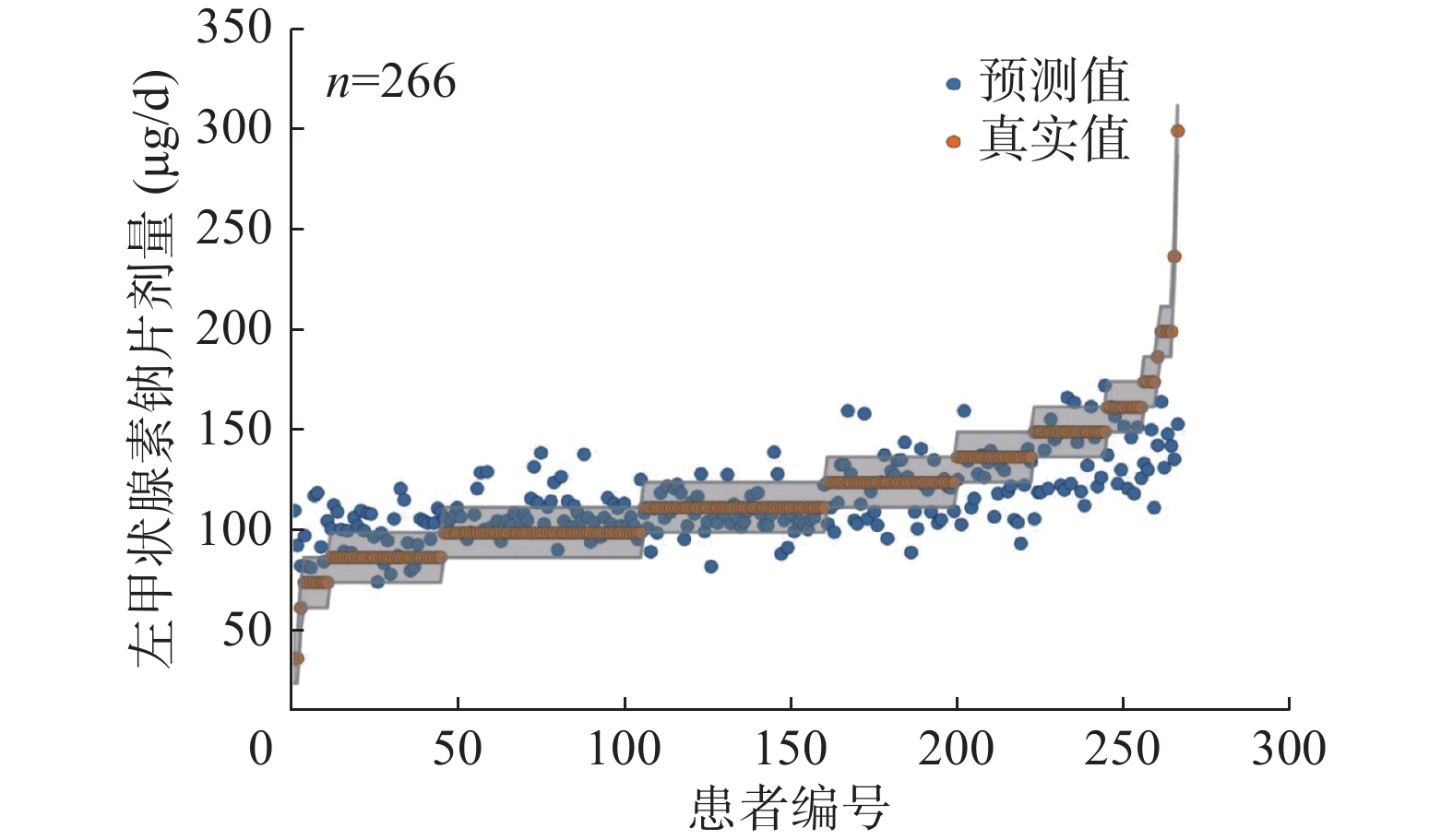
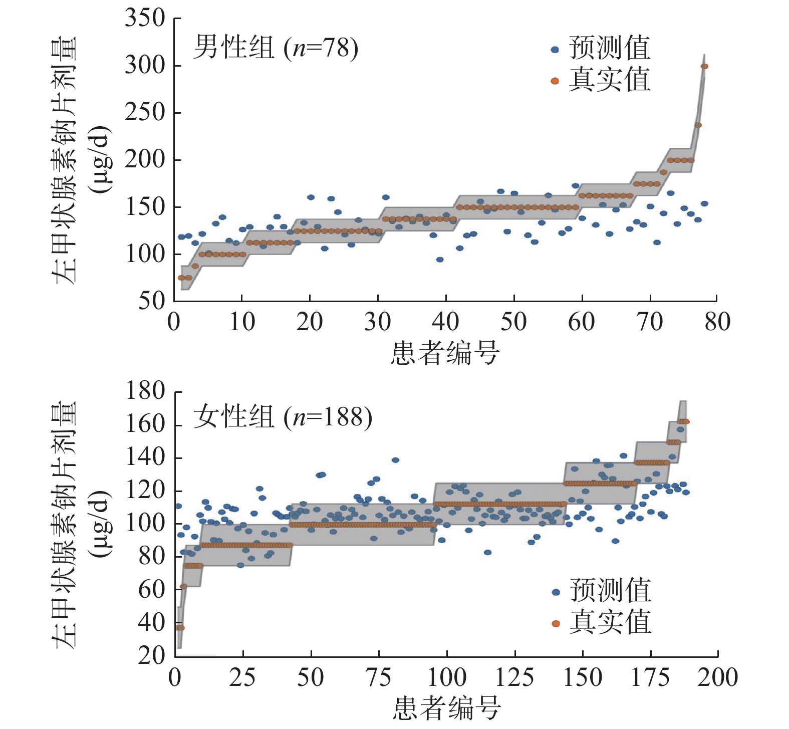
Objective To construct a prediction model for the optimal initial dose of levothyroxine sodium tablets in patients with differentiated thyroid cancer (DTC) after 131I treatment by machine learning. Methods A total of 266 DTC patients (78 males (male group) and 188 females (female group), aged 18 to 70 (40.0+11.5) years old) who received 131I treatment followed by thyroid stimulating hormone (TSH) suppressive therapy in the Department of Nuclear Medicine, Konggang Hospital, Tianjin Cancer Hospital between November 2019 and November 2020 were retrospectively analyzed for final compliance. A total of 16 clinical and biochemical indicators and data related to thyroid function were obtained, and each adjusted dose of levothyroxine sodium tablets was collected from patients with regular post-discharge rechecks. The indicators strongly correlated with the optimal dose of levothyroxine sodium tablets were screened by calculating random forest feature importance. A wide variety of regression models were constructed with the selected indicators and optimal dose of levothyroxine sodium tablets as independent and dependent variables, respectively. Selected the most accurate model using the cross-validation method. Counting data were compared between male and female groups using the chi-square test of independence. Results Body weight, height, body mass index, body surface area, hemoglobin, mean corpuscular volume, systolic/diastolic blood pressure, postoperative parathyroid hormone, and the reaching levothyroxine sodium tablets dose of 266 patients were (68.4±12.9) kg, (165.8±12.8) cm, 24.6±3.5, (1.9±0.2) m2, (140.1±19.1) g/L, (88.6±5.5) fl, (125.7±18.9) mm Hg/(82.7±12.4) mm Hg, (4.1±2.2) pmol/L, and (117.0±30.1) μg/d, respectively. Six indicators with a strong correlation with levothyroxine sodium tablets dose were screened using the feature selection method. According to the order of importance, the six indicators were body surface area, body weight, hemoglobin, height, body mass index, and age. Their average random forest importances were 0.2805, 0.1951, 0.1315, 0.1252, 0.1080 and 0.0819 respectively. The support vector regression (SVR) model using radial basis kernel had the highest accuracy (53.4%, 142/266) by cross-training validation. In addition, in this study, SVR's accuracy was significantly higher than the first success rate of empirical administration of levothyroxine sodium tablets (15.0%, 40/266). Moreover, the SVR model's accuracy was compared by dividing the patients into different subgroups according to gender. The results showed that the female patient group's accuracy was significantly higher than that of the male group (60.6% (114/188) vs. 35.9% (28/78)), with a statistically significant difference (χ2=13.51, P<0.001). Conclusions The SVR model is constructed based on machine learning and is expected to improve the first success rate of levothyroxine sodium tablets in DTC patients after being treated with 131I. It is more pronounced in female patients and helps to improve the quality of life and prognosis among DTC patients.
2022, 46(4): 203-209.
doi: 10.3760/cma.j.cn121381-202102028-00170
Abstract:
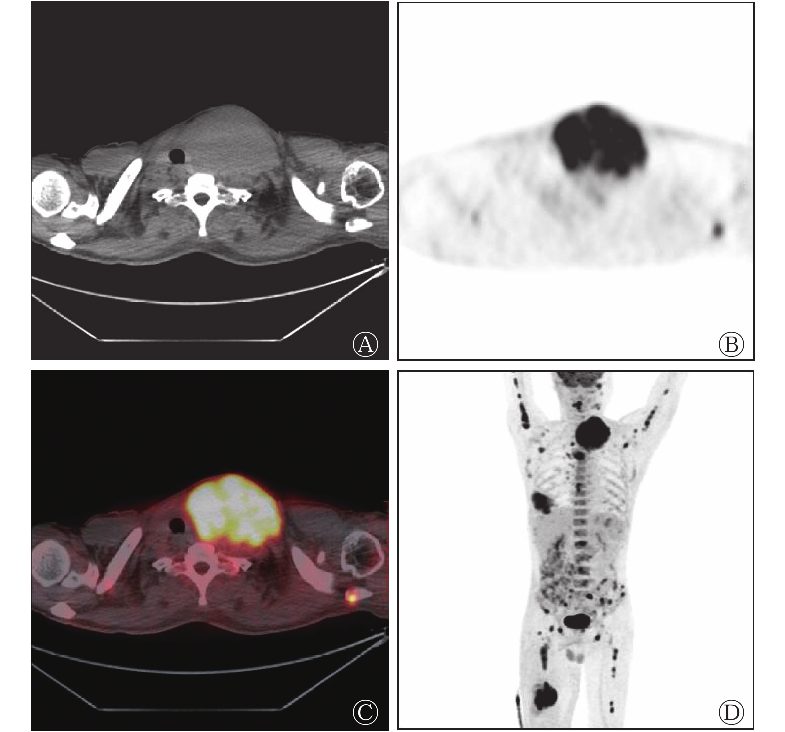
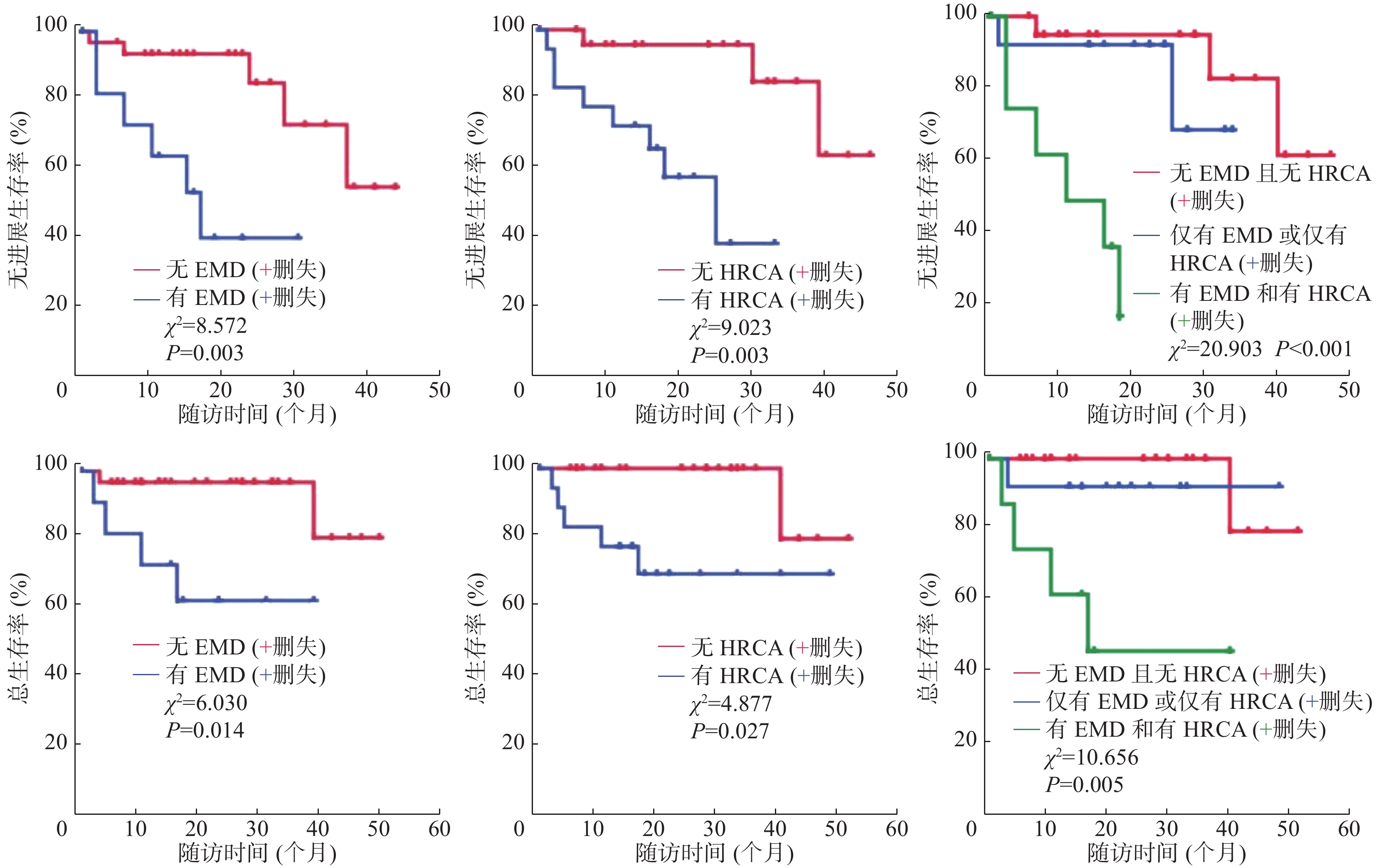
Objective To investigate the correlation between 18F-fluorodeoxyglucose (FDG) PET/CT imaging findings and high-risk cytogenetic abnormalities (HRCA) in patients newly diagnosed with multiple myeloma (MM), and the value of both combined applications in evaluating the prognosis of patients with MM. Methods The clinical and imaging data of 44 patients with MM diagnosed by bone marrow histopathology and laboratory examination and who underwent 18F-FDG PET/CT imaging before treatment in Shantou Central Hospital from June 2016 to November 2020 were retrospectively analyzed, including 23 males and 21 females, aged 38–91 (61.1±9.6) years old. Patients were divided into the HRCA group and the non-HRCA group according to the result of fluorescence in situ hybridization. Patients were divided into stage Ⅰ+Ⅱ group and stage Ⅲ group according to the Revised-International Staging System (R-ISS) issued by the International Myeloma Working Group. Patients were divided into two groups, a standard-risk group, and a high-risk group according to the Mayo Stratification of Myeloma and Risk-adapted Therapy (mSMART) 3.0 risk stratification criteria. Through the analysis of the 18F-FDG PET/CT imaging data, patients were divided into ≤3 groups and >3 groups according to the number of focal lesions (FLs), divided into ≤4.2 groups and >4.2 groups according to maximum standardized uptake value (SUVmax), divided into extramedullary disease (EMD) group and non-EMD group according to the presence of EMD lesions, respectively. Gather data on progression-free survival (PFS) and overall survival (OS) begins from the first follow-up. Imaging findings with clinical features, HRCA, and prognostic stages were compared using the χ2 test. The independent risk factors of HRCA and stages were analyzed using the multivariate logistic regression analysis. The differences between PFS and OS among the groups were compared using the Kaplan-Meier method and Log-rank test. The independent risk factors of PFS and OS were analyzed using the Cox proportional hazards regression model. Results FLs≤3 or >3 varied among groups of R-ISS, mSMART 3.0, and HRCA (χ2=4.919, 8.472, 8.167; all P<0.05). EMD or non-EMD varied among groups of mSMART 3.0 and HRCA (χ2=4.061, 6.808; both P<0.05). FLs>3 were independent risk factors for HRCA, R-ISS, and mSMART 3.0 (OR=10.952, 5.000, 10.714; 95%CI: 1.195–100.393, 1.127–22.181, 2.269–50.598; all P<0.05). PFS and OS varied among groups of EMD and HRCA (PFS: χ2=8.572, 9.023; both P<0.01 and OS: χ2=6.030, 4.877; both P<0.05). EMD was an independent poor prognosis factor for both PFS and OS (OR=4.466, 6.520; 95%CI: 1.084–18.396, 1.174–36.211; both P<0.05). HRCA was an independent poor prognosis factor for PFS (OR=8.458, 95%CI: 1.671–42.812, P<0.05). By the end of follow-up, patients without EMD and HRCA or only one of them had not reached median PFS and median OS; median PFS for patients with both EMD and HRCA was 11 months (χ2=20.903, P<0.001) and median OS were 17 months (χ2=10.656, P<0.01). Conclusion There is a significant correlation between 18F-FDG PET/CT imaging findings and HRCA in patients newly diagnosed with MM, and the combination of both has a certain predictive value for the prognosis of patients with MM.
2022, 46(4): 210-216.
doi: 10.3760/cma.j.cn121381-202105002-00173
Abstract:
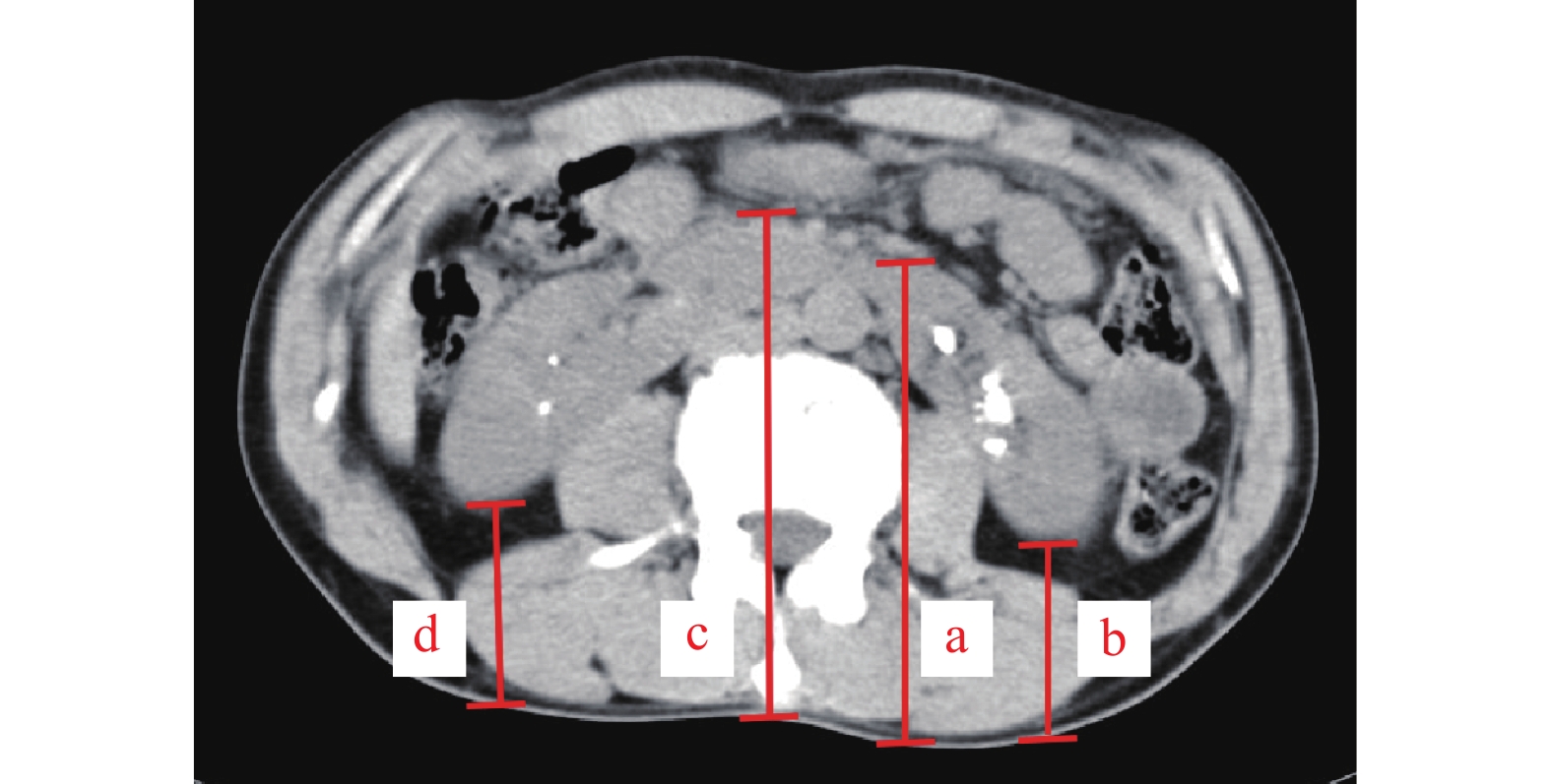
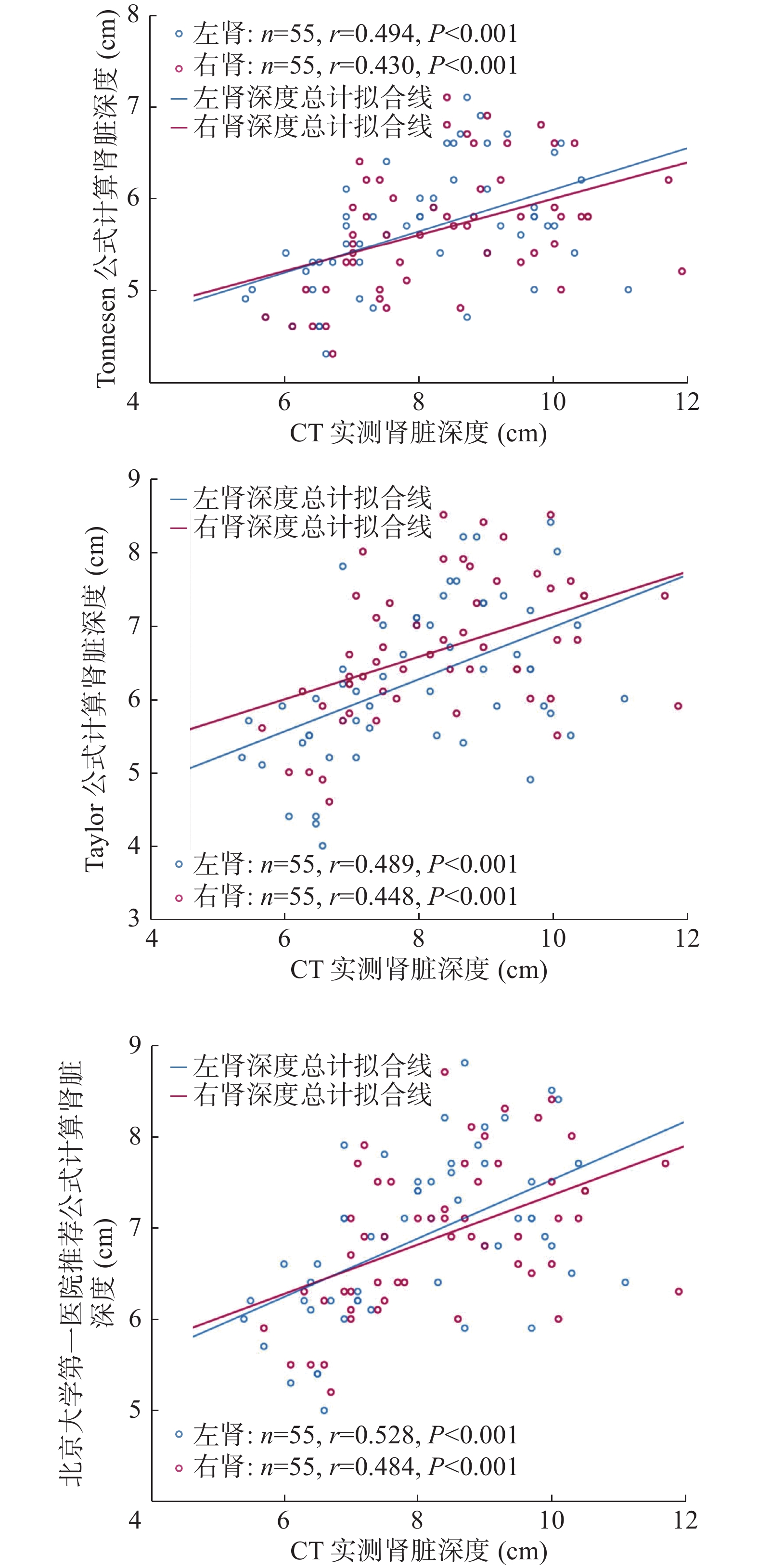
Objective To validate the accuracy of different estimation formulas for measuring renal depth in patients with horseshoe kidney using CT actual measured renal-depth as a reference standard. Methods The clinical data of 55 patients with horseshoe kidney who underwent 99Tcm-diethylene-triaminepentaacetic acid renal dynamic imaging in the First Affiliated Hospital of Chongqing Medical University from January 2015 to December 2020 were analyzed retrospectively. These patients included 33 males and 22 females aged 19–80 (42.2±16.3) years. The vertical distance between the farthest and nearest points of the renal hilum and the skin of both kidneys were selected respectively, and the average value was taken as the renal-depth. The gender, age, height, and weight of the patients were recorded. The estimated renal-depths were obtained using the Tonnesen, Taylor, and Li Qian formulas, respectively. Paired t test, Pearson correlation analysis, and Bland-Altman analysis were performed between the estimated and CT measured renal-depths. Results In 55 patients, the renal-depths calculated by the Tonnesen, Taylor, and Li Qian formulas were all lower than the CT measured renal-depths, and the differences were all statistically significant (left kidney: t=−14.04 to −6.85, all P<0.01; right kidney: t=−15.19 to −8.47, all P<0.01). A significant correlation existed between formulas estimated and CT measured renal-depths (r=0.430−0.528, all P<0.001), but the Li Qian formula correlated better than the Tonnesen and Taylor formula, where the correlation coefficient was (r=0.528, P<0.001) for the left kidney and (r=0.484, P<0.001) for the right kidney. All formulas underestimated the renal-depth; the estimated error increased with increased renal-depth, and the difference was statistically significant (95%CI: Conclusions The accuracy of renal-depth in patients with horseshoe kidney calculated using Tonnesen, Taylor, and Li Qian formulas were not as good as the actual CT measurement. Therefore, to accurately assess glomerular filtration rate, CT is recommended to measure the renal-depth of patients with horseshoe kidney.

2022, 46(4): 217-222.
doi: 10.3760/cma.j.cn121381-202009013-00163
Abstract:
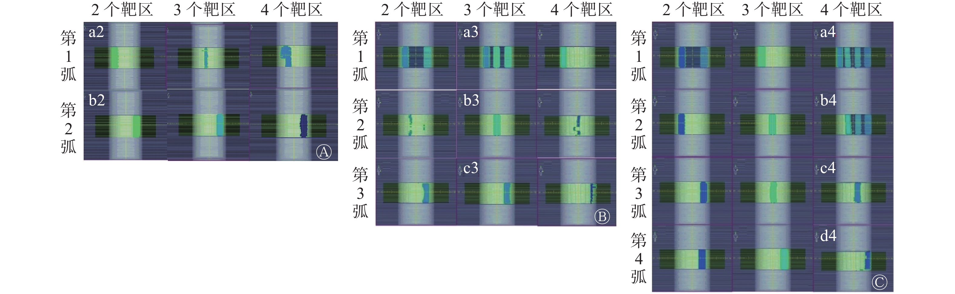
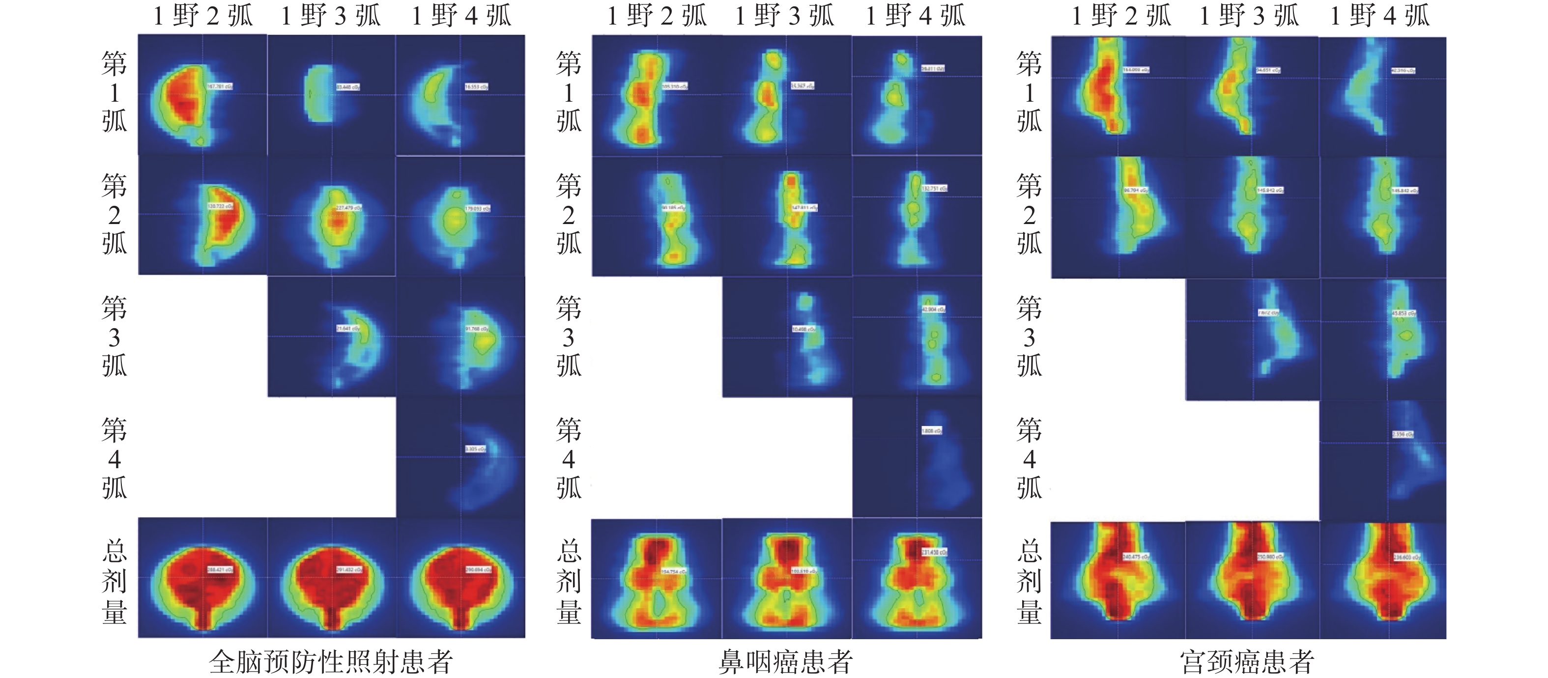
Objective To study the specific splitting mode of fluence in Monaco planning system with single-beam multi-arc. Methods (1) Simulation plan: two, three, and four cylindrical structures were drawn along the x direction on the CT image of the cylindrical Delta4 phantom with uniform density to simulate three cases that need to irradiate two, three, and four isolated target volumes at the same time, respectively, to observe the relationship between the shape of the irradiation fields and the target volumes and analyze their regularity. (2) Patient irradiation: the following past cases in Jilin Cancer Hospital were completely random selected: one case of whole brain prophylactic irradiation, one case of nasopharyngeal carcinoma, and one case of cervical cancer. Three volume-modulated arc therapy plans were designed for each patient. A 360° beam was used in all plans, and the difference is that the numbers of arcs were two, three, and four, respectively. The gantry angles of all real case plans were set to 0°, the real patient plans were delivered on the accelerator, and the Matrixx dose verification system was used to measure the total dose of the plans and the partial dose per arc. Compare the positional relationship between the total dose for each plan and its dose per arc. Results The results of simulation plan and patient irradiation study showed that on the beam's eye view (BEV), the fluence was divided along the x-axis, and the number of fluence divisions was equal to the number of arcs in the plan. Each arc was illuminated from left to right in the BEV direction in sequence. Conclusion The fluence splitting mode in Monaco planning system was elucidated, providing guidance for its practical application in clinic.

2022, 46(4): 223-229.
doi: 10.3760/cma.j.cn121381-202110017-00167
Abstract:
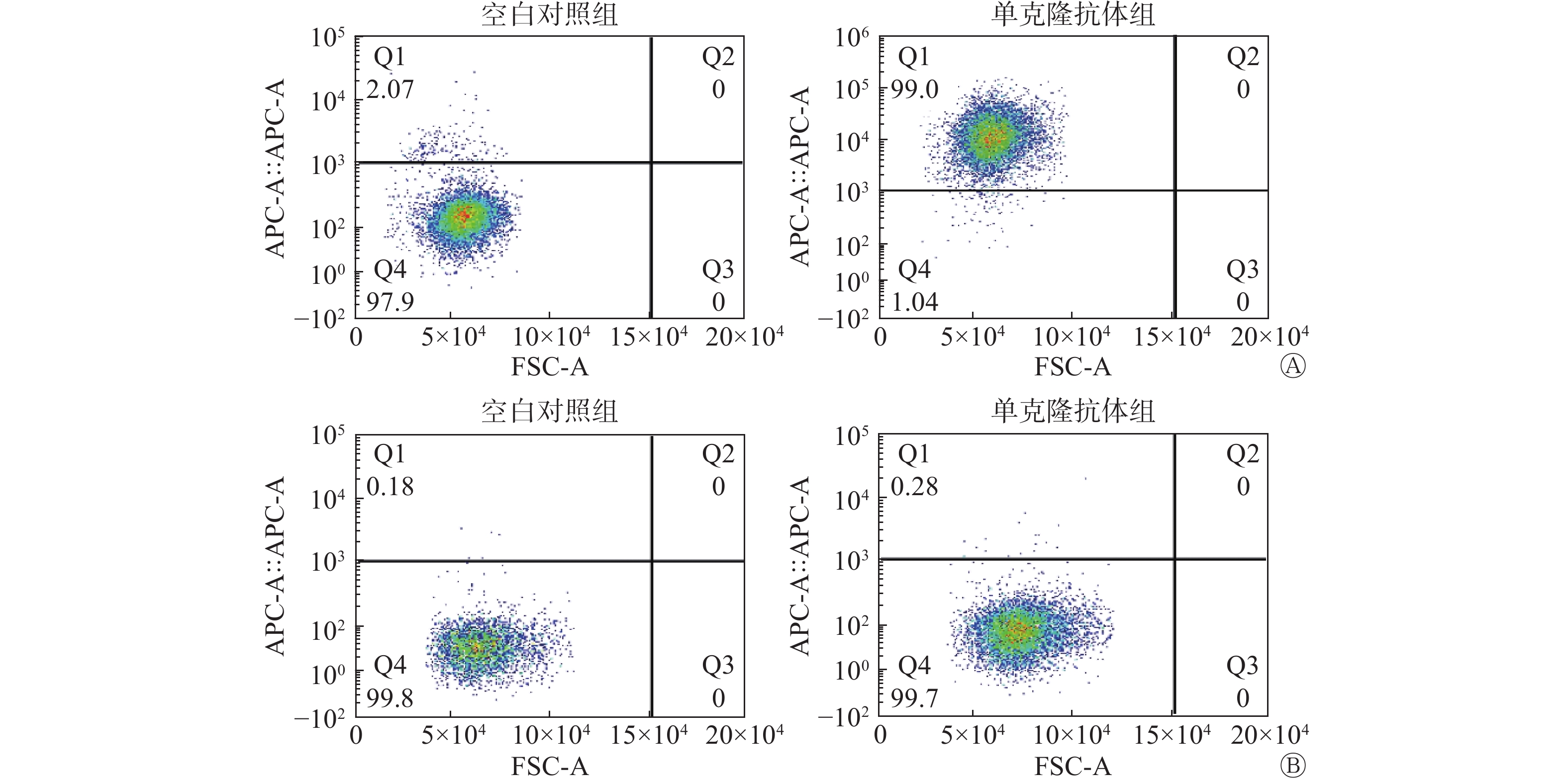
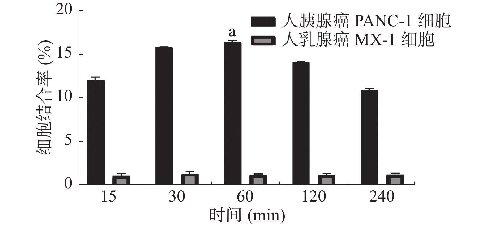
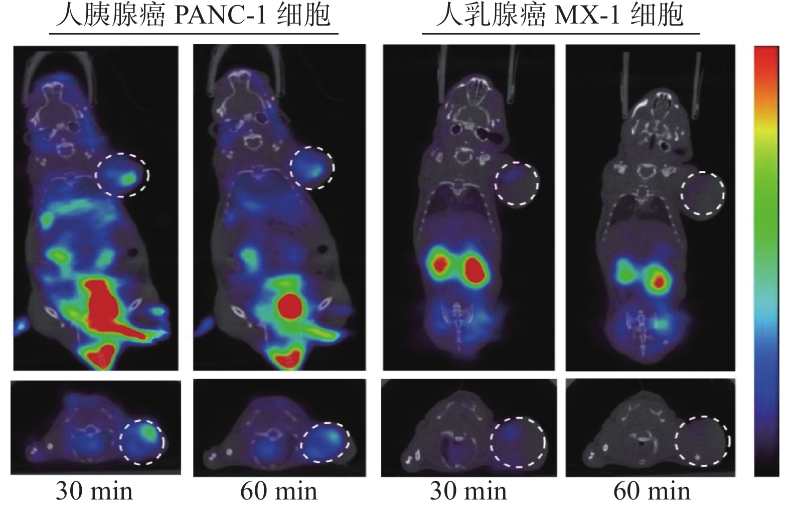
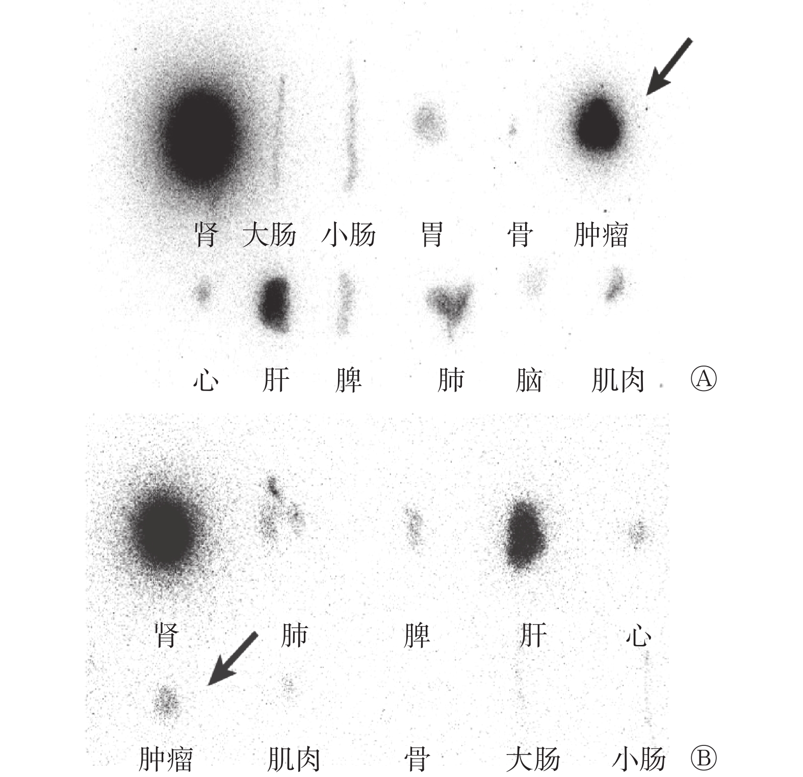
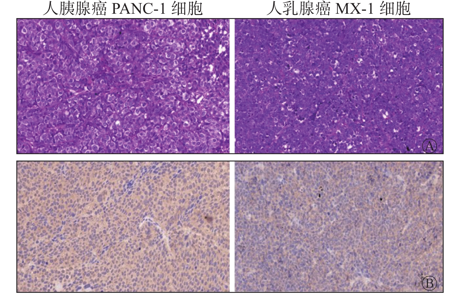
Objective To develop a radiolabeled peptide molecular tracer 99Tcm-His-Arg-Pro-Tyr-Ile-Ala-His (99Tcm-T7) targeting transferrin receptor and evaluate its micro SPECT/CT imaging effect in tumor-bearing nude mice models. Methods The peptide probe 99Tcm-T7 was developed by indirect labeling method with the coordination of co-ligands N-tri (hydroxymethyl) methylglycine and ethylenediamine diacetate. The expression levels of TfR on the surface of human pancreatic PANC-1 tumor cells and human breast MX-1 tumor cells were measured through flow cytometry. Cell binding and competitive blocking assays was conducted to analyze the binding affinity and specificity of 99Tcm-T7 in vitro. Micro SPECT/CT imaging and biodistribution after the establishment of mouse xenograft models were performed in vivo to evaluate the affinity and feasibility of noninvasive tumor imaging. Radio-autograph assay and immunohistochemical staining were conducted to validate the correlation between the uptake of 99Tcm-T7 and expression of TfR in tumor tissues. Independent sample t-test was used for the comparison between the two groups. Results The radiolabeled probe 99Tcm-T7 was successfully synthesized with a radiolabeling yield of greater than 95%. It exhibited great stability in vitro, with radiochemical purities of (95.3±0.3)% and (90.6±0.4)% after incubation in normal saline and fetal bovine serum for 4 hours, respectively. The results of flow cytometry showed that PANC-1 tumor cells overexpressed TfR on the surface with a high tendency to bind TfR monoclonal antibody ((98.9±0.1)%), whereas MX-1 tumor cells showed low TfR expression on the membrane( (0.2±0.1)%). In vitro cell binding assay results showed that the binding rate of PANC-1 cells to 99Tcm-T7 reached a peak ((16.12±0.01)%) after 60 minutes of incubation, which was higher than that of MX-1 cells ((1.20±0.01)%), and the difference between them was statistically significant (t=28.67, P<0.001). The results of cell competition inhibition experiment showed that the binding rate of PANC-1 blocking group to 99Tcm-T7 decreased to (2.40±0.01)%, which was significantly different from that of PANC-1 experimental group(t=26.91, P<0.001). The results of micro SPECT/CT imaging in nude mice bearing tumor showed that 99Tcm-T7 could be quickly cleared from the blood and mainly eliminated from the kidneys. PANC-1 tumor-bearing nude mice models showed clear tumor contour 30 minutes after injection of 99Tcm-T7, with a tumor-to-muscle ratio of 2.80±0.22. The results of biological distribution experiments showed that the uptake of 99Tcm-T7 by tumors and organs (percentage injection dose rate (%ID/g) per gram of tissue) was consistent with the imaging results, and the uptake of 99Tcm-T7 in PANC-1 cells ((0.55±0.18)%ID/g) was higher than that in MX-1 cells ((0.16±0.11)%ID/g), and the difference was statistically significant (t=6.42, P<0.001). The radio-autograph assay showed that PANC-1 cells significantly absorbed 99Tcm-T7 compared with MX-1 cells 30 minutes after injection of 99Tcm-T7. The highest uptake in normal organs was observed in the kidney, followed by the liver. Hematoxylin-eosin and immunohistochemical staining revealed no obvious necrosis in the tumor parenchyma. The PANC-1 cells overexpressed TfR, and whereas the MX-1 cells had low TfR expression. Conclusion A specific polypeptide molecular probe 99Tcm-T7 targeting TfR was successfully prepared, which has excellent imaging efficiency in tumor-bearing nude mice models, and is expected to provide a new imaging method for monitoring the expression of tumor TFR in vivo.
2022, 46(4): 230-234.
doi: 10.3760/cma.j.cn121381-202104021-00169
Abstract:
Rheumatoid arthritis (RA) is a chronic autoimmune disease characterized by synovitis with unknown etiology, which often involves the facet joints of hands and feet, showing symmetrical and invasive arthritis changes, resulting in joint deformities and even loss of function. Different from traditional imaging methods, PET provides an imaging method at the cellular level and is a potential and highly sensitive method for the evaluation of synovitis. In this paper, clinical application and research progress of PET molecular probes in RA were reviewed in order to provide ideas for the diagnosis of RA.
Rheumatoid arthritis (RA) is a chronic autoimmune disease characterized by synovitis with unknown etiology, which often involves the facet joints of hands and feet, showing symmetrical and invasive arthritis changes, resulting in joint deformities and even loss of function. Different from traditional imaging methods, PET provides an imaging method at the cellular level and is a potential and highly sensitive method for the evaluation of synovitis. In this paper, clinical application and research progress of PET molecular probes in RA were reviewed in order to provide ideas for the diagnosis of RA.
2022, 46(4): 235-239.
doi: 10.3760/cma.j.cn121381-202103032-00164
Abstract:
Pancreatic cancer is a malignant tumor with high malignant degree, easy metastasis, rapid progression and poor prognosis, so early diagnosis, accurate staging and timely efficacy evaluation are crucial for patients with pancreatic cancer. As a novel multimodality imaging technology, integrated PET/MRI combines with high resolution for soft-tissue, multi-sequences, multi-parameters of MRI and high sensitivity of PET metabolic imaging, it has potential application value in tumor staging, efficacy evaluation, prognosis prediction, and recurrence monitoring of pancreatic cancer patients. Therefore, this paper reviews the advantages of 18F-fluorodeoxyglucose (FDG) PET/MRI and its clinical application and new progress in pancreatic cancer.
Pancreatic cancer is a malignant tumor with high malignant degree, easy metastasis, rapid progression and poor prognosis, so early diagnosis, accurate staging and timely efficacy evaluation are crucial for patients with pancreatic cancer. As a novel multimodality imaging technology, integrated PET/MRI combines with high resolution for soft-tissue, multi-sequences, multi-parameters of MRI and high sensitivity of PET metabolic imaging, it has potential application value in tumor staging, efficacy evaluation, prognosis prediction, and recurrence monitoring of pancreatic cancer patients. Therefore, this paper reviews the advantages of 18F-fluorodeoxyglucose (FDG) PET/MRI and its clinical application and new progress in pancreatic cancer.
2022, 46(4): 240-244.
doi: 10.3760/cma.j.cn121381-202103002-00165
Abstract:
The easy Z-score imaging system (eZIS) is a statistical analysis method based on statistical parametric mapping and three-dimensional stereotactic surface projection to assist the automatic diagnosis of cerebral blood perfusion SPECT imaging, which can be used in the diagnosis of nervous system diseases such as dementia, Parkinson disease, ataxia and motor neuron disease. Compared with other computer-aided analysis methods, eZIS can provide a normal database, and realize data sharing among different institutions through image conversion program, so that the future multi-center, large sample clinical research is possible. In this review, the clinical application of eZIS in the diagnosis of neurodegenerative dementia by cerebral blood perfusion SPECT is reviewed in order to improve clinicians' understanding of eZIS and promote its application in clinical and scientific research work.
The easy Z-score imaging system (eZIS) is a statistical analysis method based on statistical parametric mapping and three-dimensional stereotactic surface projection to assist the automatic diagnosis of cerebral blood perfusion SPECT imaging, which can be used in the diagnosis of nervous system diseases such as dementia, Parkinson disease, ataxia and motor neuron disease. Compared with other computer-aided analysis methods, eZIS can provide a normal database, and realize data sharing among different institutions through image conversion program, so that the future multi-center, large sample clinical research is possible. In this review, the clinical application of eZIS in the diagnosis of neurodegenerative dementia by cerebral blood perfusion SPECT is reviewed in order to improve clinicians' understanding of eZIS and promote its application in clinical and scientific research work.
2022, 46(4): 245-253.
doi: 10.3760/cma.j.cn121381-202103030-00168
Abstract:
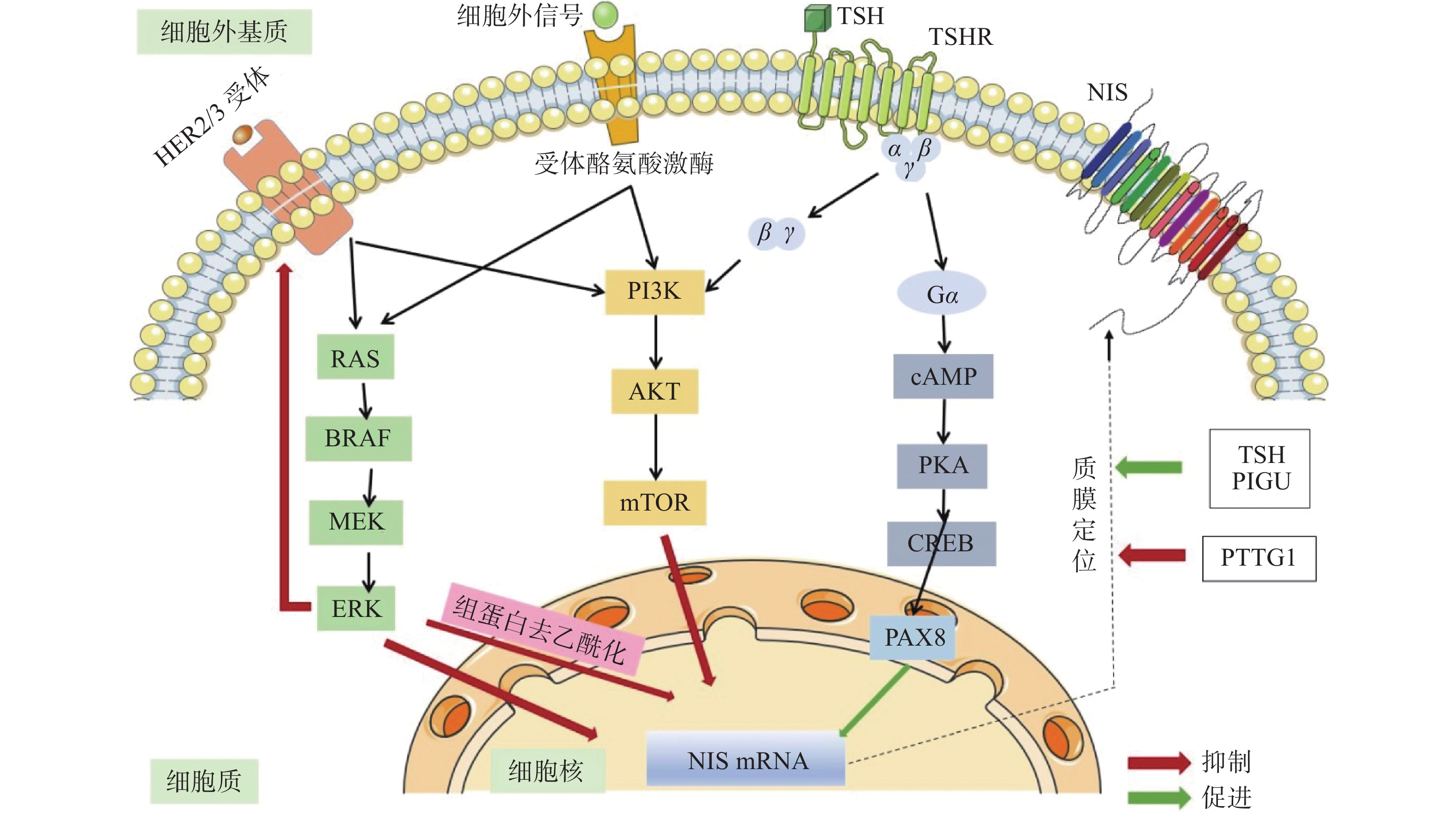
Radioactive iodine (RAI) therapy is one of the critical treatments for differentiated thyroid cancer (DTC). The therapeutic effect is mainly related to the uptake ability of tumor lesions to RAI and radiosensitivity of the tumor cells. The sensitivity to RAI therapy may be improved by the two aforementioned directions in patients who are unsatisfactory in the evaluation of response to RAI therapy. By analyzing the relevant signaling pathways and molecular mechanisms that affect RAI therapy in DTC patients in recent years, the authors summarize the relevant mechanisms affecting RAI uptake, including sodium iodide symporter (NIS) expression and plasma membrane localization, as well as the DNA damage repair mechanisms that affect the radiosensitivity of RAI. It is expected to provide direction for the basic and clinical research of internal irradiation sensitization therapy for DTC patients.
Radioactive iodine (RAI) therapy is one of the critical treatments for differentiated thyroid cancer (DTC). The therapeutic effect is mainly related to the uptake ability of tumor lesions to RAI and radiosensitivity of the tumor cells. The sensitivity to RAI therapy may be improved by the two aforementioned directions in patients who are unsatisfactory in the evaluation of response to RAI therapy. By analyzing the relevant signaling pathways and molecular mechanisms that affect RAI therapy in DTC patients in recent years, the authors summarize the relevant mechanisms affecting RAI uptake, including sodium iodide symporter (NIS) expression and plasma membrane localization, as well as the DNA damage repair mechanisms that affect the radiosensitivity of RAI. It is expected to provide direction for the basic and clinical research of internal irradiation sensitization therapy for DTC patients.
2022, 46(4): 254-258.
doi: 10.3760/cma.j.cn121381-202101032-00154
Abstract:
Strontium-89 chloride (hereinafter referred to as "89Sr") is a radiopharmaceutical for the treatment of bone metastases, especially systemic multiple bone metastases, but its clinical application is limited due to the adverse effect of myelosuppression. In addition to the radiobiological effects of 89Sr itself, bone tumor burden during 89Sr treatment, radiotherapy, chemotherapy and anti-androgen therapy before 89Sr treatment are all important factors leading to myelosuppression. The authors review the related factors of myelosuppression induced by 89Sr treatment of bone metastases.
Strontium-89 chloride (hereinafter referred to as "89Sr") is a radiopharmaceutical for the treatment of bone metastases, especially systemic multiple bone metastases, but its clinical application is limited due to the adverse effect of myelosuppression. In addition to the radiobiological effects of 89Sr itself, bone tumor burden during 89Sr treatment, radiotherapy, chemotherapy and anti-androgen therapy before 89Sr treatment are all important factors leading to myelosuppression. The authors review the related factors of myelosuppression induced by 89Sr treatment of bone metastases.
2022, 46(4): 259-261.
doi: 10.3760/cma.j.cn121381-202103017-00157
Abstract:
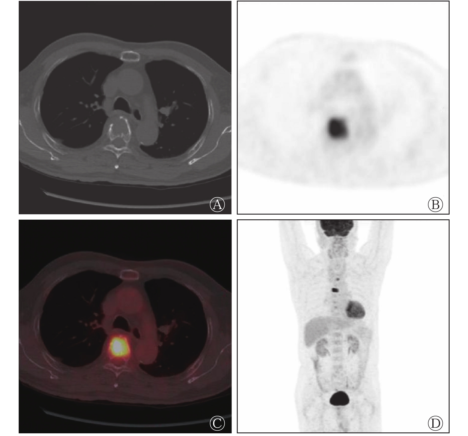
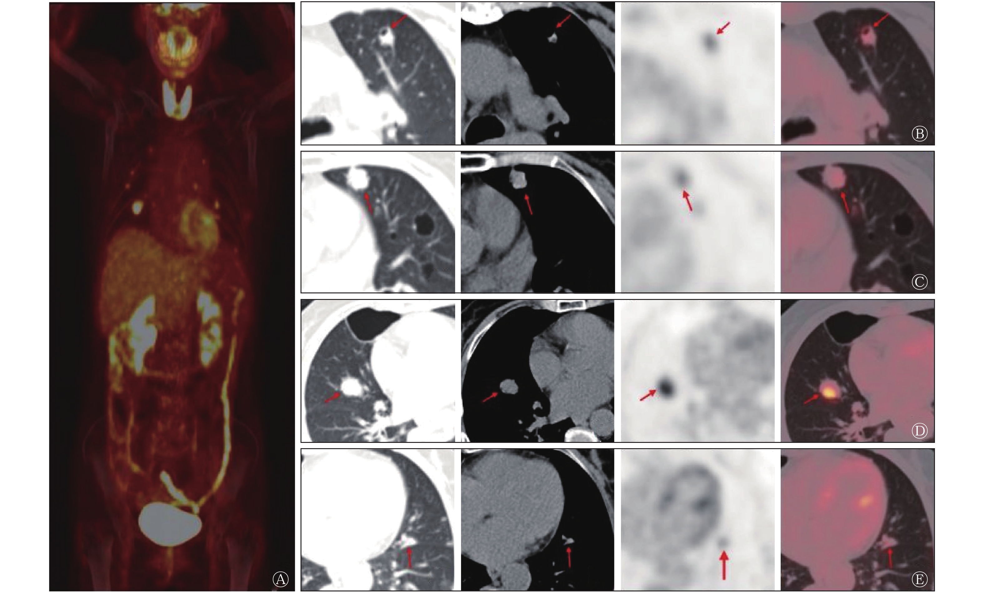
A case of double lung multiple nodular amyloidosis with 18F-fluorodeoxyglucose (FDG) PET/CT imaging was reported. The characteristics of the disease were analyzed from clinical symptoms, imaging manifestations and pathological diagnosis. The understanding of pulmonary amyloidosis was deepened through literature review. The incidence of pulmonary nodular amyloidosis is low, the clinical symptoms are atypical, and it is difficult to differentiate from common lung tumors. It is suggested that the possibility of pulmonary nodular amyloidosis should be considered in the differential diagnosis of pulmonary nodules. Therefore, 18F-FDG PET/CT imaging is very important to evaluate the extent of double lung multiple nodular amyloidosis.
A case of double lung multiple nodular amyloidosis with 18F-fluorodeoxyglucose (FDG) PET/CT imaging was reported. The characteristics of the disease were analyzed from clinical symptoms, imaging manifestations and pathological diagnosis. The understanding of pulmonary amyloidosis was deepened through literature review. The incidence of pulmonary nodular amyloidosis is low, the clinical symptoms are atypical, and it is difficult to differentiate from common lung tumors. It is suggested that the possibility of pulmonary nodular amyloidosis should be considered in the differential diagnosis of pulmonary nodules. Therefore, 18F-FDG PET/CT imaging is very important to evaluate the extent of double lung multiple nodular amyloidosis.

 Submit
 Author Login
 Referees
 Editor-in-Chief
 Editor Center
 Email alert
 RSS
RSS