2020 Vol. 44, No. 6
Display Method:
2020, 44(6): 0-0.
 [PDF 2914KB]
Abstract:
2020, 44(6): 339-344.
doi: 10.3760/cma.j.cn121381-201904009-00028
Abstract:
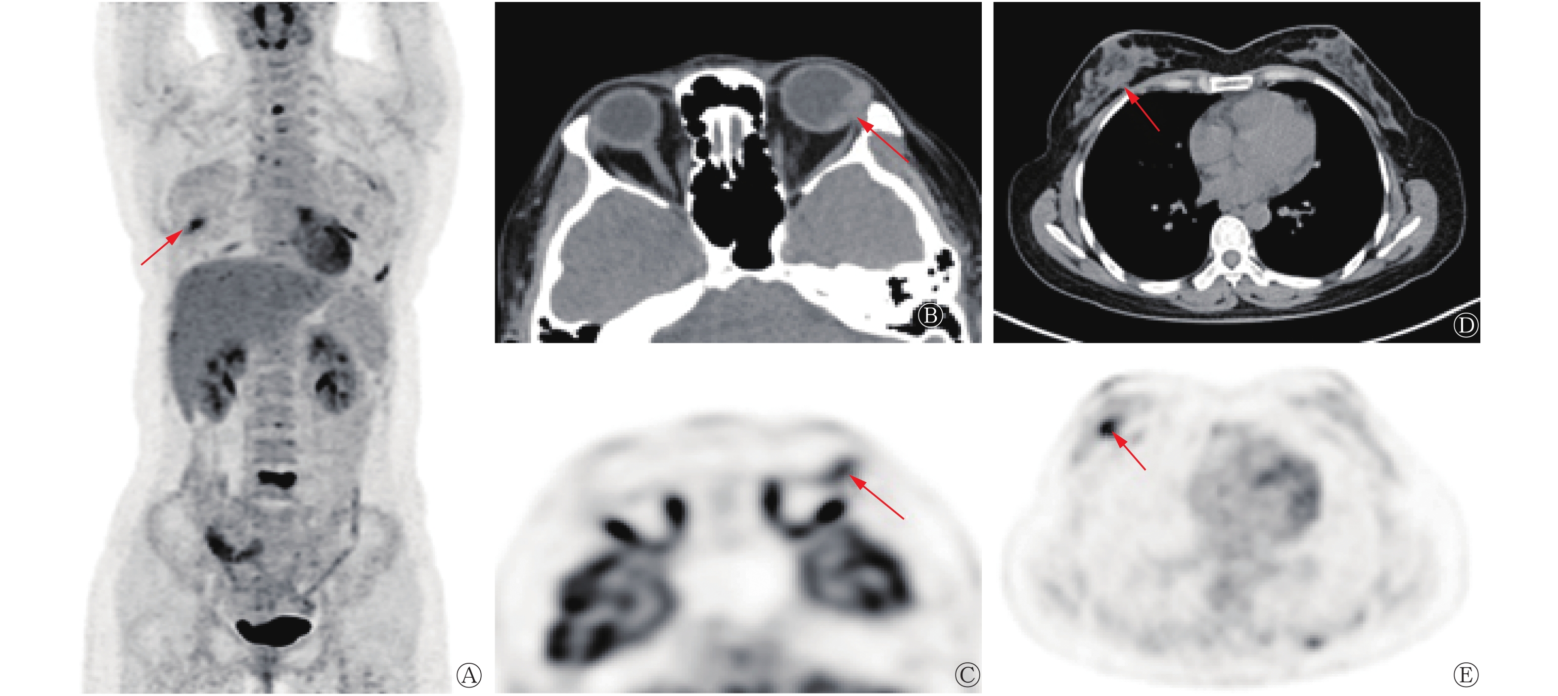
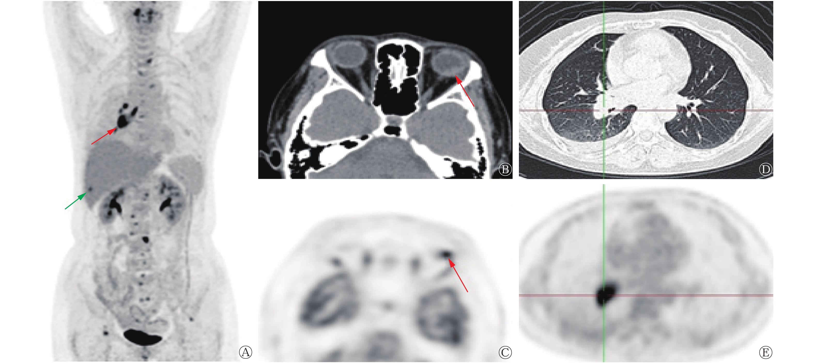
Objective To analyze the features and application value of 18F-FDG PET/CT in detecting intraocular metastases. Methods A total of 53 patients with intraocular metastasis (54 eyes, including 1 case of binocular metastasis) diagnosed by pathology or clinical assessment at the Beijing Hospital and Beijing Tongren Hospital, Capital Medical University from March 2011 to February 2019 were enrolled in this retrospective study. The patients included 23 males and 30 females aged 22–73(52.7±11.6) years. All patients underwent 18F-FDG PET/CT, and the features and parameters of the resulting images, including maximum (SUVmax) and average (SUVmean) standardized uptakes, were analyzed. The value of PET/CT for the detection of primary tumors and metastases was then assessed. Analysis of variance was used to compare the data of multiple groups, and Pearson's correlation analysis was used to determine correlations. Results Intraocular metastases showed a variety of unique CT features, the most common of which was spindle-like soft tissue shadows. No significant linear relationship between the CT value and SUVmax or SUVmean (both r=−0.252; both P=0.088) among 47 cases of measurable intraocular metastasis was detected. SUVmax and SUVmean were positively correlated with their long, short, upper, and lower diameters (r=0.631–0.791; all P=0.000). The detection rate of the primary focus by PET/CT among 43 patients with no prior history of cancer was 97.7% (42/43). Among the cases of intraocular metastasis, 36 originated from lung cancer, 2 were from breast cancer, 1 was from esophageal cancer, 1 was from gastric cancer, 1 was from nasopharyngeal cancer, and 1 was from prostate cancer. PET/CT revealed 51 patients (51/53, 96.2%) with more than two metastatic lesions, 81.1% (43/53) with lymph node metastasis, and 79.2% (42/53) with bone metastasis. However, PET/CT may result in false negative findings for small intraocular and brain metastases. Conclusions Intraocular metastases exhibit a variety of distinct CT features, and only the SUV is related to the tumor size. 18F-FDG PET/CT is useful for the diagnosis of intraocular metastases, exploration of primary tumors, and discovery of metastases in other sites.
2020, 44(6): 345-351.
doi: 10.3760/cma.j.cn121381-201910034-00029
Abstract:
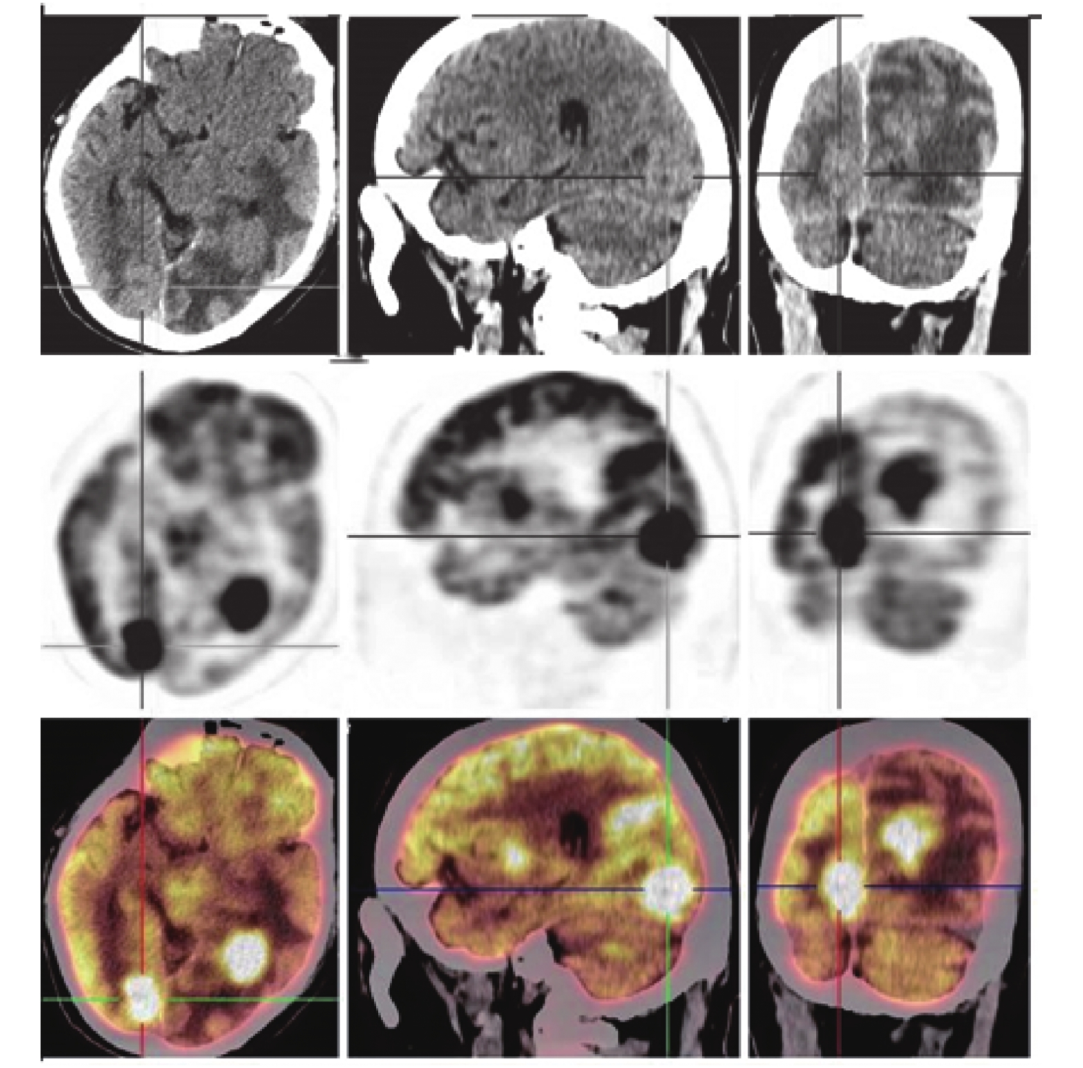
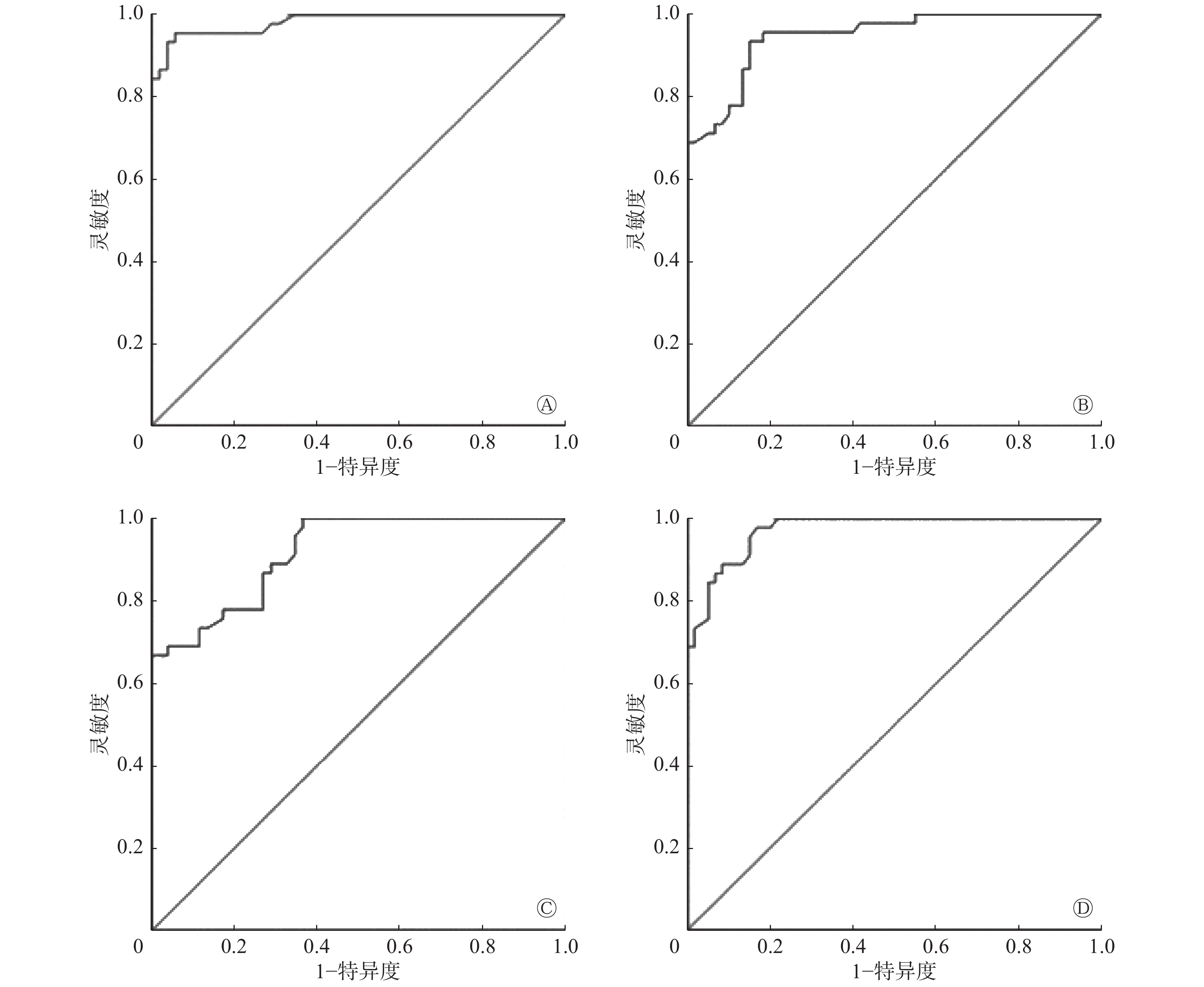
Objective To investigate the diagnostic value of fluorine-18 fluorodeoxyglucose (18F-FDG) PET/CT visual and semi-quantitative analyses in intracranial primary central nervous system lymphoma (PCNSL). Methods PET/CT images of 45 patients with PCNSL who underwent 18F-FDG PET/CT examination in the Department of Nuclear Medicine of the First Affiliated Hospital of Zhengzhou University from May 2011 to December 2018 (26 males and 19 females, 57.49±2.54 years old) were retrospectively reviewed and compared with 52 cases of gliomas and 60 cases of brain metastases to evaluate the value of 18F-FDG PET/CT in the diagnosis of intracranial PCNSL. The lesion distribution and morphological characteristics of the 3 groups of patients were visually analyzed, and the maximum standardized uptake value (SUVmax) and the ratio of SUVmax of tumor to white matter (T/WM) were semi-quantitatively analyzed. The mean comparison between the two groups was performed using independent sample t test and adjusted t test. The comparison of the diagnostic efficacy between the two groups and the judgment of the differential diagnosis threshold were performed using receiver operating characteristic (ROC) curve analysis. Results Visually, intracranial PCNSL showed a very high uptake of 18F-FDG in single, focal nodule or mass lesions mostly located in the supratentorial brain. The space-occupying effects of edema, as well as cystic degeneration, were not obvious in PCNSL. Semi-quantitative analysis showed that intracranial PCNSL had the highest SUVmax (gliomas: 9.96±0.48, brain metastases: 11.97±0.58, PCNSL: 26.42±1.17) and T/WM (gliomas: 2.99±0.09, brain metastases: 2.60±0.08, PCNSL: 4.37±0.10) among the three types of tumors with statistical differences (t=13.02 and 11.07, t=10.13 and 13.88, all P=0.000). In the differential diagnosis of intracranial PCNSL and glioma, the area under the ROC curve (AUC) analysis reached the largest value at the SUVmax of 15.8. The AUC for PCNSL and metastatic tumor peaked at the SUVmax of 16.8. The T/WMs of 3.395 and 3.220 were considered the optimal thresholds for the differential diagnosis of intracranial PCNSL from gliomas and brain metastases, respectively. Conclusion 18F-FDG PET/CT imaging can effectively complement the traditional diagnosis of intracranial PCNSL, especially in the differential diagnosis of PCNSL from gliomas and brain metastases.
2020, 44(6): 352-358.
doi: 10.3760/cma.j.cn121381-201903012-00037
Abstract:
Objective To investigate the effect of parathyroid lesion weight on the diagnostic sensitivity of 99Tcm-MIBI dual-phase plane imaging and 99Tcm-MIBI SPECT/CT tomography fusion imaging. Methods A total of 22 patients with hyperparathyroidism that was confirmed via operation and pathology in the First People's Hospital of Kunshan were collected from February 2017 to October 2018. The patients included 9 males and 13 females aged 28–73 (50.77±8.79) years old. All patients underwent 99Tcm-MIBI biphasic imaging and 99Tcm-MIBI SPECT/CT fusion imaging in the early stage. The gold standard was postoperative pathological results. All resected lesions were divided into two groups: group A, wherein lesion weight ≤1.00 g, and group B, wherein lesion weight >1.00 g. χ2 test was used to analyze the diagnostic efficacy of the two imaging methods in the different weight groups. Results A total of 58 lesions were removed from the 22 patients via surgical operation. The diagnostic sensitivity values of 99Tcm-MIBI dual-phase plane imaging for groups A and B were 47.83% (11/23) and 84.00% (21/25) respectively, and were statistically significantly different between the two groups (χ2=7.05, P=0.008). The diagnostic sensitivity values of 99Tcm-MIBI SPECT/CT early tomography for groups A and B were 78.26% (18/23) and 85.19% (23/27), respectively, and did not show significant differences between the two groups (χ2=0.40, P=0.525). In group A, the diagnostic sensitivity of 99Tcm-MIBI SPECT/CT early tomography was higher than that of dual-phase plane imaging; this difference was statistically significant (χ2=4.57, P=0.033). In group B, the diagnostic sensitivity of 99Tcm-MIBI SPECT/CT early tomography was higher than that of dual-phase plane imaging; however, no significant difference was observed (χ2=0.01, P=0.906). Conclusions The weight of parathyroid lesions has an effect on dual-phase planar imaging. Specifically, sensitivity decreased when the parathyroid glands were light. However, lesion weight had no significant effect on the diagnostic sensitivity of early tomographic fusion imaging.


2020, 44(6): 359-364.
doi: 10.3760/cma.j.cn121381-201903046-00040
Abstract:
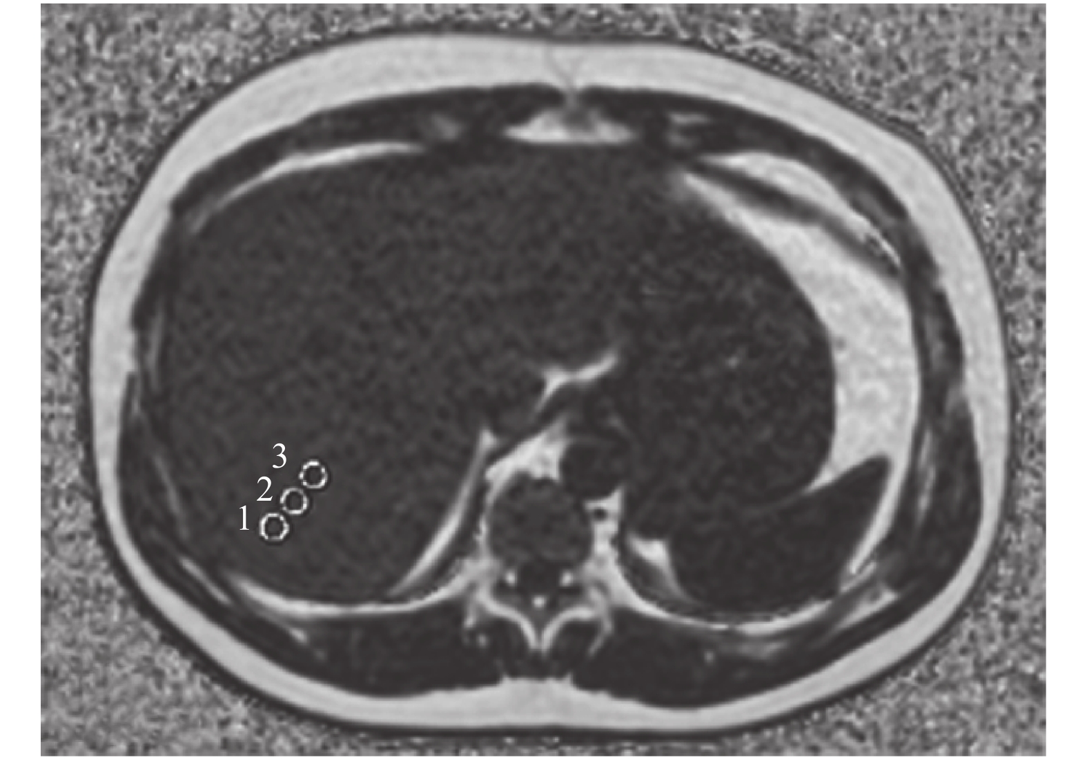
Objective To explore the effect of radiologists' qualifications and region of interest (ROI) settings on the evaluation of liver proton density fat fraction (PDFF) by iterative decomposition of water and fat with echo asymmetry and least-squares estimation and image quantification (IDEAL-IQ). Methods A total of 43 patients(39 males and 4 females, 40.37±14.65 years old) underwent 3D IDEAL-IQ scanning by a GE Discovery MR750W 3.0 T MRI scanner at the First People's Hospital of Foshan. PDFFs were determined on fat fraction maps by three different radiologists with ROIs of 10, 25, and 50 mm2. When the measurement data showed a normal distribution and the variance was homogeneous, one-way ANOVA was used for statistical analysis. The consistency of the same group of data measured repeatedly by the same researcher according to the ROI of different areas with that measured by different radiologists was evaluated via reliability analysis by using SPSS 17.0. Intra-group correlation coefficients ( Results The PDFFs measured with three different ROIs by the same doctor were (14.17±8.40)%, (13.49±8.42)%, and (13.25±8.39)%, respectively. There was not significant difference (F=0.138, P=0.871).The PDFFs measured by three doctors according to the same ROI respectively were (14.10±8.81)%, (12.75±8.48)% and (14.06±8.22)%, respectively. The difference was not statistically significant (F=0.352, P=0.704). The same radiologists determined the same group of data on the fat fraction maps with different ROIs, and reliability analysis indicated alpha>0.8 and ICC>0.75. These results indicate that the determination of PDFFs with different ROIs has high reliability and repeatability. The three radiologists then measured the same group of data on fat fraction maps with the same ROI, and reliability analysis indicated alpha>0.8 and ICC>0.75. Differences between the PDFFs measured at 10, 20, and 30 mm involved the vertical distance between the center of the ROI and the hepatic envelope. Conclusions Radiologists' qualifications and ROI settings have no effect on the IDEAL-IQ sequence evaluation of liver PDFF. Measurements made under different qualifications and ROIs by using the IDEAL-IQ technique have high reliability and repeatability.

2020, 44(6): 365-373.
doi: 10.3760/cma.j.cn121381-202002028-00032
Abstract:
Objective To analyze the quality control acceptance test results of partial types of medical digital radiography equipment, evaluate their application quality and provide references for the subsequent detection. Methods According to the Specifications of the quality control testing of medical X-ray diagnostic equipment (WS 76-2017) and the Specifications of quality control testing in medical digital radiography (DR) systems (WS 521-2017), 17 types (19 sets in total) of newly installed medical digital radiography equipment in Tianjin and Inner Mongolia from January 2018 to June 2019 were selected as the research objects. General and special test items, such as the deviation of tube voltage indication, output repeatability, dark noise, limited spatial resolution and low contrast details, and so on, were tested. Finally, the test results were recorded and analyzed. Results In addition to dark noise, detector dose indication, limited spatial resolution, low contrast detail detection, automatic exposure control (AEC) sensitivity, and AEC tube voltage change consistency to establish the baseline value, the detection results of other medical digital radiography equipment met the requirements of WS 76-2017 and WS 521-2017. The deviations of 60 and 80 kV tube voltage indication ranged from −3.61% to 2.71%, whereas the deviations of 100, 117, 120 and 121 kV tube voltage indication ranged from −2.9 to 4.9 kV. Meanwhile, the repeatability of output ranged from 0.0290% to 1.4700%, and the useful harness half value layers corresponding to 80 and 81 kV were between 2.5 and 5.9 mmAl. The deviations of exposure time indication in the range of ≥100 ms and <100 ms were −5.19% to 2.40% and −7.15% to 2.80%, respectively. There were no residuals and artifacts found in the imaging. The baseline values established by dark noise detection ranged from 14.2 to 5262.0, whereas the baseline values established by the detector dose indication were between 188 and 60,280. The baseline values of AEC sensitivity and those of the AEC tube voltage change consistency ranged from 0.67 to 6.90 mAs and from 2.20 to 6.69 μGy, respectively. The measurement results of response uniformity were between 0.11% and 4.70%. The ranging error were −0.40%−1.45%. The consistency between AEC ionization chambers were −8.70%−5.15%. The results of the limit spatial resolution in the horizontal and vertical directions ranged from 2.2 to 3.7 lp/mm and 2.2 to 3.4 lp/mm, respectively. Finally, the determination coefficients of the fitting formulas of signal transmission characteristics were all greater than 0.98. Conclusions The quality control acceptance test results of the medical digital radiography equipment met the requirements of the WS 76-2017 and WS 521-2017 standards. Clear and complete test conditions and acceptance test results can provide reference for the subsequent tests.
2020, 44(6): 374-380.
doi: 10.3760/cma.j.cn121381-201903043-00033
Abstract:
Radiotherapy is one of the important means of combined therapy for treating tumors. However, radioresistance is a serious issue that affects the curative effect and prognosis of radiotherapy for tumor patients. Given the complex mechanism of radioresistance in tumor cells, the specific switch molecules that can regulate radiosensitivity are not yet discovered. Circular RNA (circRNA) is a kind of closed circular RNA molecules that covalently bind the 3'-end and 5'-end by trans-splicing with high abundance, stable structure, and strong specificity. CircRNA is involved in tumorigenesis, development, invasion, and metastasis and can be used as a novel tumor molecular marker and potential therapeutic target. In addition, circRNA is differentially expressed in irradiated tumor cells and can serve as a sponge to regulate microRNA and its downstream signaling pathways that are related to tumor radioresistance. Therefore, the research on circRNA might be a promising breakthrough toward overcoming tumor radioresistance. In this study, we reviewed the progress in the research on circRNAs as novel tumor markers, as well as the research prospects regarding the application of radiotherapy.
Radiotherapy is one of the important means of combined therapy for treating tumors. However, radioresistance is a serious issue that affects the curative effect and prognosis of radiotherapy for tumor patients. Given the complex mechanism of radioresistance in tumor cells, the specific switch molecules that can regulate radiosensitivity are not yet discovered. Circular RNA (circRNA) is a kind of closed circular RNA molecules that covalently bind the 3'-end and 5'-end by trans-splicing with high abundance, stable structure, and strong specificity. CircRNA is involved in tumorigenesis, development, invasion, and metastasis and can be used as a novel tumor molecular marker and potential therapeutic target. In addition, circRNA is differentially expressed in irradiated tumor cells and can serve as a sponge to regulate microRNA and its downstream signaling pathways that are related to tumor radioresistance. Therefore, the research on circRNA might be a promising breakthrough toward overcoming tumor radioresistance. In this study, we reviewed the progress in the research on circRNAs as novel tumor markers, as well as the research prospects regarding the application of radiotherapy.
2020, 44(6): 381-385.
doi: 10.3760/cma.j.cn121381-201906003-00039
Abstract:
Radiotherapy is one of the three conventional methods for the treatment of malignant tumors. However, its expected effect is not often achieved because of some limitations, such as normal tissue damage by high radiation dose and the radiation resistance of tumor cells. Thus, exploring new strategies for new radiosensitizers and radio-chemotherapy agents has become a research focus to improve the efficacy of radiotherapy and reduce its side effects on normal tissues. Polymer nanomaterials have broad application prospects in improving the effect of radiotherapy because of their excellent biocompatibility and physiological stability. This article reviews the research progress on polymer nanomaterials for radiosensitization.
Radiotherapy is one of the three conventional methods for the treatment of malignant tumors. However, its expected effect is not often achieved because of some limitations, such as normal tissue damage by high radiation dose and the radiation resistance of tumor cells. Thus, exploring new strategies for new radiosensitizers and radio-chemotherapy agents has become a research focus to improve the efficacy of radiotherapy and reduce its side effects on normal tissues. Polymer nanomaterials have broad application prospects in improving the effect of radiotherapy because of their excellent biocompatibility and physiological stability. This article reviews the research progress on polymer nanomaterials for radiosensitization.
2020, 44(6): 386-393.
doi: 10.3760/cma.j.cn121381-201903023-00030
Abstract:
Metastatic renal cell carcinoma (mRCC) is insensitive to both radiotherapy and chemotherapy; hence, the prognosis of patients with mRCC is poor. With the development of molecular targeted therapy, various targeted drugs, such as sunitinib and sorafenib, have greatly improved the prognosis of mRCC. However, targeted therapy is not effective for some patients, and targeted drugs may also cause related adverse reactions. Therefore, early non-invasive assessment of tumor response to targeted drugs is particularly important to allow patients and physicians to decide on the best course of treatment. However, researchers worldwide have not yet found an ideal biomarker for mRCC. As a functional imaging technology, the clinical value of PET/CT has been recognized in diagnosing numerous tumors. In recent years, the application of PET/CT in mRCC has gradually increased. This review focuses on the application of PET/CT in patients with mRCC. In particular, this review discusses the value and limitations of the application of PET/CT in evaluating targeted treatment response and prognosis.
Metastatic renal cell carcinoma (mRCC) is insensitive to both radiotherapy and chemotherapy; hence, the prognosis of patients with mRCC is poor. With the development of molecular targeted therapy, various targeted drugs, such as sunitinib and sorafenib, have greatly improved the prognosis of mRCC. However, targeted therapy is not effective for some patients, and targeted drugs may also cause related adverse reactions. Therefore, early non-invasive assessment of tumor response to targeted drugs is particularly important to allow patients and physicians to decide on the best course of treatment. However, researchers worldwide have not yet found an ideal biomarker for mRCC. As a functional imaging technology, the clinical value of PET/CT has been recognized in diagnosing numerous tumors. In recent years, the application of PET/CT in mRCC has gradually increased. This review focuses on the application of PET/CT in patients with mRCC. In particular, this review discusses the value and limitations of the application of PET/CT in evaluating targeted treatment response and prognosis.
2020, 44(6): 394-398.
doi: 10.3760/cma.j.cn121381-201903064-00041
Abstract:
Myocardial perfusion imaging plays an important role in the diagnosis and treatment of patients with known or suspected coronary artery disease. The cadmium-zinc-telluride (CZT)-based dedicated cardiac SPECT (called CZT-SPECT) with high resolution and photon sensitivity reduces radiation exposure and imaging time while improving image quality and enhancing the value of SPECT myocardial perfusion imaging in clinical practice. The review elaborates the clinical progress of CZT-SPECT.
Myocardial perfusion imaging plays an important role in the diagnosis and treatment of patients with known or suspected coronary artery disease. The cadmium-zinc-telluride (CZT)-based dedicated cardiac SPECT (called CZT-SPECT) with high resolution and photon sensitivity reduces radiation exposure and imaging time while improving image quality and enhancing the value of SPECT myocardial perfusion imaging in clinical practice. The review elaborates the clinical progress of CZT-SPECT.
2020, 44(6): 399-404.
doi: 10.3760/cma.j.cn121381-202003043-00043
Abstract:
Right heart hemodynamics play a crucial role in the circulatory system. Biochemical indicators and clinical trials can only evaluate the right ventricular function indirectly. Imaging techniques, such as echocardiography, nuclide blood pool imaging, and cardiac magnetic resonance imaging, increase the objectivity, accuracy, and comprehensiveness of the evaluation of the right ventricular function. To obtain a comprehensive understanding of the technology for evaluating the right ventricular function, the author reviews the application of the abovementioned inspection methods in such evaluations.
Right heart hemodynamics play a crucial role in the circulatory system. Biochemical indicators and clinical trials can only evaluate the right ventricular function indirectly. Imaging techniques, such as echocardiography, nuclide blood pool imaging, and cardiac magnetic resonance imaging, increase the objectivity, accuracy, and comprehensiveness of the evaluation of the right ventricular function. To obtain a comprehensive understanding of the technology for evaluating the right ventricular function, the author reviews the application of the abovementioned inspection methods in such evaluations.
 Submit
 Author Login
 Referees
 Editor-in-Chief
 Editor Center
 Email alert
 RSS
RSS