2020 Vol. 44, No. 1
Display Method:
 [PDF 2965KB]
2020, 44(1): 0-0.
Abstract:
2020, 44(1): 2-4.
doi: 10.3760/cma.j.issn.1673-4114.2020.01.002
Abstract:
In the field of medical application, artificial intelligence in medical image has made rapid progress in the aspect of industry, education and research. The research of artificial intelligence in medical imaging has produced good research and development situation and broad clinical application, and its the progress is exhibited in medical imaging equipment, image diagnosis and intelligent service. Academic exchanges are becoming increasingly active in exploring the academic progress and the development of artificial intelligence. Authoritative reports on the development of medical artificial intelligence have also been released. This issue focuses on a number of research articles on artificial intelligence in medical imaging, and shows the latest research findings on artificial intelligence and product application from different angles.
In the field of medical application, artificial intelligence in medical image has made rapid progress in the aspect of industry, education and research. The research of artificial intelligence in medical imaging has produced good research and development situation and broad clinical application, and its the progress is exhibited in medical imaging equipment, image diagnosis and intelligent service. Academic exchanges are becoming increasingly active in exploring the academic progress and the development of artificial intelligence. Authoritative reports on the development of medical artificial intelligence have also been released. This issue focuses on a number of research articles on artificial intelligence in medical imaging, and shows the latest research findings on artificial intelligence and product application from different angles.
2020, 44(1): 5-10.
doi: 10.3760/cma.j.issn.1673-4114.2020.01.003
Abstract:
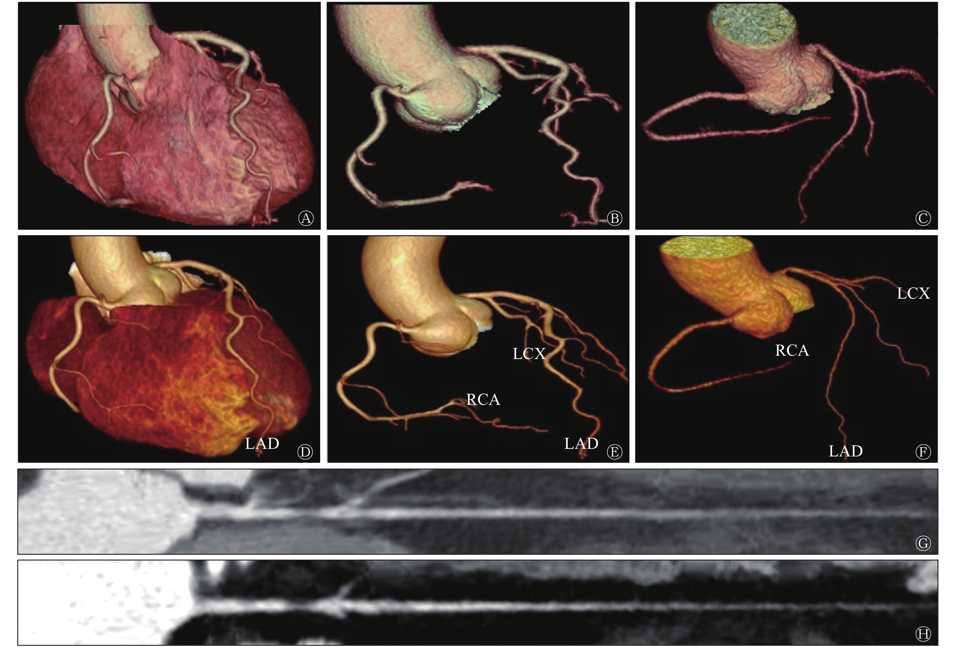
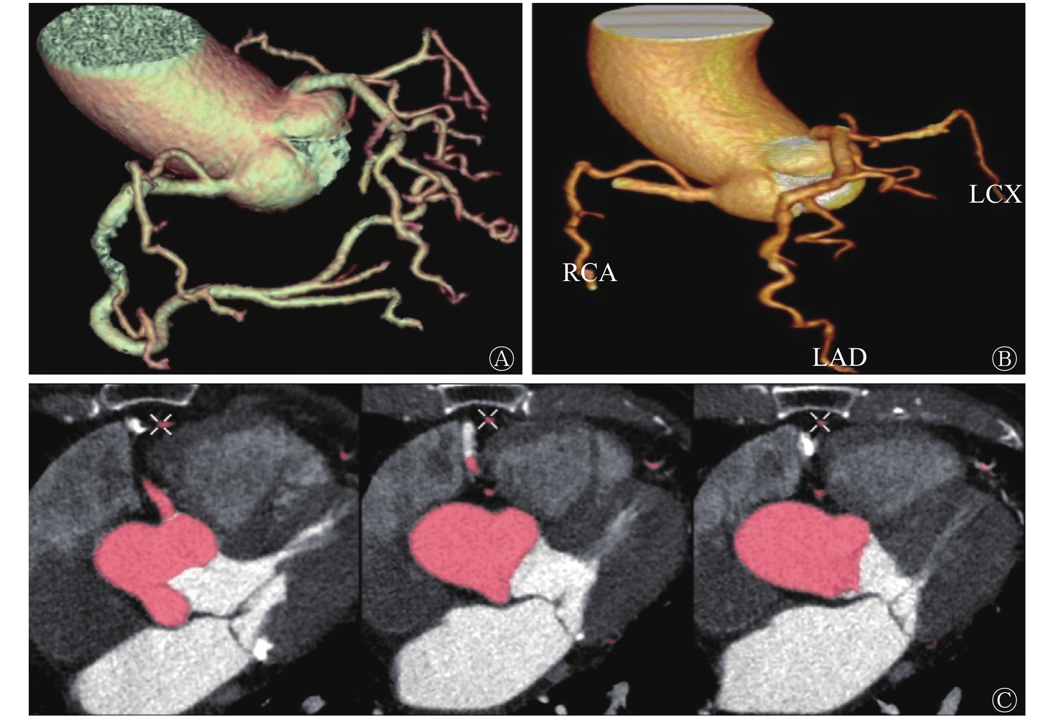
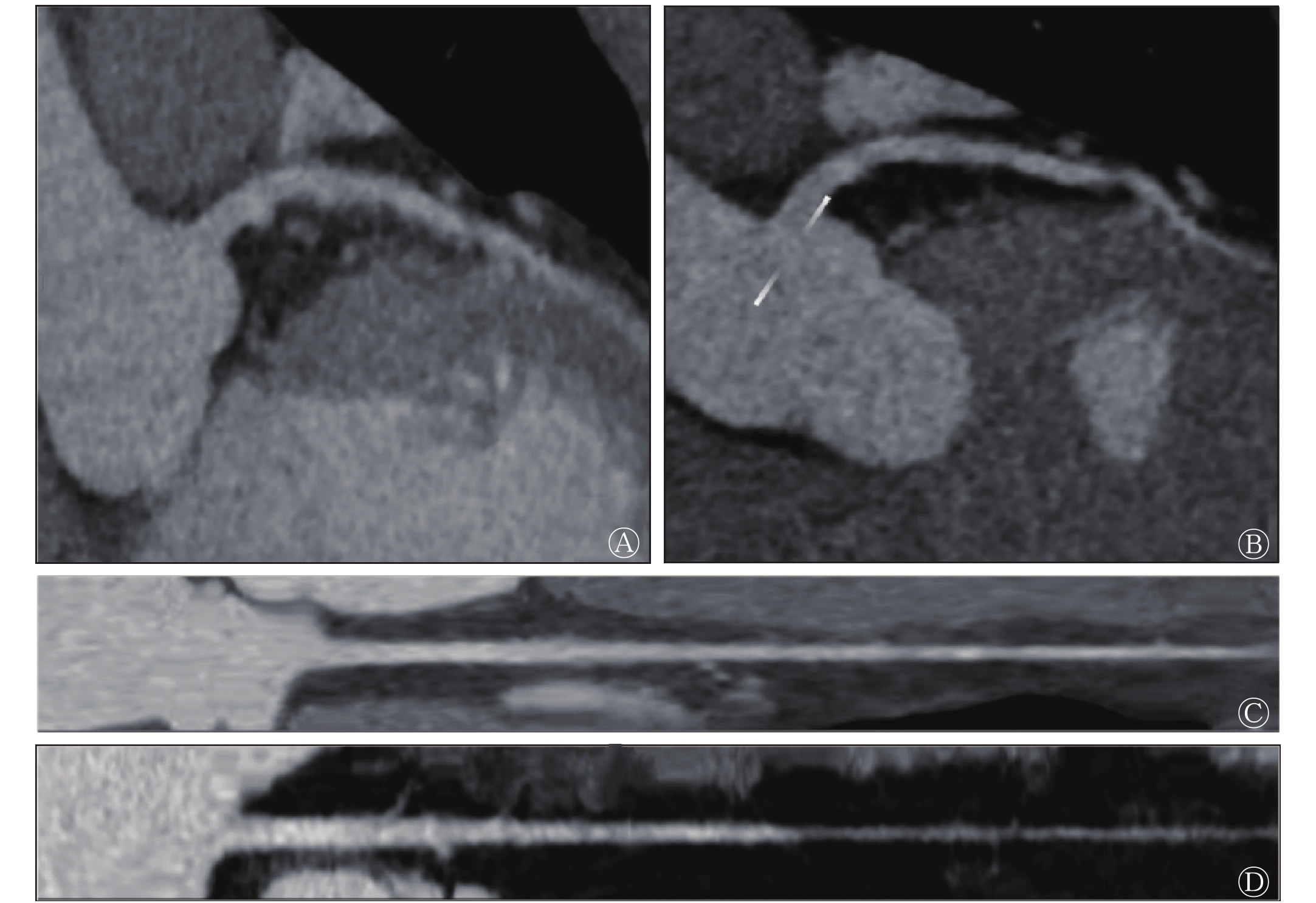
Objective To explore the value of coronary artificial intelligence (AI) in the post-processing and diagnosis of coronary CT angiography (CCTA). Methods Sixty-four patients with suspected coronary heart disease who were admitted to Third Affiliated Hospital of Chongqing Medical University from April to July 2019, including 40 males and 24 females, aged (62.16±14.13) years, were randomly selected. All patients underwent coronary CT angiography. The original image quality was scored in accordance with the Likert scoring standard, and artificial and AI image post-processing were carried out. The time, qualified rate, time of the diagnosis report, and diagnostic efficiency of the two were compared. Results The post-processing time of AI images of the coronary arteries was about 3 min, and the time of artificial post-processing was 20−30 min after CCTA. The qualified rate of AI post-processing of the coronary arteries was 92.2% (59/64). Compared with manual processing, the AI images of the coronary arteries after processing were smoother, had more small branches and clearer vessel contrast, and can automatically identify coronary artery stenosis. The diagnosis report of coronary artery AI images was completed immediately after image reconstruction (< 1 min), whereas the artificial diagnosis report was about 15 min. The sensitivity of AI plaque in the coronary artery was almost the same as that of artificial detection (i.e., 93.3% and 92.0%, respectively). The specificity of the artificial diagnosis report was 100% and that of AI was 93.8%. Conclusion Coronary AI has certain advantages in image post-processing speed, image quality and efficiency of reporting diagnosis, and is expected to be an effective auxiliary tool for CCTA analysis.
2020, 44(1): 11-15.
doi: 10.3760/cma.j.issn.1673-4114.2020.01.004
Abstract:
With the advent of the era of big data, artificial intelligence (AI) has emerged and rapidly developed in the field of medicine. The application of AI has huge potential in achieving prompt and accurate analysis of tumor information aggregation. Moreover, AI can reflect the distribution of imaging data in the real environment by using key technologies, such as automatic image segmentation and extraction. Accordingly, tumor diagnosis can change from subjective perception into objective science. Therefore, AI can assist doctors in efficiently and accurately diagnosing the presence of tumors and providing a solid foundation for the formulation of an appropriate diagnosis plan and informed judgment of the prognosis. This paper reviews the key AI technologies and their current applications in tumor diagnosis.
With the advent of the era of big data, artificial intelligence (AI) has emerged and rapidly developed in the field of medicine. The application of AI has huge potential in achieving prompt and accurate analysis of tumor information aggregation. Moreover, AI can reflect the distribution of imaging data in the real environment by using key technologies, such as automatic image segmentation and extraction. Accordingly, tumor diagnosis can change from subjective perception into objective science. Therefore, AI can assist doctors in efficiently and accurately diagnosing the presence of tumors and providing a solid foundation for the formulation of an appropriate diagnosis plan and informed judgment of the prognosis. This paper reviews the key AI technologies and their current applications in tumor diagnosis.
2020, 44(1): 16-21.
doi: 10.3760/cma.j.issn.1673-4114.2020.01.005
Abstract:
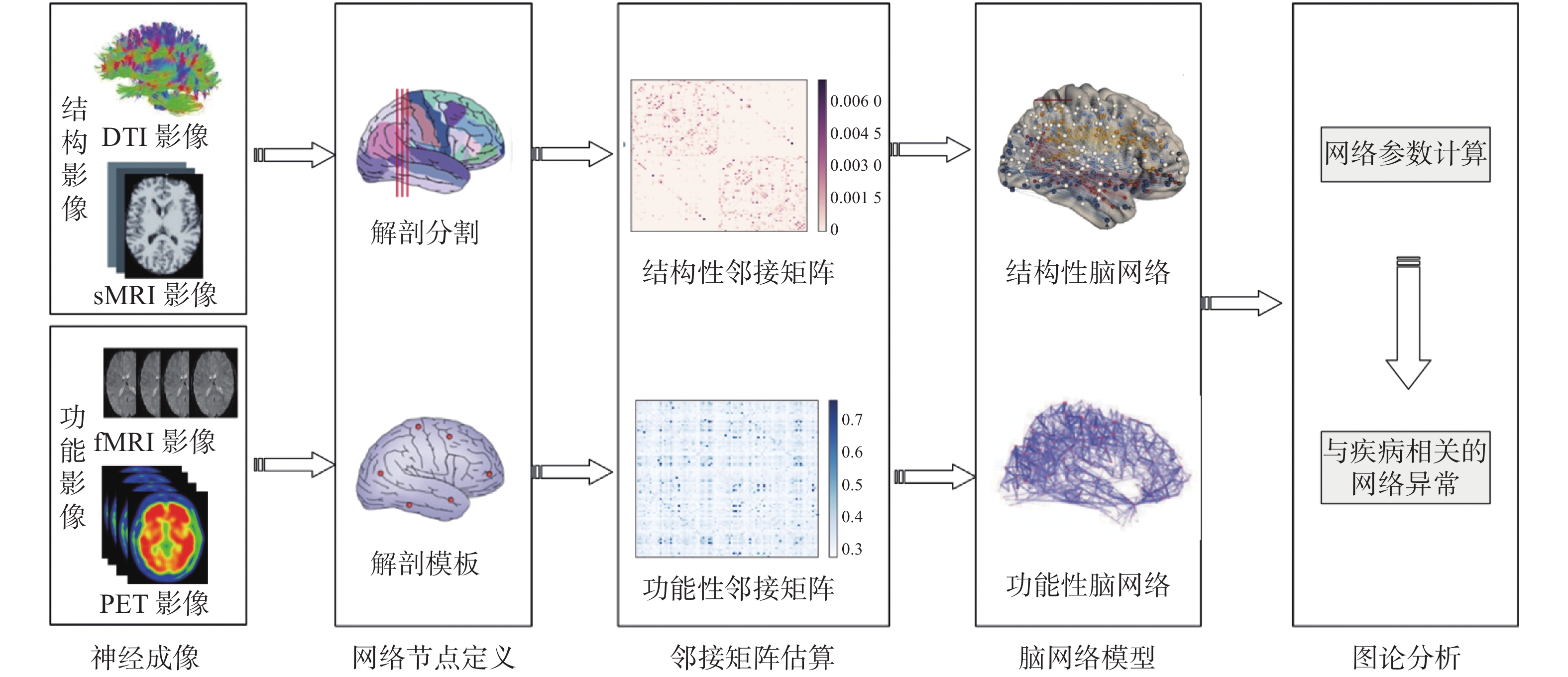
Artificial intelligence techniques have been widely applied in computer-aided diagnosis and disease mechanism studies for Alzheimer's disease (AD). Graph-based complex network analysis is one of the common data mining methods. A combination of complex network analysis technology and multimodal brain imaging information from neuroimaging methods, such as structural magnetic resonance imaging, functional magnetic resonance imaging, and positron emission computer imaging, could identify the abnormal changes of topological properties in brain structure and functional networks. This result provided new ideas on achieving early diagnosis and mechanism research in patients with AD. In this paper, the clinical application of complex network analysis method in structure and functional AD brain imaging was discussed, and its development trend was prospected.
Artificial intelligence techniques have been widely applied in computer-aided diagnosis and disease mechanism studies for Alzheimer's disease (AD). Graph-based complex network analysis is one of the common data mining methods. A combination of complex network analysis technology and multimodal brain imaging information from neuroimaging methods, such as structural magnetic resonance imaging, functional magnetic resonance imaging, and positron emission computer imaging, could identify the abnormal changes of topological properties in brain structure and functional networks. This result provided new ideas on achieving early diagnosis and mechanism research in patients with AD. In this paper, the clinical application of complex network analysis method in structure and functional AD brain imaging was discussed, and its development trend was prospected.
2020, 44(1): 22-26.
doi: 10.3760/cma.j.issn.1673-4114.2020.01.006
Abstract:
Chest CT scan is the primary medical imaging method performed for the early screening and diagnosis of lung cancer. Deep-learning based computer aided diagnosis (CAD) system for chest CT imaging is helpful for detecting and classifying pulmonary nodules. Deep-learning techniques can improve the performance of CAD systems, especially in enhancing the accuracy of pulmonary nodule detection and reducing false-positive rates. This article reviewed the current application status of deep-learning models in CAD systems and the progress that has been achieved in using these systems for imaging pulmonary nodules.
Chest CT scan is the primary medical imaging method performed for the early screening and diagnosis of lung cancer. Deep-learning based computer aided diagnosis (CAD) system for chest CT imaging is helpful for detecting and classifying pulmonary nodules. Deep-learning techniques can improve the performance of CAD systems, especially in enhancing the accuracy of pulmonary nodule detection and reducing false-positive rates. This article reviewed the current application status of deep-learning models in CAD systems and the progress that has been achieved in using these systems for imaging pulmonary nodules.
2020, 44(1): 27-31.
doi: 10.3760/cma.j.issn.1673-4114.2020.01.007
Abstract:
With its application to various fields, artificial intelligence (AI) has become a research hotspot in today's society. The current shortage of personnel in the medical industry and the increased rated of medical diagnosis are crucial for AI application in the medical industry, especially in imaging diagnosis. AI-assisted diagnosis can improve the detection rate of diseases, provide effective diagnostic and treatment information for clinicians, and reduce the repetitive work of imaging physicians, thereby saving time for the research of difficult cases. In this paper, medical imaging AI is briefly introduced, and the latest and most influential research results at home and abroad are combined to explore the new development of medical imaging AI.
With its application to various fields, artificial intelligence (AI) has become a research hotspot in today's society. The current shortage of personnel in the medical industry and the increased rated of medical diagnosis are crucial for AI application in the medical industry, especially in imaging diagnosis. AI-assisted diagnosis can improve the detection rate of diseases, provide effective diagnostic and treatment information for clinicians, and reduce the repetitive work of imaging physicians, thereby saving time for the research of difficult cases. In this paper, medical imaging AI is briefly introduced, and the latest and most influential research results at home and abroad are combined to explore the new development of medical imaging AI.
2020, 44(1): 32-36.
doi: 10.3760/cma.j.issn.1673-4114.2020.01.008
Abstract:
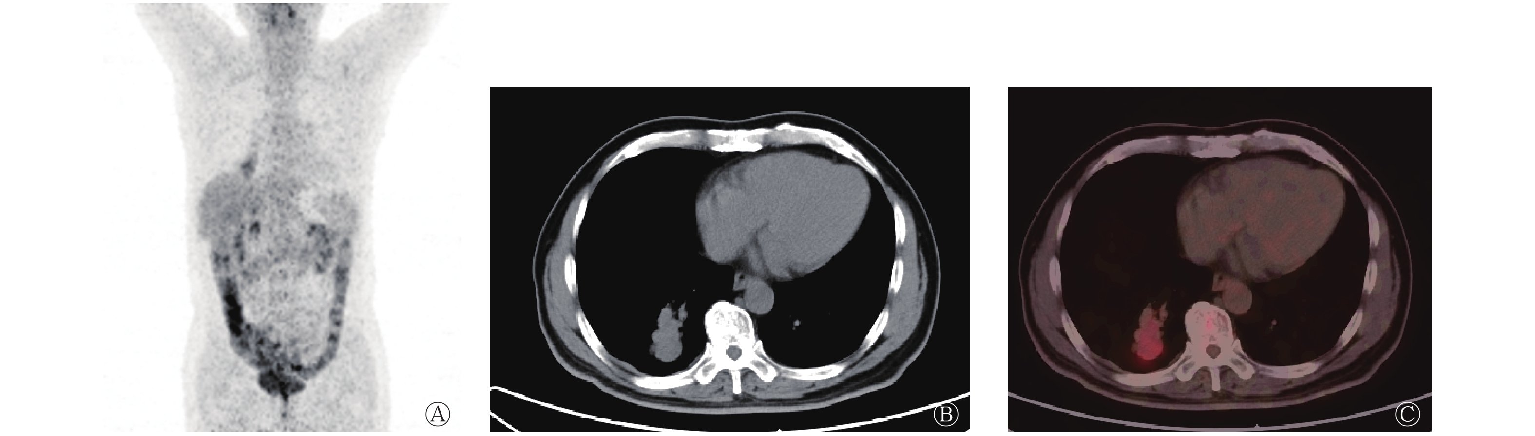
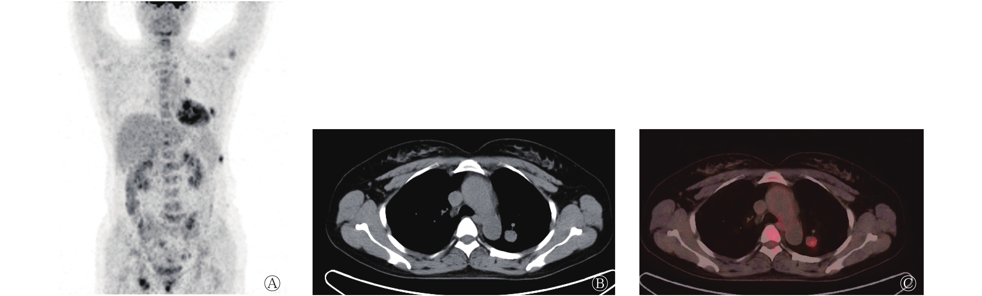
Objective To analyze the image features of lung carcinoid tumor during 18F-fluorodeoxyglucose (18F-FDG) PET/CT. Methods The 18F-FDG PET/CT image manifestations of 16 patients with lung carcinoma, as confirmed by pathology, who underwent 18F-FDG PET/CT pretreatment from March 2009 to August 2018 were reviewed. The location, shape, whole-body metastasis, and metabolism of the lesions were observed. The 16 cases included 6 males and 10 females aged 43−78 years old, with a median age of 65 years. The cases included 5 typical carcinoma (TC) and 11 atypical carcinoma (AC). The statistical difference in maximum standardized uptake value (SUVmax) between TC and AC was investigated using t test. Results All the 16 cases were solitary and showed uniform lesion density and no necrotic cystic degeneration nor calcification. The CT value was(38±7) HU. Nine and seven cases were central and peripheral types, respectively. Out of the nine central type cases, eight showed iceberg lesions, and five presented obstructive pneumonia. However, obstructive pneumonia was absent in the seven cases of peripheral pulmonary carcinoma. In total, 1 case exhibited multiple bone metastases through PET/CT, whereas the remaining 15 cases revealed no lymph node nor distant metastasis. The tumor size and SUVmax were (2.59±1.00) cm and 4.00±1.64, respectively. No correlation was found between the SUVmax and tumor size (r=0.238, P=0.375). The SUVmax of TC and AC reached 3.32±1.17 and 5.49±1.60, respectively, with significant difference between two groups (t=−3.083, P=0.008). Conclusions The 18F-FDG PET/CT manifestations of most lung carcinoid tumors were round soft tissue nodules or masses with mild 18F-FDG uptake. Iceberg sign was detected in most central lung carcinomas, whereas necrotic cystic degeneration and calcification were rare.
2020, 44(1): 37-44.
doi: 10.3760/cma.j.issn.1673-4114.2020.01.009
Abstract:
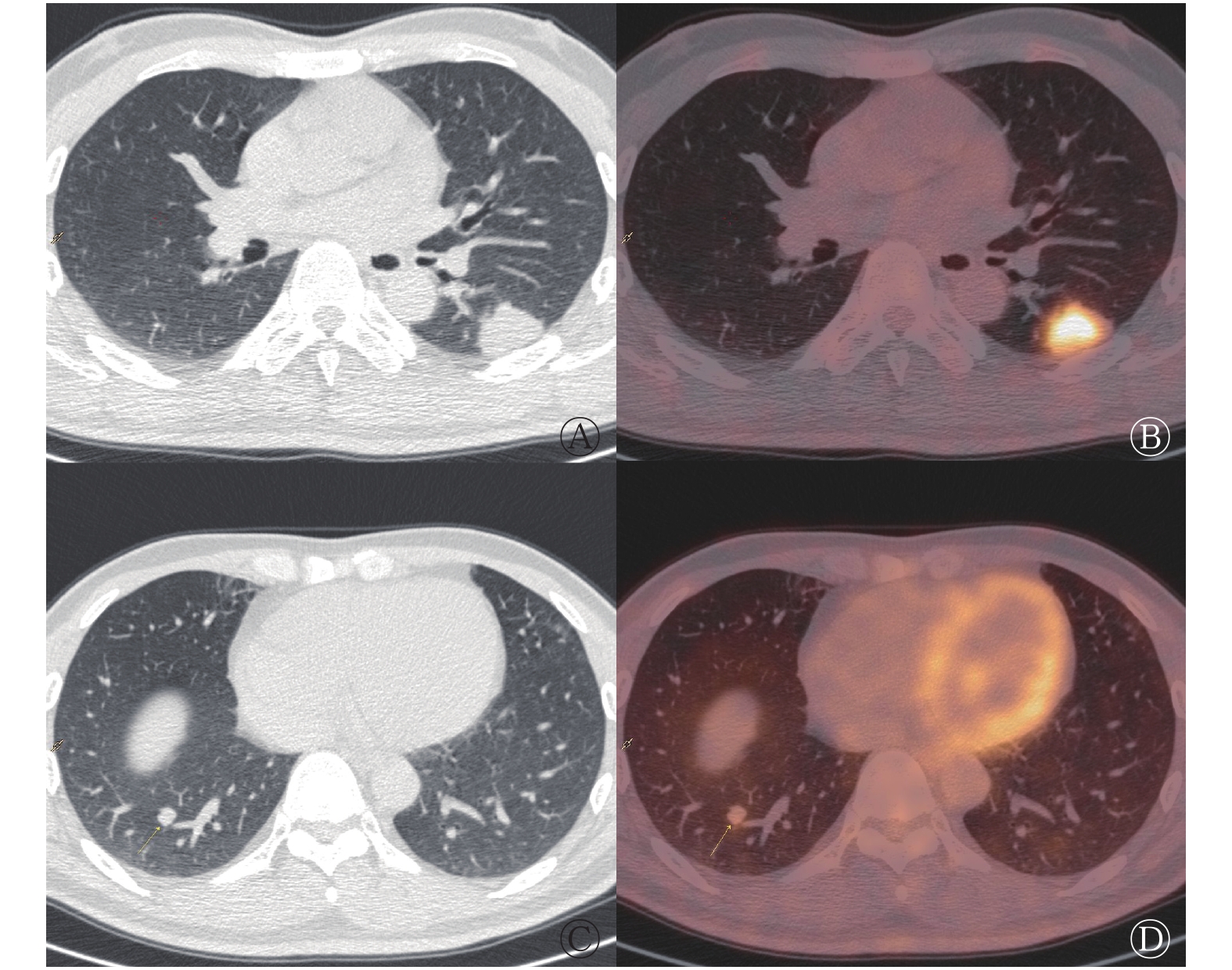
Objective To investigate the 18F-fluorodeoxyglucose (FDG) PET/CT imaging characteristics of pulmonary cryptococcosis (PC). Methods A retrospective study was performed in 22 patients with PC (17 male and 5 female; aged 54.77±7.93 years), confirmed through etiology or pathological examination from January 2011 to January 2017 in Fujian Provincial Cancer Hospital. 18F-FDG PET/CT was performed. The number, size, distribution, maximum standardized uptake value (SUVmax), and nodule sign of the nodules were analyzed. T test or Pearson correlation analysis was used in the comparison in measurement data. The differences in nodule signs among the different groups were determined by χ2 test, continuous correction χ2 test, or Fisher exact probability method. Results ① A total of 235 nodules were found by 18F-FDG PET/CT imaging in the 22 cases of PC patients with the diameter of (0.7±0.5) cm. Of these nodules, 130 showed high 18F-FDG metabolism with an SUVmax of (3.5±2.9) and a diameter of (0.9±0.5) cm. A positive correlation was found between the two (r=0.702, P=0.000). Meanwhile, 105 nodules showed no 18F-FDG metabolism with a diameter of (0.3±0.1) cm. The diameter in the group with high 18F-FDG metabolism was greater than that in the group without 18F-FDG metabolism, and the difference was statistically significant (t=13.621, P=0.000). ② The nodules were mainly found in the right lung (57.4%, 135/235), lower lobe (64.3%, 151/235), and outer zone or subpleura (80.0%, 188/235). ③ The SUVmax in the nine cases of immunocompromised host with high 18F-FDG metabolism was 5.7±4.7 higher than that in the 13 cases of non-immunocompromised host (SUVmax was 3.0±2.0), and the difference was statistically significant (t=2.731, P=0.011). The ratio of wide base posted to the pleural sign was 17.5 (10/57) of the former and higher the 6.2% (11/178) of the latter. The ratio of halo sign was 25.3% (45/178) in the latter and higher than the former of 10.5% (6/57). All the differences were statistically significant (χ2=7.911, 4.628; P=0.005, 0.031). ④ The nodules of the group with high 18F-FDG metabolism accounted for 80% (16/20) and 53% (114/215), respectively, in the each of 11 cases of one lobe or multiple lobar involvement patients. The former SUVmax was 5.6±3.4 and higher than the latter of 3.2±2.7. The difference was statistically significant (t=2.652, P=0.016). The lobulation sign (35%, 7/20), spicule sign (30%, 6/20), and pleural indentation sign (15%, 3/20) in the former were higher than the 3.3% (7/215), 0.9% (2/215) and 0.9% (2/215) in the latter, respectively. The difference was statistically significant (χ2=32.911, 47.022, 17.395, all P<0.01). ⑤ The SUVmax of the 21 nodules with high 18F-FDG metabolism in the 44 nodules found in the 11 misdiagnosed cases was 5.0±4.6, which was higher than 3.2±2.4 in the 11 other correctly diagnosed cases where 191 nodules were found, including 109 nodules with high 18F-FDG metabolism. The difference was not statistically significant (t=16.825, P=0.106). The lobulation sign, spicule sign and pleural indentation sign in the former were 15.9% (7/44), 18.2% (8/44), 6.8% (3/44) respectively, which are higher than the 3.7% (7/191), 0.0% (0/191), and 1.0% (2/191) in the latter. The difference was statistically significant (χ2=9.570, 35.951, 5.720; all P<0.05). The halo sign was 26.2% (50/191) in the latter, and higher than the 2.3% (1/44) in the former. The difference was statistically significant (χ2=12.027, P=0.000). Conclusions The SUVmax of the nodules is positively correlated with diameter in cases of PC by 18F-FDG PET/CT imaging. Halo sign is a reliable sign of diagnosis. The resembling tumor signs and the high 18F-FDG metabolism, caused the 18F-FDG PET/CT imaging to misdiagnose easily in patients of PC with single lobe involvment.
2020, 44(1): 45-51.
doi: 10.3760/cma.j.issn.1673-4114.2020.01.010
Abstract:
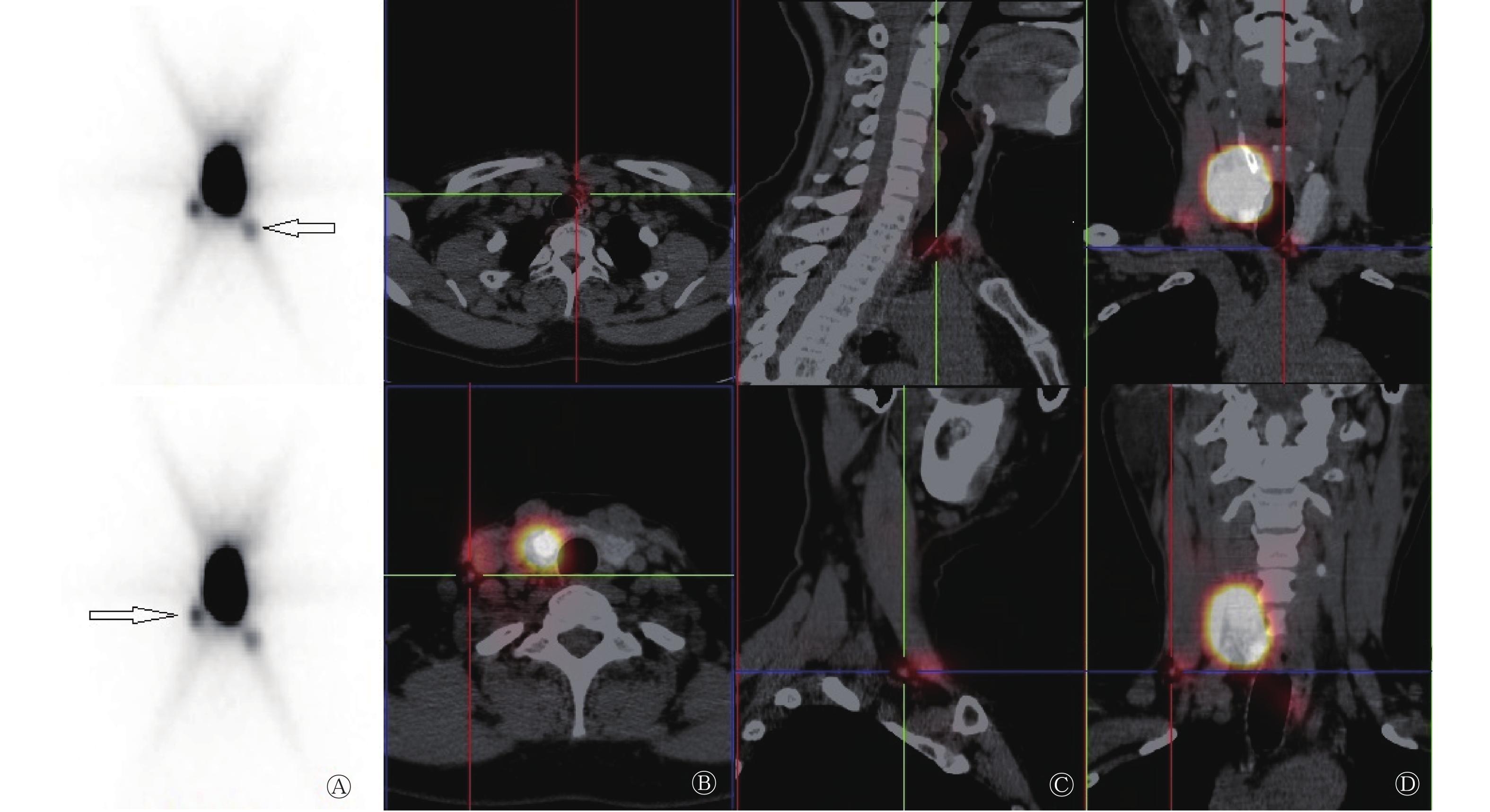
Objective To evaluate the value of SPECT/CT lymphoscintigraphy for sentinel lymph node (SLN) detection in patients with cN0 papillary thyroid carcinoma(PTC). Methods From April to November 2017, 2 male and 14 female patients with cN0 PTC, with mean age of 47.0±8.4 years, and hospitalized in the first Hospital of Shanxi Medical University, were enrolled in this study. Planar and SPECT/CT SLN imaging were performed before operation. All patients underwent intraoperative lymphatic mapping with a handheld gamma probe. All specimens were sent to a pathology laboratory. Results Among the 16 patients, 14 cases of SLN were found, with a detection rate of 87.50% (14/16). Preoperative planar and SPECT/CT fusion images showed 31 and 35 SLNs, respectively. With the application of intraoperative gamma probe, a total of 37 SLNs were detected in 13/16 patients (81.25%). The number of SLNs detected by gamma detector was inconsistent with the SPECT/CT lymphography in 4 cases and consistent in 12 cases. The coincidence rate of the two methods was 75% (12/16). Fifteen patients (93.75%) had lymph node metastasis. The sensitivity, accuracy, and false negative rate of SLN detection were 86.67%, 81.25%, and 13.33%, respectively. Conclusion Preoperative SPECT/CT lymphoscintigraphy can not only enables precise localization of SLN but also detects the cervical lymph node metastasis in patients with cN0 papillary thyroid carcinoma.

2020, 44(1): 52-58.
doi: 10.3760/cma.j.issn.1673-4114.2020.01.011
Abstract:
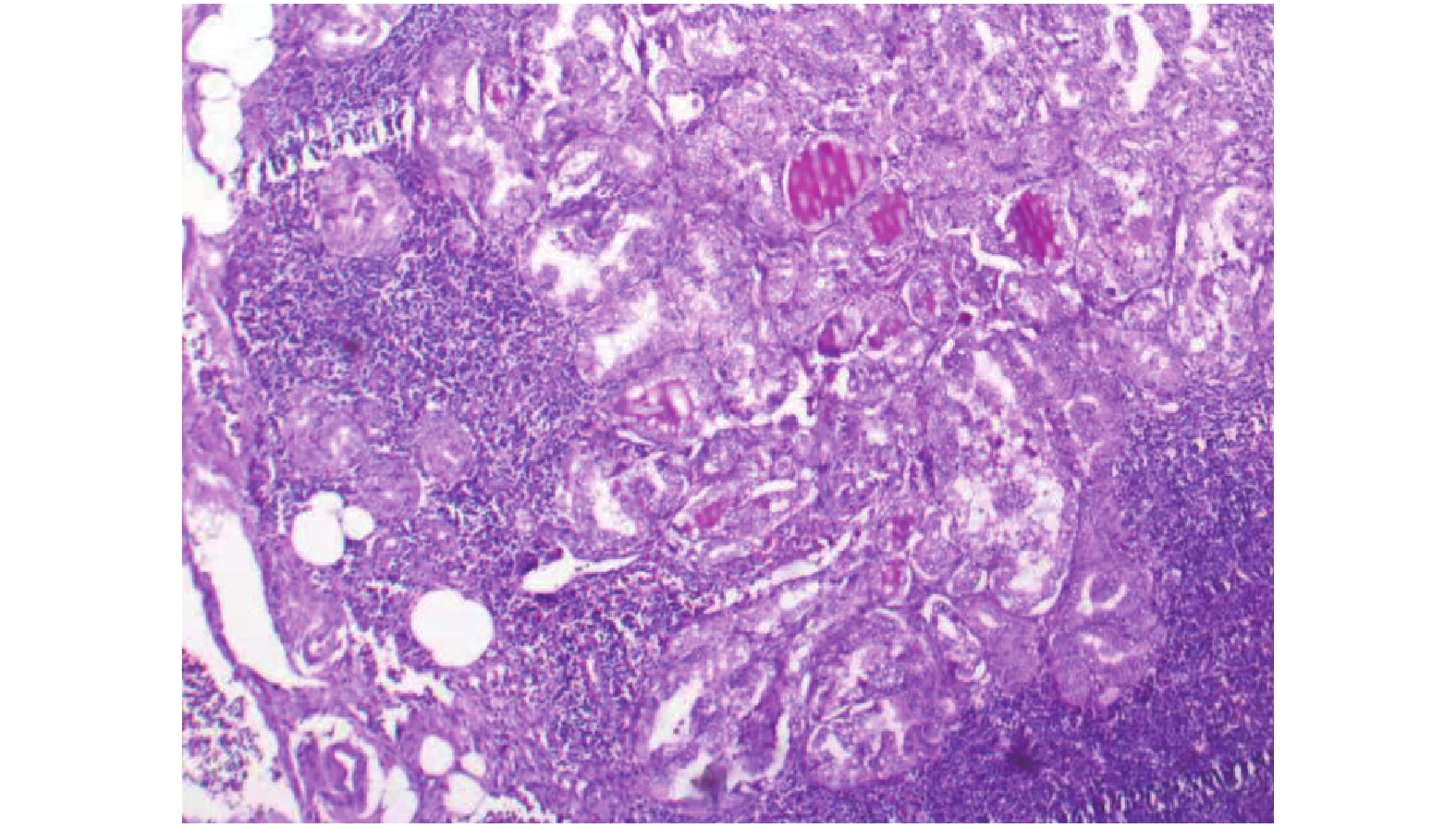
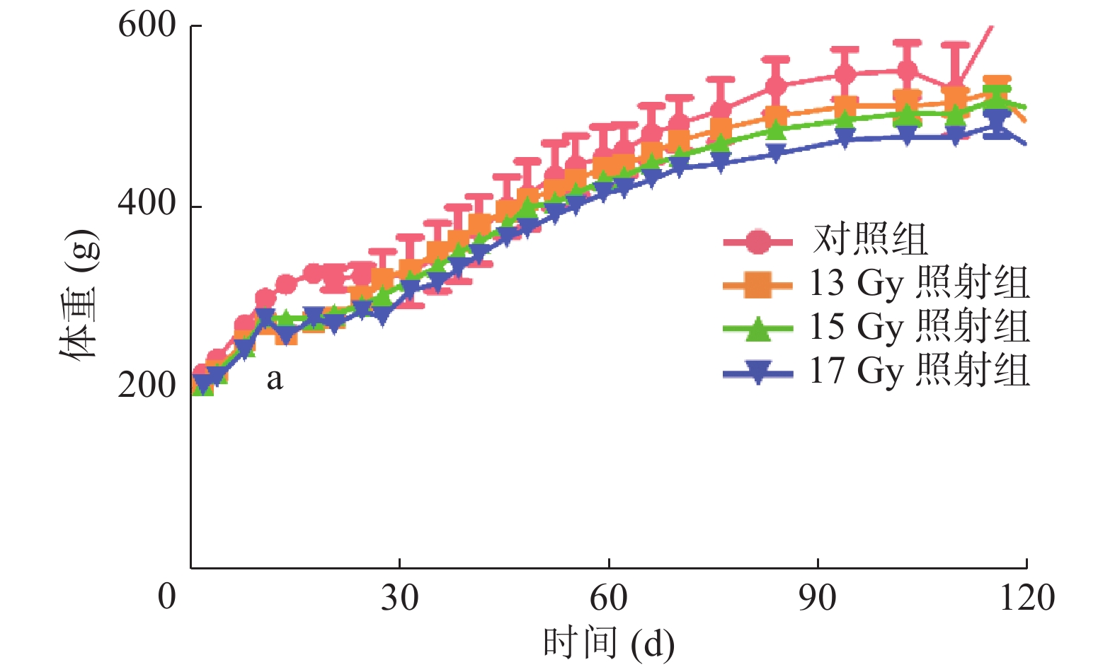
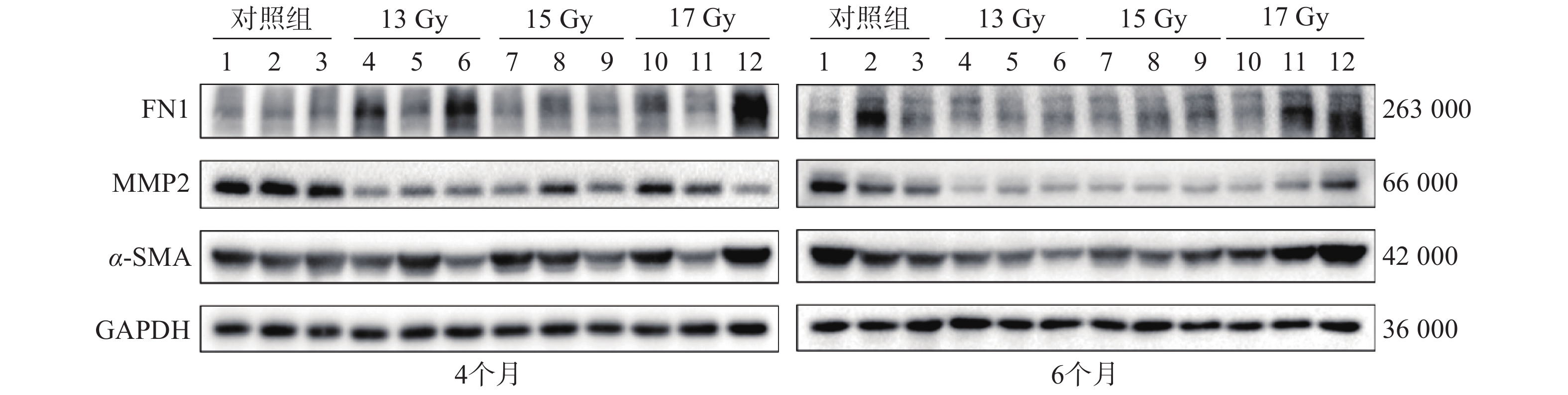
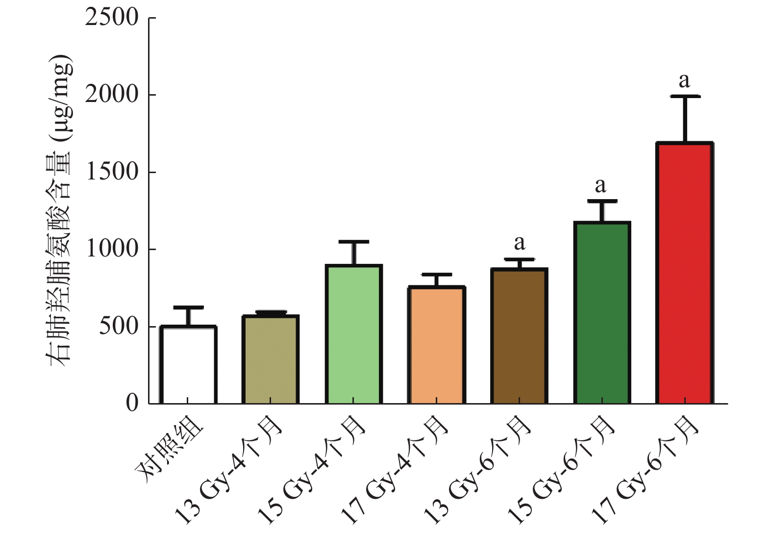
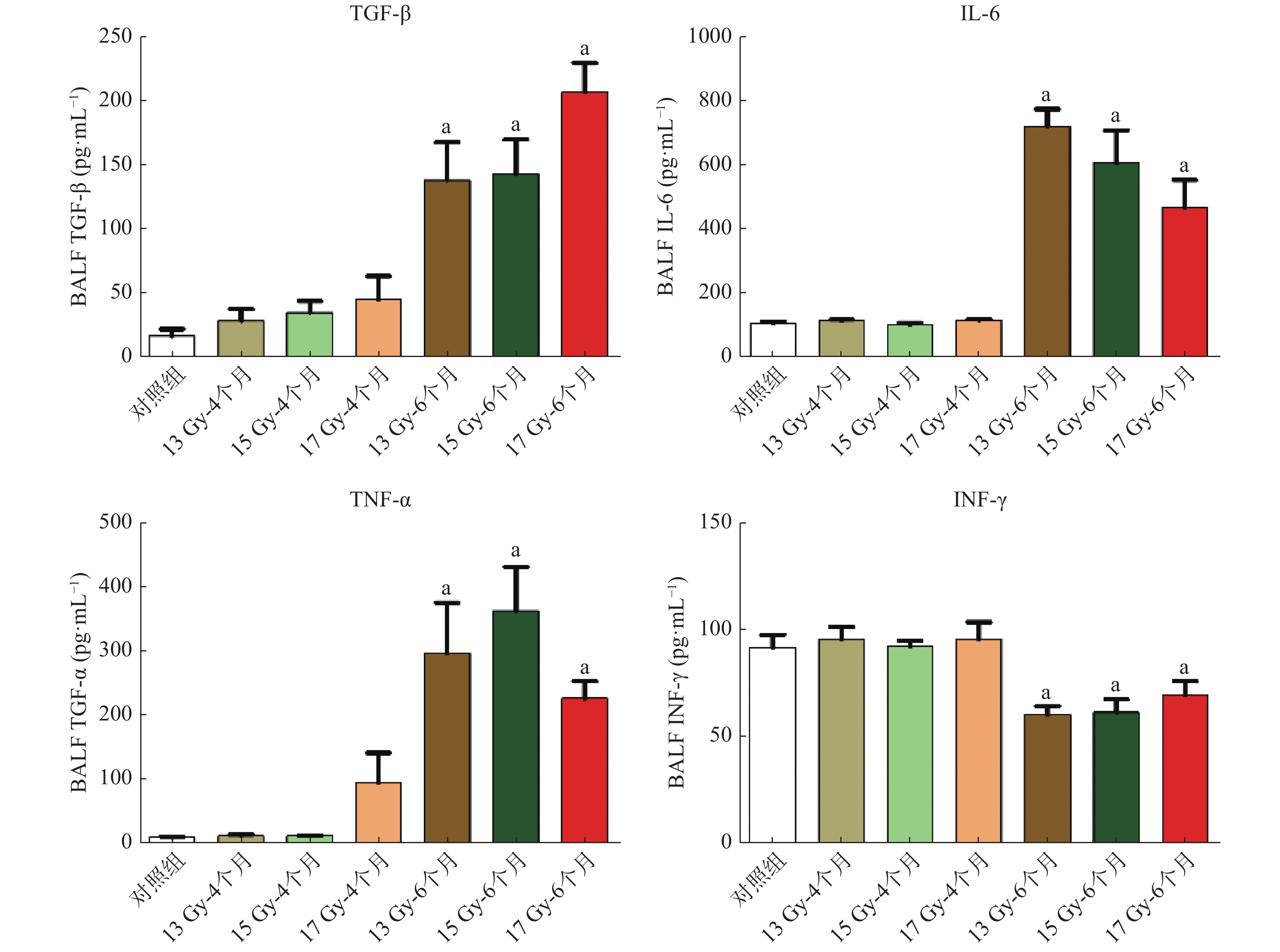
Objective A radioactive pulmonary fibrosis model of SD rats was established to explore the application of fibrosis-related proteins and cytokines as the evaluation degree of tissue fibrosis and serve as the basis for studies on radioactive pulmonary fibrosis. Method Thirty-seven male SD rats weighing 180-200 g were randomly divided into the control (n=5) and irradiation groups (n=32). The irradiation groups were irradiated by an X-ray line characterized by a single exposure dose of 13 Gy (n=10), 15 Gy (n=10), and 17 Gy (n=12). After exposure for 4 and 6 months, hematoxylin-eosin staining and Masson staining were used to evaluate pulmonary fibrosis in the rats. Fibronectin 1 (FN1), matrix metalloproteinase 2 (MMP2), and α-smooth muscle actin (α-SMA) expression levels in lung tissues were detected by Western blot analysis. Hydroxyproline content in the lung tissue were evaluated the lung tissue collagen protein expression levels. ELISA was employed to detect the change of cytokines, such as the transforming growth factor β (TGF-β), interleukin 6, tumor necrosis factor α (TNF-α), and interferon γ (INF-γ), in the bronchoalveolar lavage fluid. Independent sample t-test was used for the intergroup comparison. Results Compared with the control group, the model group developed pulmonary fibrosis after 4-6 months, and the degree of fibrosis increased with the irradiation dose and time. The protein expression level of FN1 and α-SMA in the lung tissues of the model group was higher than that of the control group, and the protein expression level of MMP2 was lower than that of the control group. The hydroxyproline content in the 6-month model group was higher than that of the control group, increased from (514.19±282.20) μg/mg to (886.13±145.01), (1188.70±273.84), (1700.70±590.95) μg/mg respectively (t=2.621, 3.609, 4.004, all P<0.05). The TGF-β, interleukin 6, and TNF-α levels in the bronchoalveolar lavage fluid of the model group were up-regulated compared with those in the normal control group (t=4.030–12.780, all P<0.05), whereas the IFN-γ was down-regulated (t=2.498–4.303, all P<0.05). Conclusions The SD rat model of radiation pulmonary fibrosis was successfully constructed. The fibrin content of the rat lung tissues failed to reflect the degree of lung tissue fibrosis. The hydroxyproline content in the lung tissue and the cytokines in the bronchoalveolar lavage fluid can be used as evaluation indexes in severe fibrosis.

2020, 44(1): 59-64.
doi: 10.3760/cma.j.issn.1673-4114.2020.01.012
Abstract:
A high level of thyroid stimulating hormone (TSH) is required to stimulate sufficient radioiodine uptake for diagnostic imaging or therapy. The current methods for improving TSH level include thyroid hormone withdrawal and recombinant human TSH. However, acute hypothyroidism caused by thyroid hormone withdrawal (THW) before radioactive iodine remnant ablation/treatment or diagnostic scanning significantly influences lipid metabolism, renal function, cardiovascular and neuropsychiatric diseases, and quality of life. This review aimed to summarize these methods and their effect on the clinical and quality of life in patients with differentiated thyroid cancer.
A high level of thyroid stimulating hormone (TSH) is required to stimulate sufficient radioiodine uptake for diagnostic imaging or therapy. The current methods for improving TSH level include thyroid hormone withdrawal and recombinant human TSH. However, acute hypothyroidism caused by thyroid hormone withdrawal (THW) before radioactive iodine remnant ablation/treatment or diagnostic scanning significantly influences lipid metabolism, renal function, cardiovascular and neuropsychiatric diseases, and quality of life. This review aimed to summarize these methods and their effect on the clinical and quality of life in patients with differentiated thyroid cancer.
2020, 44(1): 65-70.
doi: 10.3760/cma.j.issn.1673-4114.2020.01.013
Abstract:
With its rapid development, 3D printing technology has been widely used and achieved breakthrough progress in the medical field, especially in orthopedics, oral and maxillofacial surgery, organ transplantation, and other aspects. As a major cancer treatment, radiation therapy combined with 3D printing technology provides a powerful guarantee for the precise radiotherapy of tumors. This review presents the application and prospects of 3D printing technology in tumor radiotherapy.
With its rapid development, 3D printing technology has been widely used and achieved breakthrough progress in the medical field, especially in orthopedics, oral and maxillofacial surgery, organ transplantation, and other aspects. As a major cancer treatment, radiation therapy combined with 3D printing technology provides a powerful guarantee for the precise radiotherapy of tumors. This review presents the application and prospects of 3D printing technology in tumor radiotherapy.
 Submit
 Author Login
 Referees
 Editor-in-Chief
 Editor Center
 Email alert
 RSS
RSS