


 下载:
下载:
-
颅内血管外皮细胞瘤(hemangiopericytoma,HPC)是起源于脑膜间质的恶性肿瘤,发生率较低,约占所有原发性中枢神经系统肿瘤的0.4%[1],术后可发生颅外转移,常见部位是骨、肺及肝,目前国内报道较少。
-
患者男性,59岁,主因“左下肢乏力半年,左髋部疼痛3个月”于2014年3月15日入院。6年前曾因“颅内占位病变,怀疑脑膜瘤”于北京天坛医院行双额顶开颅肿瘤切除术,术后病理示间变性血管外皮细胞瘤(anaplastic hemangiopericytoma,AHPC),术后放疗1个月,后规律复查颅脑MRI,1年前发现肿瘤复发,行伽马刀治疗,术后口服中药。既往病史无特殊。
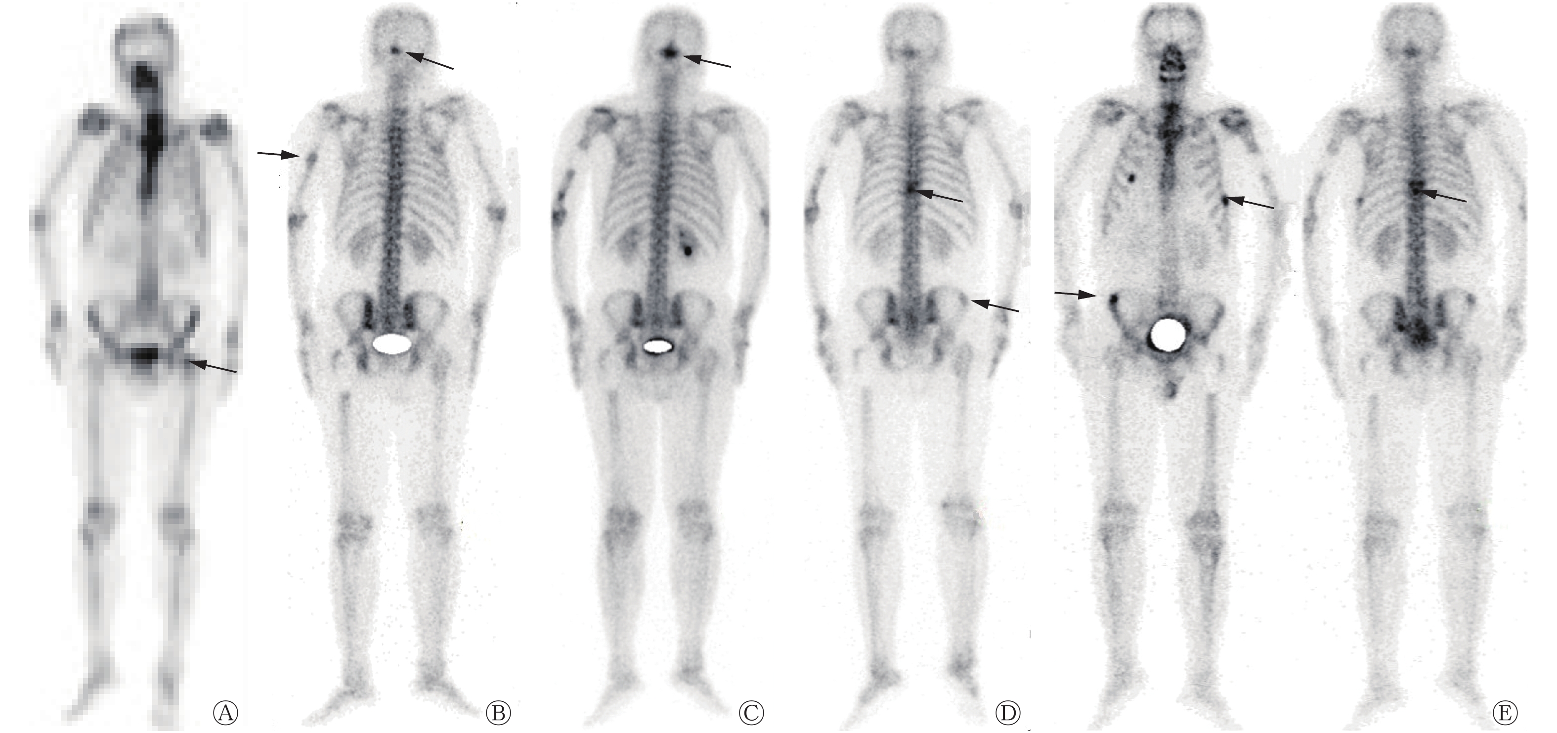
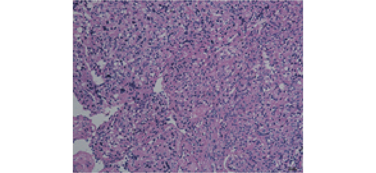
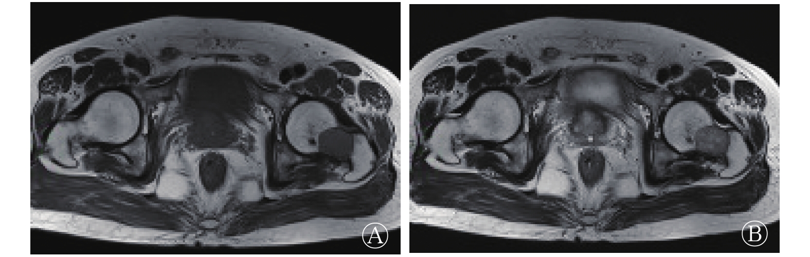
患者此次入院后髋关节MR提示左股骨颈部骨肿瘤(图1),性质待查。全身骨显像提示左股骨颈部局部骨代谢增高,不排除骨肿瘤性病变(图2中A)。PET/CT检查结果提示左侧股骨颈局灶性骨质结构破坏伴FDG代谢增高,考虑骨肿瘤性病变。查体:左髋部无明显畸形,局部皮肤无异常改变,局部无触压痛,无骨盆挤压痛,左髋关节活动正常,左侧下肢肌力稍减弱,肌张力正常,末梢感觉及血运良好,足背动脉搏动良好,病理征阴性。其余肢体检查未见明显异常。入院诊断考虑左股骨颈病变(肉瘤)或颅内AHPC转移。入院给予止痛等对症治疗,缓解不明显。为进一步治疗,患者于2014年4月2日就诊于北京大学人民医院,完善术前相关辅助检查后行左股骨上段瘤段截除+肿瘤型股骨上段假体置换术,手术顺利,术后病理结果提示AHPC骨转移。1年后(2015年6月18日),患者行全身骨显像,结果示枕骨骨代谢增高(图2中B),建议进一步检查;左侧肱骨中段骨代谢增高,考虑脑细胞瘤骨转移。1个月后(2015年7月10日)行左侧肱骨中段瘤段截除术,病理结果提示AHPC骨转移。半年后(2015年12月11日),患者复查骨显像,结果示枕骨骨代谢较之前增高(图2中C),考虑转移灶;左侧肱骨下段术后改变。给予枕骨区局部放疗,9个月后(2016年9月11日)复查,结果示原枕骨区域骨代谢较之前降低;右侧第4前肋、左侧第7肋骨、胸8、9椎体及右侧髂骨骨代谢增高(图2中D),建议进一步检查。3个月后(2016年12月19日),骨显像结果示右侧第4前肋骨代谢较之前降低,考虑为损伤所致;胸8、9椎体及右侧髂骨骨代谢较之前无明显变化,左侧第7肋骨骨代谢较之前增高(图2中E),结合患者病史,考虑胸8、9椎体、右侧髂骨及左侧第7肋骨骨转移。胸椎MR示胸8附件骨异常信号,符合转移瘤的影像学特征。患者于2016年12月22日在济宁市第一人民医院全麻下行胸椎后路胸8占位切除减压固定术,术后病理结果提示血管外皮瘤(图3)。之后患者反复出现咳嗽、咳痰、发热等重症肺炎症状,胸部CT提示肺及肝转移,患者于2018年3月16日因“中枢性呼吸循环衰竭”去世。
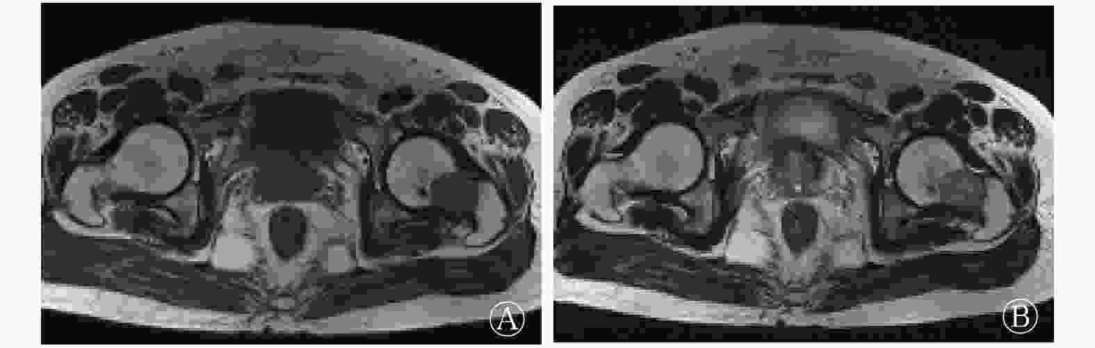
图 1 颅内间变性血管外皮细胞瘤伴骨转移患者双侧髋关节MRI图(2014年3月15日)
Figure 1. Bilateral hip joint MRI in a patient with bone metastasis secondary to intracranial anaplastic hemangiopericytoma
图 2 颅内间变性血管外皮细胞瘤伴骨转移患者骨显像图像(患者男性,59岁)
Figure 2. Bone imaging in a patient with bone metastasis secondary to intracranial anaplastic hemangiopericytoma (male, 59 years old)
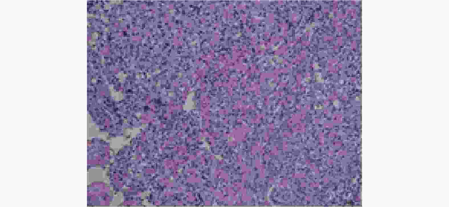
图 3 颅内间变性血管外皮细胞瘤伴骨转移患者病理图
Figure 3. Pathology in a patient with bone metastasis secondary to intracranial anaplastic hemangiopericytoma
-
血管外皮细胞瘤是一种少见的软组织肿瘤,来源于毛细血管壁外的周细胞,常见于骨骼系统,发生于颅内者更为少见。据报道,颅内HPC可发生于任何年龄,总体以中年常见[2],年龄多在45岁以下,男性较女性多见。颅内HPC多为单发,好发部位为颅底、矢状窦、大脑镰旁、小脑幕或静脉窦附近。该例患者发病部位为矢状窦。颅内HPC常见症状为头痛及因肿瘤发生部位不同而逐渐出现的颅内压增高和脑部受压体征,肌力减弱和癫痫少见。该例患者6年前因头痛起病,肌力减弱不明显,未出现癫痫。该病病程短,进展快,就诊时肿瘤多较大,肿瘤血供极为丰富,与脑膜瘤相比,全切率低,复发率高。有文献报道,颅内HPC 5年存活率为67%~96%,10年存活率40%~75%,平均局部复发时间为40~108个月,15年转移率高达64%~79%,最常见的转移部位依次为骨、肺[3]及肝[4]。本例患者术后1年内复发,术后6年后陆续出现股骨、肱骨及胸椎等骨骼转移及肺、肝转移,术后生存时间约10年。
恶性肿瘤骨转移指原发于骨外器官或组织的恶性肿瘤,通过血液循环或淋巴系统转移到骨骼并继续生长,形成转移灶,临床较常见,其直接影响肿瘤分期,诊断不及时会严重影响肿瘤患者的生活质量。骨骼病变的确诊需行活检,操作起来较其他部位更复杂,可能对患者造成伤害,全身骨显像可作为筛查骨转移的重要影像手段,操作简便[5-6],一次成像可发现全身骨骼病变,费用低,可以较X射线检查提前3~6个月发现骨转移病灶,阳性率为33.4%~60%[7],因此肿瘤患者需要定期行该检查判断有无骨转移。虽然全身骨显像灵敏度高,但其特异度低,骨转移征象不明显时很难鉴别,需要借助其他影像学检查,如MRI、PET/CT和血清肿瘤标志物[8]及病灶靶/非靶值[9-10]来判定。骨转移常见的肿瘤包括前列腺癌、乳腺癌、肺癌等,发生于颅内肿瘤者较少见,因此较难判断。骨转移性肿瘤病灶的分布部位[11]发生率由高至低依次为胸部(主要集中于肋骨)、脊椎、骨盆和四肢+颅骨,本研究的病例首次行骨显像时,股骨部位病变较难定性的原因在于其为孤立病灶,显像剂浓聚程度不高,因此需借助手术明确诊断。在骨显像随访时,结合之前的病史,诊断上能有所侧重。后来患者出现了肱骨转移,最后出现脊柱转移,与常见的骨转移特点正好相反,可能与此类肿瘤的性质相关。本研究中病例的骨转移病灶均显示显像剂浓聚,国外有报道显示此类病变骨扫描图像上表现为显像剂浓聚区内出现显像剂缺损,可作为此类肿瘤骨显像的特征性表现[12]。AHPC病灶在18F-FDG PET/CT图像上呈低代谢[13],68Ga-PMSA PET可以显示高代谢病变,但PET检查费用昂贵,不适合临床随访。因此建议恶性肿瘤患者,尤其是少见骨转移者定期行骨显像检查,必要时可行PET/CT检查,这对于早期发现骨转移、确定临床分期及治疗方案的选择等至关重要。
利益冲突 本研究由署名作者按以下贡献声明独立开展,不涉及任何利益冲突。
作者贡献声明 苑丽丽负责研究命题的提出、设计及研究过程的实施、论文起草及最终版本修订;谭建负责论文的审阅及修改;吴德坤负责数据的获取、提供与分析;朱雁冬及朱亚丽负责数据的分析及研究过程的实施;黄承明负责论文的审阅及修改。
颅内间变性血管外皮细胞瘤伴骨转移一例
Intracranial anaplastic hemangiopericytoma with bone metastasis: a case report
-
摘要: 笔者报道了1例颅内间变性血管外皮细胞瘤(AHPC)伴骨转移的病例,从临床表现、影像学检查尤其是全身骨显像检查以及术后病理综合分析了该病特点。并通过文献复习加深了对AHPC骨转移的认识。多数恶性肿瘤常见骨转移,而颅内AHPC是一种少见的中枢神经系统肿瘤,较少发生骨转移。通过全身骨显像发现了该例患者多处骨骼病变,术后病理提示颅内AHPC骨转移,因此提示恶性肿瘤尤其是少见骨转移者应把骨显像检查作为随访的一个重要检查,对于早期发现骨转移、确定临床分期及治疗方案的选择等都是至关重要的。Abstract: A case of bone metastase secondary to intracranial anaplastic hemangiopericytoma (AHPC) was reported. The characteristics of the disease were analyzed on the basis of clinical symptoms and imaging examinations, especially bone imaging examination and pathology. The understanding of intracranial AHPC bone metastasis was deepened by literature review. Bone metastases are common in most malignant tumors, but intracranial AHPC, which is a rare central nervous system tumor, has an extremely low bone metastasis potential. In the present case, the multiple lesions of bone metastasis from intracranial AHPC were detected through a whole body bone imaging examination. Thus, whole body bone scans should be regarded as important examination tools for patients with malignant tumors during follow-up, especially patients with rare bone metastasis, because the scans have a key value in early detection, clinical staging, and treatment decision.
-
Key words:
-
图 1 颅内间变性血管外皮细胞瘤伴骨转移患者双侧髋关节MRI图(2014年3月15日)
Figure 1. Bilateral hip joint MRI in a patient with bone metastasis secondary to intracranial anaplastic hemangiopericytoma
图 2 颅内间变性血管外皮细胞瘤伴骨转移患者骨显像图像(患者男性,59岁)
Figure 2. Bone imaging in a patient with bone metastasis secondary to intracranial anaplastic hemangiopericytoma (male, 59 years old)
-
[1] Kleihues P, Cavenee WK. 神经系统肿瘤病理学和遗传学[M]. 李青, 徐庆中, 译. 北京: 人民卫生出版社, 2006: 234−237.
Kleihues P, Cavenee WK. Pathology & genetics tumours of the nervous system[M]. Li Q, Xu QZ, trans. Beijing: People's Medical Publishing House, 2006: 234−237.[2] 孙新国, 李福增, 姚军, 等. 颅内血管外皮细胞瘤的临床特点[J]. 临床神经病学杂志, 2017, 30(4): 297−299. DOI: 10.3969/j.issn.1004−1648.2017.04.019.
Sun XG, Li FZ, Yao J, et al. Clinical characteristic of intracranial hemangiopericytoma[J]. J Clin Neurol, 2017, 30(4): 297−299. DOI: 10.3969/j.issn.1004−1648.2017.04.019.[3] 马春华, 张学斌, 姜镕, 等. 颅内间变性血管外皮瘤合并肺部多发性转移一例[J]. 中国现代神经疾病杂志, 2016, 16(2): 107−112. DOI: 10.3969/j.issn.1672−6731.2016.02.009.
Ma CH, Zhang XB, Jiang R, et al. Intracranial anaplastic hemangiopericytoma with pulmonary metastases: one case report[J]. Chin J Contemp Neurol Neurosurg, 2016, 16(2): 107−112. DOI: 10.3969/j.issn.1672−6731.2016.02.009.[4] Soyuer S, Chang EL, Selek U, et al. Intracranial meningeal hemangiopericytoma: The role of radiotherapy: report of 29 cases and review of the literature[J]. Cancer, 2004, 100(7): 1491−1497. DOI: 10.1002/cncr.20109. [5] Abikhzer G, Gourevich K, Kagna O, et al. Whole-body bone SPECT in breast cancer patients: the future bone scan protocol[J]. Nucl Med Commun, 2016, 37(3): 247−253. DOI: 10.1097/MNM.0000000000000427. [6] 陈少亮. 临床核医学进展: SPECT-CT与PET-CT技术与应用[M]. 北京: 科学出版社, 2017: 282.
Chen SL.Advances in clinical nuclear medicine: technology and application of SPECT-CT and PET-CT[M].Beijing: Science Press, 2017: 282.[7] 安锐, 黄钢. 核医学[M]. 3版. 北京: 人民卫生出版社, 2015: 247.
An R, Huang G. Nuclear medicine[M]. 3rd ed. Beijing: People's Medical Publishing House, 2015: 247.[8] 石海蓉, 张影, 陆颖, 等. 血清肿瘤标志物在肺癌骨转移诊断中的价值[J]. 中华核医学与分子影像杂志, 2018, 38(5): 331−335. DOI: 10.3760/cma.j.issn.2095−2848.2018.05.006.
Shi HR, Zhang Y, Lu Y, et al. Diagnostic value of serum tumor makers for bone metastasis from lung cancer[J]. Chin J Nucl Med Mol Imaging, 2018, 38(5): 331−335. DOI: 10.3760/cma.j.issn.2095−2848.2018.05.006.[9] 孙斌, 刘举珍, 张飞飞. SPECT/CT断层融合显像联合血清CEA、CYFRA21-1在诊断非小细胞肺癌骨转移中的应用[J]. 国际放射医学核医学杂志, 2018, 42(3): 212−217. DOI: 10.3760/cma.j.issn.1673−4114.2018.03.004.
Sun B, Liu JZ, Zhang FF. Application of SPECT/CT tomographic fusion imaging combined with serum CEA and CYFRA21-1 in the diagnosis of bone metastasis of non-small cell lung cancer[J]. Int J Radiat Med Nucl Med, 2018, 42(3): 212−217. DOI: 10.3760/cma.j.issn.1673−4114.2018.03.004.[10] 杨志, 柴华, 李宁, 等. 发射型计算机断层显像全身骨扫描联合靶/非靶比值在骨转移瘤中的诊断价值[J]. 广西医学, 2019, 41(4): 427−430. DOI: 10.11675/j.issn.0253−4304.2019.04.07.
Yang Z, Chai H, Li N, et al. Diagnostic value of whole-body bone scintigraphy combined with target to non-target ratio of emission computed tomography for bone metastases[J]. Guangxi Med J, 2019, 41(4): 427−430. DOI: 10.11675/j.issn.0253−4304.2019.04.07.[11] 赵雨薇, 赵明. 肺癌和乳腺癌骨转移患者单光子发射计算机断层扫描全身骨显像的临床应用价值[J]. 中国医师进修杂志, 2019, 42(1): 65−67. DOI: 10.3760/cma.j.issn.1673−4904.2019.01.018.
Zhao YW, Zhao M. The Clinical application value in whole-body bone imaging with single-photon emission computed tomography in lung and breast cancer patients with bone metastases[J]. Chin J Postgrad Med, 2019, 42(1): 65−67. DOI: 10.3760/cma.j.issn.1673−4904.2019.01.018.[12] Hoshi M, Araki N, Naka N, et al. Bone metastasis of intracranial meningeal hemangiopericytoma[J]. Int J Clin Oncol, 2005, 10(3): 208−213. DOI: 10.1007/s10147−005−0476−y. [13] Patro KC, Palla M, Kashyap R. Unusual Case of Metastatic Intracranial Hemangiopericytoma and Emphasis on Role of 68 Ga-PSMA PET in Imaging[J]. Clin Nucl Med, 2018, 43(9): e331−e333. DOI: 10.1097/RLU.0000000000002203. -

 点击查看大图
图(3)
计量
- 文章访问数: 3199
- HTML全文浏览量: 2008
- PDF下载量: 11