


 下载:
下载:


-
自发性低颅压综合征(spontaneous intracranial hypotension,SIH)是一组无特殊诱因(如颅脑外伤、硬膜外穿刺等)的、脑脊液压力降低引起的、以体位性头痛为主要临床表现的,并可伴有颈部僵硬、恶心、呕吐、听觉改变、复视和视野缺损等其他症状的临床综合征[1]。该病病因尚不清楚,目前大多数学者认为自发性脊髓脑脊液漏是SIH的根本病因[2-4]。然而,临床常用的CT及MRI等神经影像学检查对脊髓脑脊液漏点的检出较为困难。本研究通过对19例临床诊断为SIH患者的放射性核素脑脊液间隙显像(radionuclide cisternography,RNC)的检查结果进行回顾性分析,描述和总结其影像学特点,分析该检查对SIH脑脊液漏的检出价值和意义。
-
回顾性分析2012年5月至2017年1月福建省立医院神经内科收治的19例SIH患者脑脊液间隙显像及神经系统MRI、CT影像学资料。19例SIH患者中,男性4例、女性15例,年龄20~63(41.7±10.5)岁。所有患者均符合2011年修订的SIH临床诊断标准[5]。纳入标准:①直立性头痛;②至少存在颅压低(≤60 mm H2O)(1 kPa≈ 102 mm H2O)、硬脊膜外血贴治疗后头痛缓解、有一个或多个活动性脑脊液漏、颅脑MRI检查提示低颅压样改变(如硬脑膜强化或脑下垂)中的一项;③近期无硬脊膜穿刺病史。排除标准:排除其他继发性因素,如头、颈部外伤、腰椎穿刺或硬膜外穿刺、手术等常见的低颅压综合征等原因。其中3例患者伴复视,1例合并慢性硬膜下血肿。患者发病初期常规行颅脑MRI+增强或头颅CT检查。所有患者行RNC前均在常规腰穿成功后测定脑脊液压力,1例患者的脑脊液压力为70 mm H2O,18例脑脊液压力<60 mm H2O,其中5例脑脊液压力为0。所有患者均于检查前签署了知情同意书。
-
仪器为美国GE公司Discovery NMCT670 双探头SPECT/CT。SPECT显像参数:配低能高分辨准直器,能峰140 keV,窗宽20%。全脊髓蛛网膜下腔采集矩阵256×1024,放大倍数0.92,扫描速度13.3 cm/min;局部静态采集矩阵128×128,放大倍数1.0,300 s/帧;SPECT断层显像矩阵64×64,放大倍数1.0,16 s/帧,1帧/6°,旋转180°,共采集64帧。CT成像参数:SPECT/CT采集完成后,仪器按SPECT的扫描视野进行CT透射扫描;管电压120 kV,管电流100 mA,矩阵512×512,扫描层厚2.5 mm,螺距为1。图像重建采用flash 三维迭代技术。通过计算机处理得到SPECT、CT图像并进行图像融合。99Tcm-DTPA由中国广东希埃医药有限公司提供,放射化学纯度>95%。
患者均取侧卧位,于第3、4腰椎间隙行腰椎穿刺,腰穿成功放出少量脑脊液后注入生理盐水,使脑脊液压力接近60 mm H2O。回抽2 mL脑脊液稀释0.5 mL99Tcm-DTPA,再将2.5 mL稀释液(185 MBq)注射入脊髓蛛网膜下腔,患者约15°角度头低脚高位平卧。分别于注射药物后30 min、1 h、2 h、3 h、6 h、24 h行前、后位脊髓段蛛网膜下腔显像及常规体位脑池显像,动态观察核素在脑脊液间隙的循环过程及放射性分布情况,异常分布处增加显像次数,加做SPECT/CT断层融合显像,并在1周内对核素显像发现脑脊液漏口处的相应部位行MRI检查。检查结果经过2位有10年以上阅片经验的主治以上核医学医师独立评估。意见不一致时通过协商达成一致。
-
SIH脊髓脑脊液漏的直接证据:脊髓硬膜外出现放射性核素异常浓聚,SPECT/CT断层融合图像见脊髓段硬膜外沿神经根走行区的短条状放射性浓聚。SIH脑脊液鼻漏的直接证据:鼻腔出现放射性核素异常浓聚,SPECT/CT断层融合图像见鼻腔点片状放射性异常浓聚。脑脊液在脊髓段硬膜外漏出的间接证据:放射性核素在蛛网膜下腔快速消失(≤4.0 h),或(和)过早(≤2.5 h)在膀胱内出现,放射性核素难以升达基底池到达脑部以及颅内的放射性浓聚程度少于正常[6-7]。
所有患者RNC检查后1个月、3个月、6个月、12个月常规电话随访其体位性头痛改善及发作情况,无1例失访。
-
采用SPSS 11.5软件对数据进行统计学分析。计量资料以
$\overline x $ ±s表示。计数资料的组间比较采用χ2检验(Fisher确切概率法)。P<0.05表示差异有统计学意义。 -
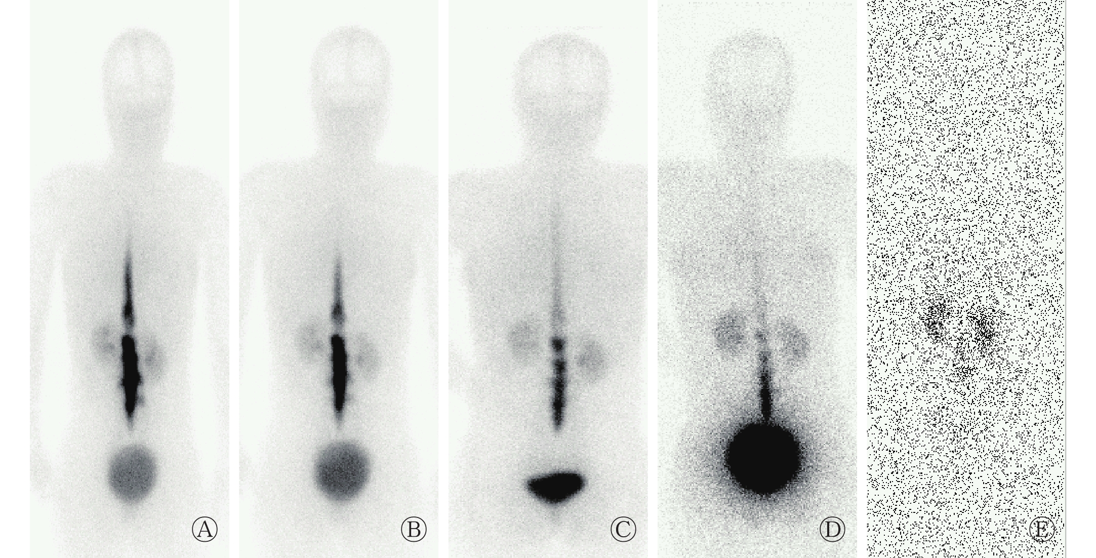
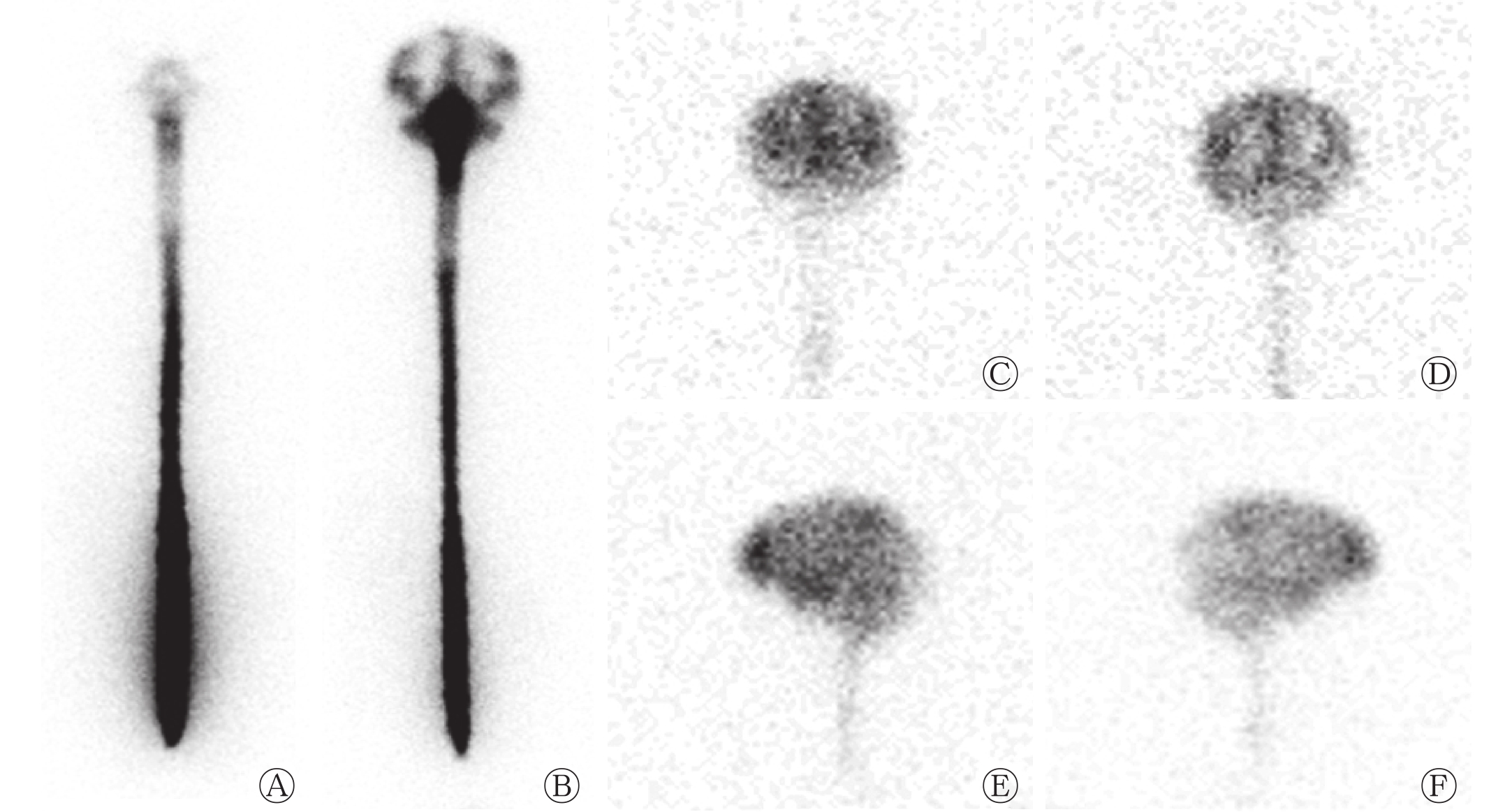
所有患者均在注射药物后30 min内出现硬膜外放射性异常浓聚,3 h内影像较清晰。此后随着脑脊液的漏出及核素衰变,漏口显像逐渐模糊。RNC检出脑脊液漏17例,阳性率89.5%(17/19)。存在直接脑脊液漏证据的12例,其中脊髓脑脊液漏10例:漏点位于颈胸交界段及胸椎旁的多发放射性异常浓聚8例(图1)、单发片状放射性异常浓聚1例;1例漏口位于腰段,且为大流量脑脊液漏,该例患者在注射99Tcm-DTPA后30 min,腰段蛛网膜下腔出现放射性核素大量漏至椎旁间隙,此后该浓聚灶逐渐减淡,胸上段以上脊髓蛛网膜下腔及各脑池均未见显影,肾脏和膀胱在注射药物后30 min即明显显影,24 h时蛛网膜下腔放射性分布同本底相仿(图2)。脑脊液鼻漏2例(图3)。17例RNC阳性患者均存在脑脊液漏间接证据,有10例放射性核素难以升达大脑凸面。未发现直接漏口仅存在脊髓脑脊液漏间接证据5例(图4)。另2例SIH患者RNC正常(图5)。19例患者RNC检查过程顺利,均未出现不良反应。
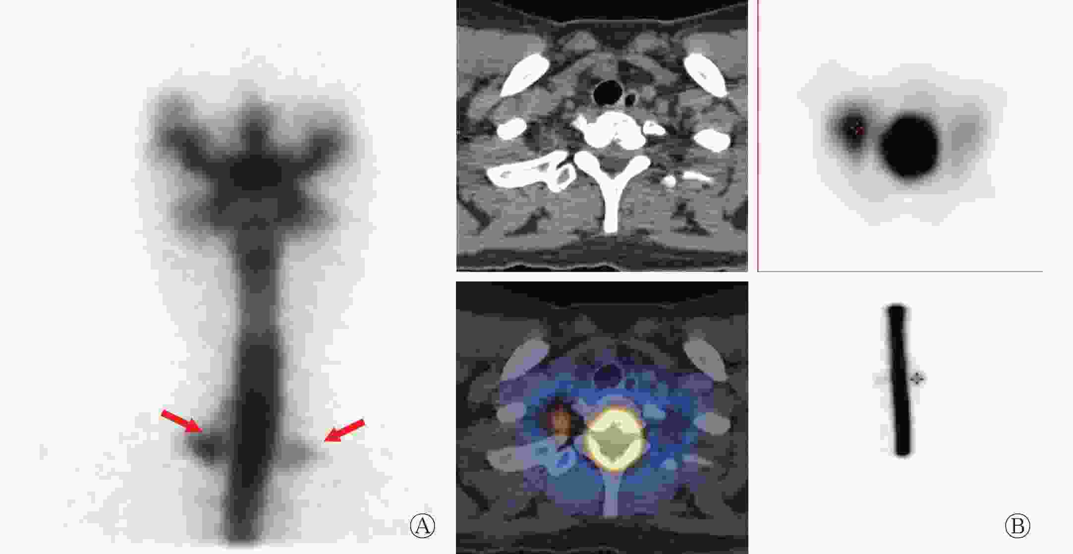
图 1 SIH患者(女性,37岁)颈胸交界处脑脊液漏的RNC和SPECT/CT断层融合图像
Figure 1. Radionuclide cisternography and SPECT/CT tomographic fusion imaging of cerebrospinal fluid leakage at cervicothoracic junction in patients with spontaneous intracranial hypotension(female,37 years old)
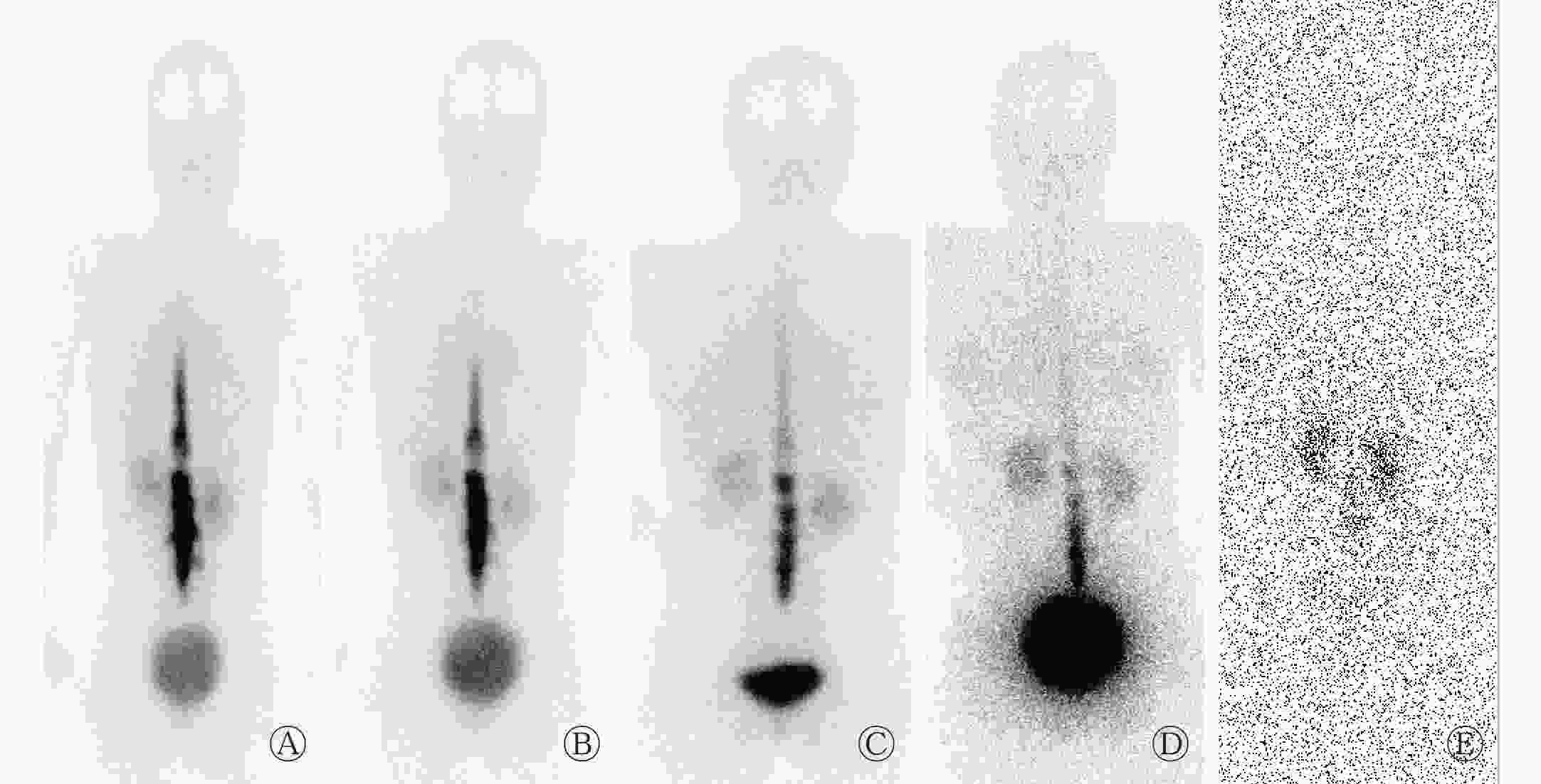
图 2 SIH患者(女性,33岁)腰段大流量脑脊液漏RNC图像
Figure 2. Radionuclide cisternography image of high flow cerebrospinal fluid leakage at lumbar spinal in patients with spontaneous intracranial hypotension(female,33 years old)
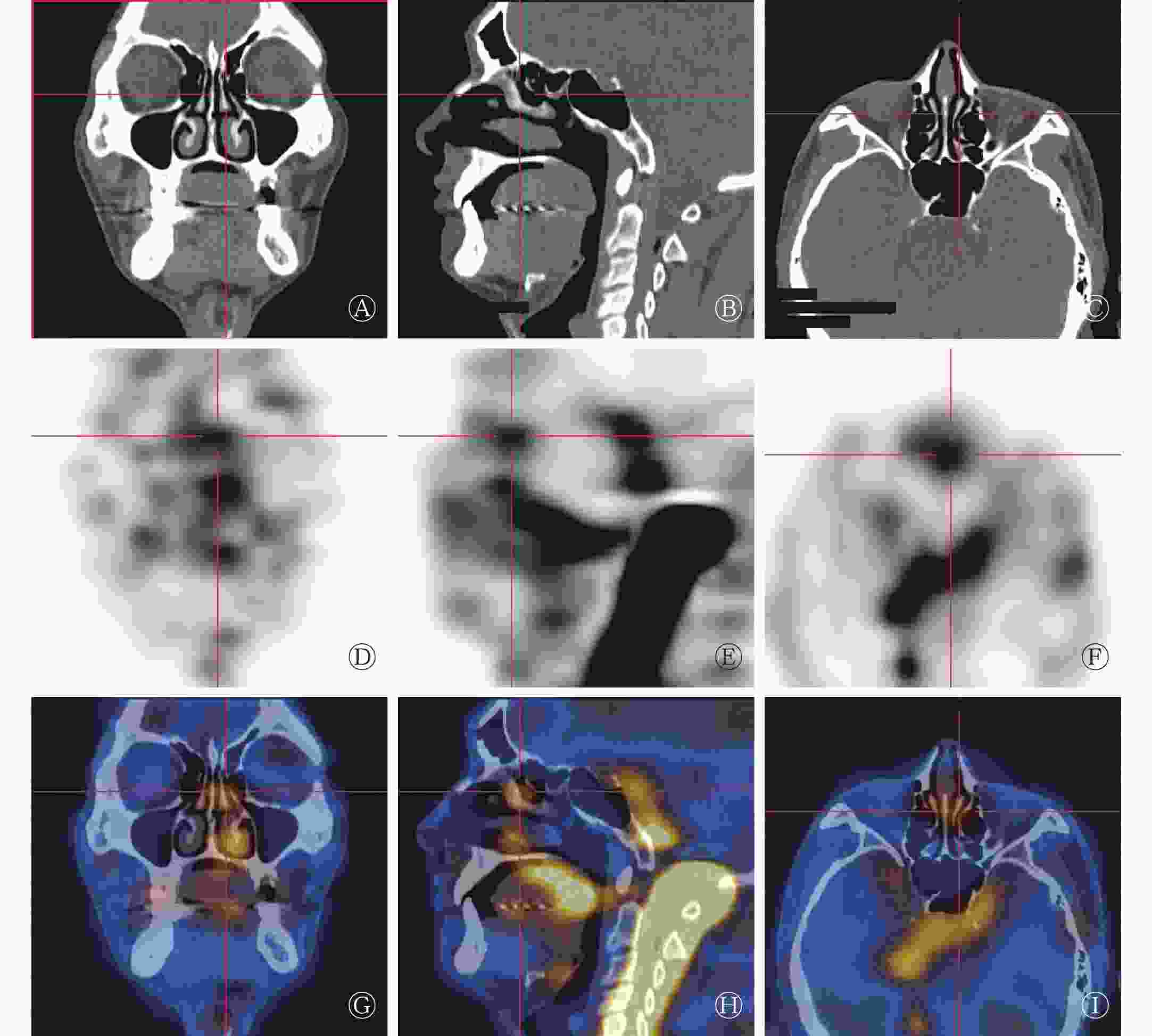
图 3 SIH患者(男性,35岁)脑脊液鼻漏SPECT/CT断层融合显像图
Figure 3. SPECT/CT tomographic fusion imaging of spinal fluid rhinorrhea in patients with spontaneous intracranial hypotension(male,35 years old)
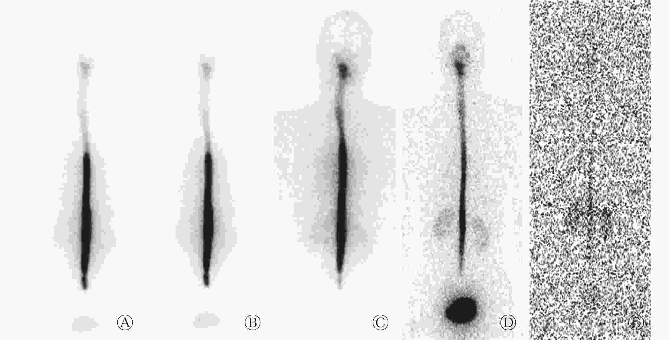
图 4 SIH患者(男性,63岁)间接性脊髓脑脊液漏RNC图像
Figure 4. Radionuclide cisternography image of indirect cerebrospinal fluid leakage in patients with spontaneous intracranial hypotension (male,63 years old)

图 5 SIH患者(男性,34岁)脑脊液循环正常RNC图像
Figure 5. Radionuclide cisternography images of normal cerebrospinal fluid circulation in patients with spontaneous intracranial hypotension(male,34 years old)
-
17例患者行头颅MRI检查,其中10例患者的MRI表现为弥漫性硬脑膜增厚并强化,阳性率58.8%(10/17);2例行头颅CT的患者中有1例表现为慢性硬膜下血肿;其余患者的神经影像学检查未见明显异常。10例RNC检出脊髓脑脊液漏的相应脊髓段的患者中,经MRI检查仅发现1例脊椎MRI在胸段脊髓处发现脊膜异常强化,双侧神经根袖鞘囊状扩张,提示脑脊液漏;MRI未能检出2例脑脊液鼻漏患者的漏口。
-
MRI诊断SIH患者低颅压的阳性率为58.8%(10/17),RNC的阳性率为88.2%(15/17),两者差异无统计学意义(χ2=0.101,P>0.05)。RNC诊断SIH患者低颅压的总阳性率为89.5%(17/19)。
-
18例患者经平卧休息、补液等保守治疗后头痛消失。1例RNC表现为高流量脑脊液漏的患者(图2)经保守治疗无效,改行硬膜外血贴(自体血注射至硬膜外间隙)治疗后头痛消失。所有患者在出院后随访半年,体位性头痛均未再发。
-
文献报道SIH的发病率约为5/100 000,发病中位年龄43岁(24~69岁),男女比约为1∶2.3[8]。本研究中,19例患者年龄20~63(41.7±10.5)岁,与文献报道相似;绝大多数患者脑脊液压力<60 mm H2O,仅1例脑脊液压力达到70 mm H2O。大部分SIH患者的临床表现多样、复杂、缺乏典型性,影像学表现也不典型,加之临床医师对该病认识不足等原因,使该病极易误诊为慢性硬膜下积液、肥厚性硬脑膜炎、小脑扁桃体下疝畸形(Arnold-Chiari畸形)、颈椎病或缺血性脑血管病等疾病。医学影像学技术对该病的诊断作用不断提升,越来越多的影像学证据表明脑脊液漏是导致SIH的主要原因[9-11]。通常认为脑脊液漏是由于硬脊膜局限发育异常,结构上存在薄弱处,或存在硬和(或)脑脊膜憩室[12],正常活动或不被察觉的轻微外伤均可引起其破裂导致脑脊液漏出。在SIH的诊治过程中,确定脑脊液漏口的位置很重要,尤其是对于保守治疗效果差、需行靶向硬膜外血贴治疗的患者有特殊意义。
目前头颅MRI平扫+增强扫描仍是诊断SIH的首选检查方法,其影像的特征性改变是硬脑膜静脉系统扩张、硬脑膜弥漫性均匀性异常强化、硬膜下积液和脑组织下沉(也称脑下垂)[11, 13-14]。但上述改变缺乏特异性,其检出机制均为低颅压的间接改变,如低颅压导致硬脑膜静脉系统扩张,硬脑膜异常强化,硬脑膜下静脉扩张可致硬膜下积液、出血;低颅压时脑脊液水垫作用减弱或消失致脑组织下垂,严重的可导致小脑扁桃体下疝。这些MRI征象可见于脑膜炎、慢性硬膜下积液、蛛网膜下腔出血、脑膜癌或Arnold-Chiari畸形等多种神经系统疾病。本研究中,MRI+增强扫描检出脑膜强化率为58.8%,提示简便、无创的头颅MRI作为神经系统疾病常规检查能间接诊断低颅压,并有助于发现其他引起头痛的神经系统疾病。但MRI对脑脊液漏口的检出存在缺陷,对低颅压的程度、脑脊液漏出量不能准确判断。CT脊髓造影是指将碘油经腰穿注入蛛网膜下腔后,从颅底开始连续薄层CT扫描全脊柱至骶椎体水平,以发现碘油沿脑脊液漏点处的神经根越过椎间孔在椎旁软组织内异常聚集的阳性表现[15-16]。CT脊髓造影能发现活动性脑脊液漏的漏口,但操作复杂、费用高;其图像分析难度大,而且存在全脊柱CT扫描辐射剂量大、造影剂有潜在过敏反应等缺点,因此临床应用价值有限。有研究结果显示,CT脊髓造影的脑脊液漏点检出率明显低于RNC[17]。
RNC操作简单、微创,不仅可以反映脑脊液循环的动力学变化,还可以显示脑脊液间隙的核素分布状况。当出现脑脊液漏时,脑脊液从漏口处外溢漏出,在SPECT图像上可见脑脊液间隙外出现异常的放射性浓聚灶,从而准确定性、定位脑脊液漏,并可通过观察放射性核素的弥散速度、分布特征及漏点浓聚程度判断脑脊液的漏出量。在显像方法上,本研究有一些改进:①在腰穿成功后注入核素前,先往脊髓蛛网膜下隙注入适量生理盐水,使脑脊液压力提高到接近60 mm H2O,从而有利于发现潜在的脑脊液漏。②提前了核素脑脊液间隙显像全脊柱扫描的时间,并增加了扫描次数,多时相动态观察,有利于漏口的检出。③对可疑病灶进行SPECT/CT断层扫描提高了检出率。
本研究中,19例患者中的17例RNC存在脑脊液漏的间接证据,其中发现脑脊液漏直接证据12例,仅存在脑脊液漏间接证据5例,未发现异常2例,总阳性率为89.5%(17/19),与Hyun等[8]文献报道的90%(27/30)相仿。本研究直接脑脊液漏口发现率为63.2%(12/19),MRI仅在RNC发现漏口的相应部位检出1例硬脊膜异常强化,神经根袖鞘囊状扩张,提示脑脊液漏。可见RNC对脑脊液漏口的检出率明显优于MRI。MRI检出率低可能受检查流程安排、预约滞后等影响,部分患者治疗干预后病情好转,影响了MRI的检出阳性率。有研究结果显示,SIH患者的影像改变呈现可逆性,病情好转后复查MRI均恢复正常[18]。本研究有5例患者未发现脑脊液漏口,仅存在脊髓脑脊液漏的间接证据,可能原因有:①漏口微小的大面积渗漏,在未发现局部异常浓聚的情况下,仅行前、后位的平面显像,漏口与脊髓脑脊液影像可能重叠而漏检;②漏口位于腰骶段,局部腰穿注射放射性核素干扰该部位脑脊液漏的检出。2例脑脊液鼻漏患者无外伤史,病因未明,追问病史,患者患有慢性鼻窦炎,有长期擤鼻习惯。有研究结果表明,长期擤鼻习惯可造成瞬间颅压骤高,在脑脊液压力波的长期反复冲击下造成颅底缺损区骨质侵蚀吸收破坏,硬膜和脑组织从薄弱处疝出,因颅内压波动变化使疝出组织反复发生“疝出-回纳”,造成硬膜连同蛛网膜撕裂而发生脑脊液鼻漏[19]。另2例患者RNC检查脑脊液循环未发现异常,考虑可能与脑脊液漏口极微小、脑脊液漏的量极少或检查时处于漏出的间歇期有关[7]。对常见的漏出部位行SPECT/CT断层融合显像可能有助于脑脊液漏点的检出。
对RNC图像的特征进行分析,发现脑脊液漏以脊髓段多见,多位于颈胸交界处和胸椎旁(9/12),与文献报道相符[20]。脊髓段脑脊液漏口放射性异常浓聚于注射药物后30 min即可显影,3 h显影较清晰,此后漏点放射性浓聚程度下降,该动态变化的原因可能为:①脑脊液持续漏出后,脑脊液压力持续下降,导致漏出减少;②随着显像时间延长,放射性核素逐渐衰变,放射性活度下降;③脑脊液漏存在间歇性可能。
SIH一般以保守治疗为主,经充分补液后,患者的症状明显减轻、消失,预后良好[21-22]。本研究中,大部分检出脑脊液漏的患者经保守治疗后临床痊愈。1例大流量脑脊液漏患者常规治疗无效后,经硬膜外血贴治疗后痊愈出院。RNC通过对漏出部位的核素放射性浓聚程度、分布情况及肾脏、膀胱显影的快慢进行分析,可以客观地判断脑脊液的漏出量,且RNC精准提供了脑脊液漏的位置,提高了硬膜外血贴治疗的靶向性及成功率[23]。
总之,RNC具有可动态、多时相观察脑脊液循环状况的优势,能够通过直接发现脑脊液漏点和(或)间接脑脊液循环障碍明确SIH的病因,在脑脊液漏口的检出、漏出流量鉴别上明显优于CT和MRI等常规检查,可为大流量脑脊液漏的干预治疗提供准确信息,在SIH患者的诊疗中有着重要的应用价值。但由于RNC属微创检查,反复腰穿也是诱发脑脊液漏的危险因素,患者痊愈后,难以配合复查,因此本研究缺乏治疗后的对照显像。
利益冲突 本研究由署名作者按以下贡献声明独立开展,不涉及任何利益冲突。
作者贡献声明 林志毅负责研究过程的实施、数据的收集与分析、论文的起草及最终版修订;陈文新负责研究命题的提出、设计、方法的建立、论文的审阅;葛华、余明钿、张艳敏参与研究过程的实施。
自发性低颅压综合征脑脊液间隙显像的影像学特征分析
Analysis the imaging characteristics of radionuclide cisternography in spontaneous intracranial hypotension
-
摘要:
目的 探讨自发性低颅压综合征(SIH)患者放射性核素脑脊液间隙显像(RNC)的影像学特点及其临床意义。 方法 回顾性分析2012年5月至2017年1月福建省立医院收治的19例SIH患者,均经腰椎穿刺脊髓蛛网膜下腔注射99Tcm-DTPA,行多时相脑脊液间隙显像,对99Tcm-DTPA异常分布处加做SPECT/CT断层融合显像及MRI扫描。综合分析RNC的影像学特点。计数资料的组间比较采用 χ 2 检验。 结果 RNC可通过显示脊髓或鼻部脑脊液漏点等直接征象诊断SIH;也可通过观察脑脊液循环过程异常,如:显像剂上升缓慢,难以抵达脑池、大脑凸面、上矢状窦等及膀胱、肾脏早期显像等间接征象诊断SIH。RNC检出脑脊液漏17例。直接脑脊液漏患者12例,其中颈、胸段9例,脑脊液鼻漏2例,大流量脑脊液漏(腰段)患者1例。RNC正常者2例。MRI[阳性率为58.8%(10/17)]与RNC[阳性率为88.2%(15/17)]对SIH低颅压诊断的差异无统计学意义(χ2=0.101,P>0.05);12例RNC检出漏口的相应部位MRI仅检出1例。 结论 RNC可通过直接或间接征象协助SIH的病因诊断,在脑脊液漏口检出上明显优于MRI,并可反映脑脊液漏的流量,便于个体化治疗,在SIH的诊疗中有重要的应用价值。 -
关键词:
- 脑脊液漏 /
- 放射性核素脑脊液间隙显像 /
- 自发性低颅压综合征
Abstract:Objective To investigate the imaging features and clinical significance of radionuclide cisternography(RNC) in patients with spontaneous intracranial hypotension(SIH). Methods Lumbar puncture was made on 19 patients with SIH(4 males and 15 females), and 99Tcm-DTPA was injected into their spinal subarachnoid space. Multitemporal RNC was also performed. SPECT/CT tomography and MRI were added to examine abnormal distribution, and RNC image features were comprehensively analyzed. χ2-test was conducted to compare the enumeration data between groups. Results RNC could be applied to diagnose SIH by showing direct signs such as spinal cord or nasal cerebrospinal fluid leak points or by revealing the abnormal circulation of the cerebrospinal fluid (CSF), such as a slow increase in imaging agents; difficulty in reaching the cistern, the cerebral convex surface, and the superior sagittal sinus; and early imaging of the bladder and the kidney. RNC could also be used to detect the CSF leaks in 17 cases and reveal a direct sign of CSF leaks in 12 cases. Its detection rate was 63.2%. Of these cases, 9 were located in the cervicothoracic junction or the thoracic region, 1 was found in the lumbar, and 2 were observed to have CSF rhinorrhea. A case of high-flow CSF leakage (lumbar segment) occurred in 1 patient. When conservative treatment was ineffective, the patients were treated with targeted epidural blood patch. Two more cases had a normal RNC. A comparison of the MRI findings of 17 patients showed no significant differences between MRI (positive rate of 58.8% [10/17]) and RNC (positive rate of 88.2% [15/17]) in the diagnosis of low intracranial pressure (P > 0.05). However, RNC was superior to MRI in detecting CSF leakage. Of the 12 cases subjected to RNC, only 1 case was found to have CSF leaks through MRI. Conclusions RNC can help diagnose SIH by determining direct or indirect signs and is superior to MRI in terms of detecting CSF leakage. RNC can also be used to show the amount of CSF leakage and facilitate individualized treatment, which is important for the diagnosis and treatment of SIH. -
图 1 SIH患者(女性,37岁)颈胸交界处脑脊液漏的RNC和SPECT/CT断层融合图像
Figure 1. Radionuclide cisternography and SPECT/CT tomographic fusion imaging of cerebrospinal fluid leakage at cervicothoracic junction in patients with spontaneous intracranial hypotension(female,37 years old)
图 2 SIH患者(女性,33岁)腰段大流量脑脊液漏RNC图像
Figure 2. Radionuclide cisternography image of high flow cerebrospinal fluid leakage at lumbar spinal in patients with spontaneous intracranial hypotension(female,33 years old)
图 3 SIH患者(男性,35岁)脑脊液鼻漏SPECT/CT断层融合显像图
Figure 3. SPECT/CT tomographic fusion imaging of spinal fluid rhinorrhea in patients with spontaneous intracranial hypotension(male,35 years old)
图 4 SIH患者(男性,63岁)间接性脊髓脑脊液漏RNC图像
Figure 4. Radionuclide cisternography image of indirect cerebrospinal fluid leakage in patients with spontaneous intracranial hypotension (male,63 years old)
-
[1] Spears RC. Low-Pressure/Spinal Fluid Leak Headache[J]. Curr Pain Headache Rep, 2014, 18(6): 425. DOI: 10.1007/s11916−014−0425−4. [2] De Andres J, Esparza MJ, Ferrer-Rebolleda J, et al. Radioisotope cisternography: a potentially useful tool for headache diagnosis in patients with an IDD pump[J]. Pain Physician, 2014, 17(1): E89−E94. [3] Wang YF, Lirng JF, Fuh JL, et al. Heavily T2-weighted MR myelography vs. CT myelography in spontaneous intracranial hypotension[J]. Neurology, 2009, 73(22): 1892−1898. DOI: 10.1212/WNL.0b013e3181c3fd99. [4] Orcajo-Rincón J, Alonso-Farto JC, Mari-Hualde A, et al. Spontaneous intracranial hypotension syndrome: usefulness of direct and indirect findings of cerebrospinal fluid leakage in isotope cisternography[J]. Rev Neurol, 2011, 52(8): 509−511. [5] Schick U, Musahl C, Papke K. Diagnostics and treatment of spontaneous intracranial hypotension[J]. Minim Invasive Neurosurg, 2010, 53(1): 15−20. DOI: 10.1055/s−0030−1247552. [6] Mokri B. Headaches caused by decreased intracranial pressure: diagnosis and management[J]. Curr Opin Neurol, 2003, 16(3): 319−326. DOI: 10.1097/01.wco.0000073933.19076.c0. [7] Wang XS, Chen XY, Wang YZ, et al. Radionuclide Cisternography Findings of Spontaneous Intracranial Hypotension Syndrome Patients with Different Types of Cerebrospinal Fluid Leakage[J]. J Med Imaging Health Inform, 2015, 5(7): 1558−1562. DOI: 10.1166/jmihi.2015.1567. [8] Hyun SH, Lee KH, Lee SJ, et al. Potential value of radionuclide cisternography in diagnosis and management planning of spontaneous intracranial hypotension[J]. Clin Neurol Neurosurg, 2008, 110(7): 657−661. DOI: 10.1016/j.clineuro.2008.03.014. [9] 丰岩清, 罗柏宁, 张成, 等. 脑脊液漏致自发性低颅压综合征的诊断和治疗(1例报道及文献复习)[J]. 中国神经精神疾病杂志, 2004, 30(6): 460−461. DOI: 10.3969/j.issn.1002−0152.2004.06.022.
Feng YQ, Luo BN, Zhang C, et al. The diagnosis and treatment of spontaneous intracranial hypotension resulted from spontaneous cerebrospinal fluid leaks: 1 case report and literatures review[J]. Chin J Nerv Ment Dis, 2004, 30(6): 460−461. DOI: 10.3969/j.issn.1002−0152.2004.06.022.[10] Kranz PG, Luetmer PH, Diehn FE, et al. Myelographic Techniques for the Detection of Spinal CSF Leaks in Spontaneous Intracranial Hypotension[J]. AJR Am J Roentgenol, 2016, 206(1): 8−19. DOI: 10.2214/AJR.15.14884. [11] 王丽敏, 刘中霖, 王丽娟, 等. 自发性低颅压综合征的磁共振脊髓水成像研究[J]. 中国神经精神疾病杂志, 2012, 38(1): 47−49. DOI: 10.3969/j.issn.1002−0152.2012.01.012.
Wang LM, Liu ZL, Wang LJ, et al. Research of spontaneous intracranial hypotension syndrome with magnetic resonance myelography[J]. Chin J Nerv Ment Dis, 2012, 38(1): 47−49. DOI: 10.3969/j.issn.1002−0152.2012.01.012.[12] Schievink WI, Jacques L. Recurrent spontaneous spinal cerebrospinal fluid leak associated with "nude nerve root" syndrome: case report[J]. Neurosurgery, 2003, 53(5): 1216−1219. DOI: 10.1227/01.NEU.0000089483.30857.11. [13] 李德刚, 李启, 李在军. 原发性低颅压综合征的临床特点及MRI诊断价值[J]. 中国临床医学影像杂志, 2012, 23(6): 417−419. DOI: 10.3969/j.issn.1008−1062.2012.06.012.
Li DG, Li Q, Li ZJ. Clinical characteristics and diagnostic value of MRI for spontaneous intracranial hypotension syndrome[J]. Chin Clin Med Imaging, 2012, 23(6): 417−419. DOI: 10.3969/j.issn.1008−1062.2012.06.012.[14] 陈锦华, 田为中, 陈小云, 等. 自发性低颅压综合征的MRI诊断[J]. 中国医学影像学杂志, 2009, 17(5): 361−364. DOI: 10.3969/j.issn.1005−5185.2009.05.013.
Chen JH, Tian WZ, Chen XY, et al. MR imaging diagnosis of spontaneous intracranial hypotension[J]. Chin J Med Imaging, 2009, 17(5): 361−364. DOI: 10.3969/j.issn.1005−5185.2009.05.013.[15] Yoshida H, Takai K, Taniguchi M. Leakage detection on CT myelography for targeted epidural blood patch in spontaneous cerebrospinal fluid leaks: calcified or ossified spinal lesions ventral to the thecal sac[J]. J Neurosurg Spine, 2014, 21(3): 432−441. DOI: 10.3171/2014.5.SPINE13549. [16] Albes G, Weng H, Horvath D, et al. Detection and treatment of spinal CSF leaks in idiopathic intracranial hypotension[J]. Neuroradiology, 2012, 54(12): 1367−1373. DOI: 10.1007/s00234−012−1055−3. [17] Hashizume K, Watanabe K, Kawaguchi M, et al. Comparison between computed tomography-myelography and radioisotope-cisternography findings in whiplash-associated disorders suspected to be caused by traumatic cerebrospinal fluid leak[J]. Spine (Phila Pa 1976), 2012, 37(12): E721−E726. DOI: 10.1097/BRS.0b013e31824ae5d1. [18] 史立信, 臧颖卓, 何斌, 等. 自发性低颅压综合征42例[J]. 脑与神经疾病杂志, 2016, 24(6): 361−368.
Shi LX, Zang YZ, He B, et al. Spontaneous intracranial hypotension syndrome to 42 cases[J]. J Brain Nerv Dis, 2016, 24(6): 361−368.[19] 延鹏翔. 擤鼻与脑脊液鼻漏[J]. 国外医学. 神经病学神经外科学分册, 1992, 19(4): 213−214. DOI: 10.16636/j.cnki.jinn.1992.04.029.
Yan PX. Blowing the nose and cerebrospinal fluid rhinorrhea[J]. J Int Neurol Neurosurg, 1992, 19(4): 213−214. DOI: 10.16636/j.cnki.jinn.1992.04.029.[20] Yoo HM, Kim SJ, Choi CG, et al. Detection of CSF leak in spinal CSF leak syndrome using MR myelography: correlation with radioisotope cisternography[J]. AJNR Am J Neuroradiol, 2008, 29(4): 649−654. DOI: 10.3174/ajnr.A0920. [21] 韩燕飞, 谢聃, 许春伶. 自发性低颅压综合征12例分析并文献复习[J]. 临床和实验医学杂志, 2017, 16(7): 636−638. DOI: 10.3969/j.issn.1671−4695.2017.07.004.
Han YF, Xie D, Xu CL. A report of 12 patients with spontaneous intracranial hypotension syndrome and review of literatures[J]. J Clin Exp Med, 2017, 16(7): 636−638. DOI: 10.3969/j.issn.1671−4695.2017.07.004.[22] 杨洵哲, 王含, 钱敏, 等. 自发性低颅压综合征临床表现、影像学特点及预后分析[J]. 脑与神经疾病杂志, 2015, 23(4): 269−272.
Yang XZ, Wang H, Qian M, et al. Clinical analysis of spontaneous intracranial hypotension headache[J]. J Brain Nerv Dis, 2015, 23(4): 269−272.[23] 王瑾, 王莉, 何非方, 等. 靶向硬膜外血贴治疗自发性颅内低压价值探讨[J]. 中华医学杂志, 2009, 89(21): 1477−1479. DOI: 10.3760/cma.j.issn.0376−2491.2009.21.012.
Wang J, Wang L, He FF, et al. Targeted epidural blood patch: an effective treatment for spontaneous intracranial hypotension[J]. Natl Med J China, 2009, 89(21): 1477−1479. DOI: 10.3760/cma.j.issn.0376−2491.2009.21.012. -

 点击查看大图

计量
- 文章访问数: 6015
- HTML全文浏览量: 4898
- PDF下载量: 22
