


-
多巴胺转运体(dopamine transporter,DAT)是一种膜蛋白,属于Na+/Cl-依赖性转运蛋白,位于多巴胺能神经元突触前膜的多巴胺能再摄取位点,调控突触间隙内多巴胺的浓度。国内外研究结果显示,DAT相关基因的异常与帕金森病(Parkinson′s disease,PD)的发生相关[1-3]。DAT能间接反映黑质-纹状体通路多巴胺能神经元的数量及功能,故脑DAT被认为是多巴胺能神经元的标志物。11C-甲基-N-2β-甲基酯-3β-(4-氟-苯基)托烷(2β-carbomethoxy-3β-(4-fluorophe-nyl)-(N-11C-methyl)tropane,11C-CFT)是一种PET DAT显像剂,11C-CFT PET是早期诊断PD的有效手段。PET显像图像重建算法通常可分为解析(变换)法和代数(迭代)法,解析法中最常见的为滤波反投影法(filtered backprojection,FBP)。
有序子集最大期望值迭代法(ordered subsets expectation maximization,OSEM)与FBP在算法上有差异,但各有优势。由于两者算法的不同,导致脑部图像结果也不同。目前在临床工作中,各家医院的PET设备品牌、型号虽各不相同,但重建算法中均以FBP和OSEM为主,考虑到标准化的正常值对疾病异常诊断的重要性,笔者将重点观察FBP和OSEM对正常人11C-CFT PET显像脑内DAT分布半定量值的影响。
-
本研究纳入2014年3月至2015年6月期间41名健康受试者(正常人),纳入标准:>18岁,无精神及神经疾病,无酒精滥用及吸毒史,近期未服用精神药物;排除标准:检查前6个月内服用激素或精神类药物。其中男性18名、女性23名,年龄31~80岁,平均年龄(58.1±10.4)岁。41名受试者均行11C-CFT PET/CT显像,根据年龄分为3组,分别为A组(20~39岁)、B组(40~59岁)、C组(60~80岁)(表 1)。本研究通过了复旦大学附属华山医院伦理委员会审批(批准号:KY2013-336),所有受试者在接受检查前均签署了知情同意书。
组别 人数/名 平均年龄/岁 最小年龄/岁 最大年龄/岁 男性/名 女性/名 A组(20~39岁) 3 34.7±3.2 31 37 2 1 B组(40~59岁) 17 52.8±5.1 41 59 8 9 C组(60~80岁) 21 65.7±5.0 60 80 8 13 表 1 纳入的41名健康受试者的基本情况及分组情况
Table 1. Basic situation and grouping of 41 healthy subjects
-
对健康受试者静脉注射370 MBq 11C-CFT(由复旦大学附属华山医院PET中心制备,放化纯度>98%),注射60 min后行PET/CT扫描(德国Siemens公司Biograph 64 HD PET/CT仪),先进行CT扫描(15 s)用于衰减校正,然后采用三维模式进行头部扫描(15 min)采集PET图像。将所采集的脑PET原始数据分别进行FBP及OSEM重建并获得脑部横断面、冠状面及矢状面图像。其中,FBP使用Gaussian滤波函数重建,半高宽3.5 mm;OSEM使用Gaussian滤波函数重建,半高宽2.0 mm,3次迭代,21个子集。
-
利用MRIro图像格式转换软件将PET图像转换为Analyze7格式,在MATLAB Version 6.5.1平台(美国Mathworks公司)上,应用SPM5软件(英国Wellcome认知神经研究所)进行数据分析:用SPM5软件中的Normalize模块将PET图像拟合到标准立体空间中(模板由美国Feinstein神经科学中心提供),并用10 mm×10 mm×10 mm的半高宽对标准化后的图像进行平滑处理。应用Scanvp程序(美国Feinstein神经科学中心提供[4])选取基底节显示良好的第20~30层横断面图像,叠加并平均成1个层面,对双侧尾状核、壳核前部及后部、顶枕皮质自动勾画ROI,获得平均放射性计数,以缺乏DAT分布的顶枕皮质作为参考区,按公式(尾状核或壳核放射性计数/顶枕叶放射性计数-1)计算DAT分布的半定量值[4]。
-
使用SPSS 15.0软件进行统计学分析。各个脑区DAT分布半定量值均通过Kolmogorov-Smirnov正态性检验,P=0.155~0.200(>0.05),符合正态分布。各个脑区DAT分布半定量值均通过Levene检验,P=0.424~0.876(>0.05),符合方差齐性。DAT分布半定量值的组间比较采用配对t检验,相关性分析采用Pearson相关分析法。P < 0.05表示差异有统计学意义。
-
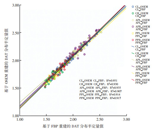
41名健康受试者经11C-CFT PET/CT显像、图像分析及计算后获得的双侧尾状核、壳核前部及后部DAT分布半定量值见表 2。基于OSEM重建的PET图像中双侧尾状核、壳核前部及后部的DAT分布半定量值均高于基于FBP重建的PET图像,且两者间的差异均有统计学意义(t=9.658~15.859,均P=0.000)(表 2)。将基于两种重建方法的PET图像中双侧尾状核、壳核前部及后部的DAT分布半定量值进行Pearson相关性分析,结果显示基于OSEM和FBP重建的PET图像的DAT分布半定量值在双侧尾状核、壳核前部及后部均呈显著正相关(R2=0.907~0.951,均P=0.000)(表 2、图 1)。
重建方法 组别 尾状核 壳核前部 壳核后部 左侧 右侧 左侧 右侧 左侧 右侧 OSEM A组 2.15±0.24 2.06±0.31 2.39±0.21 2.38±0.22 1.89±0.18 2.06±0.17 B组 1.89±0.16 1.78±0.16 2.17±0.18 2.18±0.20 1.72±0.10 1.84±0.13 C组 1.89±0.12 1.77±0.12 2.17±0.13 2.17±0.16 1.71±0.09 1.82±0.12 FBP A组 2.10±0.27 1.99±0.27 2.37±0.26 2.33±0.24 1.82±0.17 1.96±.17 B组 1.81±0.17 1.70±0.16 2.09±0.18 2.09±0.19 1.64±0.10 1.75±.13 C组 1.79±0.12 1.68±0.12 2.07±0.15 2.09±0.17 1.62±0.10 1.72±0.13 t值 14.635 12.612 11.571 9.658 15.859 14.477 P值 0.000 00.000 00.000 0.000 0.000 0.000 R2值 0.951 0.930 0.935 0.907 0.914 0.917 P值 0.000 0.000 0.000 0.000 0.000 0.000 注:表中,A组:20~39岁;B组:40~59组;C组:60~80组;FBP:滤波反投影法;OSEM:有序子集最大期望值迭代法;11C-CFT: 11C-甲基-N-2β-甲基酯-3β-(4-氟-苯基)托烷;DAT:多巴胺转运体;PET:正电子发射断层显像术。 表 2 基于FBP与OSEM重建的11C-CFT PET图像中各脑区DAT分布半定量值及比较(x±s)
Table 2. Comparison of semi-quantitative values of DAT distribution in the 11C-CFT PET images based on FBP and OSEM reconstruction(x±s)

图 1 基于FBP与OSEM重建的PET图像中各脑区DAT分布半定量值的相关性
Figure 1. Correlation of semi-quantitative values of DAT distribution in different brain regions of PET images based on FBP and OSEM reconstruction
随着年龄的增长,基于OSEM与FBP重建的PET图像中双侧尾状核、壳核前部及后部DAT分布半定量值均呈现逐渐减低的趋势(表 2)。
-
近年来PET分子影像学成为PD早期诊断的重要诊断技术之一, 包括突触后多巴胺受体显像和突触前DAT显像。对DAT进行深入广泛的研究,将有助于PD病因的阐明、早期诊断和预防药物研发。11C-CFT是目前临床常用的DAT PET显像剂之一,可以探测纹状体的多巴胺功能减退[5],在PD早期诊断方面有重要的价值。由国际运动障碍协会制订和颁布的PD诊断标准,不仅可用于相关领域的临床研究,还可在临床实践中就PD的诊断发挥指南的作用,因此该诊断标准获得了业内的广泛认可。在最新的国际运动障碍协会2015年版PD诊断标准中提到正常的突触前多巴胺能系统功能显像是PD的排除标准之一[6]。11C-CFT在临床前期就能探测到DAT分布的减少,特别是在Parkin基因(又名PARK2基因,定位于6q25.2~27,包含12个外显子,全长约1.5 Mb)突变、PINK1基因(又名PARK6基因,定位于1p36,包含8个外显子,全长约1.8 kb)突变[7]、LRRK2基因(富含亮氨酸重复序列激酶2基因)发生R1441C突变[8]、CHCHD2基因[9](一种与PD有关的基因)突变导致的早发PD家族未出现临床症状的患者中,就可以发现11C-CFT分布轻度减低,且差异有统计学意义。
11C-CFT PET脑显像可获得脑内DAT分布半定量值,而标准化的正常值对更早期地发现脑内DAT分布的异常尤为重要。笔者对41名健康受试者进行11C-CFT PET显像,计算脑内DAT分布半定量值,并研究不同图像重建方法对DAT分布半定量值的影响,结果显示基于FBP与OSEM两种重建方法的PET图像中脑内双侧尾状核、壳核前部及后部DAT分布半定量值存在显著差异,说明PET图像重建方法可以影响11C-CFT图像的半定量值。
目前PET图像重建方法中的解析法是以中心切片定理(central slice theorem)为理论基础的求逆过程,最常见的为FBP。FBP中的滤波是一维的,该法运算速度快,是最早应用于PET图像重建的算法,被认为是传统的重建方法。迭代法是从一个假设的初始图像开始,采用迭代的方法,将理论投影值同实测的投影值进行不断比较,在某种最优化准则指导下寻找最优解。在临床应用中,OSEM与FBP各有优势,OSEM相比传统的FBP大大提高了图像的质量,主要优点是减少条状噪音,增加图像的均匀性。虽然与FBP相比OSEM的对比度有所下降,但下降的程度不大。这两种图像重建方法在算法上的差异,会导致脑部图像结果的不同。在18F-FDG PET脑显像中,不同重建方法所得的PD相关脑代谢网络模式表达值存在显著差异,但是这种差异并不影响PD相关脑代谢网络模式表达值在早期诊断与判断病情严重程度等中的临床应用[10]。笔者发现在11C-CFT PET脑显像中,基于FBP与OSEM两种重建方法的正常人PET图像中各脑区DAT分布半定量值存在显著差异,但是这种差异是否会影响PD的早期诊断,还需要进一步验证。
另外,本研究结果显示20~39岁、40~59岁、60~80岁3个年龄段中出现随着年龄的增长DAT分布半定量值逐步下降的现象,并且在基于FBP与OSEM两种重建方法的PET图像中均观察到了这种趋势。因此,虽然本研究结果显示不同图像重建方法对DAT分布半定量值有影响,但是仍能显示出DAT随年龄增长而减低的趋势。国内已有研究者提出了年龄因素对于DAT分布的影响,认为随着年龄的增长会出现DAT分布的下降[11]。
本研究结果显示基于OSEM重建的半定量数值高于FBP,既往研究在18F-FDG PET显像中也得到了相似的结果[12]。造成两种方法间差异的原因主要是在FBP与OSEM的传输成像和衰减校正中使用了不同的平滑滤波器。为了在FBP重建期间获得噪声较少的图像,通常使用非定量过滤器,这可能导致SUV被低估。而在OSEM重建过程中可以使用更准确的高斯滤波器,不会影响图像质量。
由于PD患者主要为中老年人,导致本研究存在一定的缺陷,即20~39岁年龄段的入组人数较少,在今后的研究中将完善低年龄段人数。
综上所述,在正常人11C-CFT PET显像中,基于OSEM与FBP重建所得PET图像中脑内DAT分布半定量值存在显著差异。同时,在两种图像重建方法下,均出现了DAT分布随着年龄增长而减低的趋势。由于不同图像重建方法对11C-CFT PET显像的半定量值存在显著影响,因此,在临床及科研工作中,涉及多中心或纵向研究时需要保持图像重建方法的一致性,才能使图像结果更加可靠和具有可比性。
FBP和OSEM对正常人脑内多巴胺转运体分布半定量值影响的研究
Effects of the different PET image reconstruction methods on distribution of dopamine transporter in healthy human brain
-
摘要:
目的 研究不同PET重建方法对正常人脑内多巴胺转运体(DAT)分布半定量值的影响。 方法 将2014年3月至2015年6月期间41例健康受试者(正常人)的11C-甲基-N-2β-甲基酯-3β-(4-氟-苯基)托烷(11C-CFT)PET图像分别进行滤波反投影法(FBP)和有序子集最大期望值迭代法(OSEM)重建,自动勾画尾状核、壳核前部和后部等感兴趣脑区,以缺乏DAT分布的顶枕皮质作为参考区,按公式计算DAT分布的半定量值。DAT分布半定量值的组间比较采用配对t检验,相关性分析采用Pearson相关分析法。 结果 基于OSEM重建的PET图像中DAT分布半定量值分别为:尾状核(1.77~2.15)、壳核前部(2.17~2.39)、壳核后部(1.71~2.06);基于FBP重建的PET图像中DAT分布半定量值分别为:尾状核(1.68~2.10)、壳核前部(2.07~2.37)、壳核后部(1.62~1.96)。基于OSEM重建的PET图像中双侧尾状核、壳核前部及后部的DAT分布半定量值均显著高于基于FBP重建的PET图像,且两者间的差异均有统计学意义(t=9.658~15.859,均P=0.000)。Pearson相关性分析结果显示,基于OSEM与FBP重建的PET图像的DAT分布半定量值在双侧尾状核、壳核前部及后部均呈显著正相关(R2=0.907~0.951,均P=0.000)。基于OSEM与FBP重建的PET图像中双侧尾状核、壳核前部及后部DAT分布半定量值均呈现随年龄增长逐渐减低的趋势。 结论 不同PET重建方法获得的正常人脑内DAT分布半定量值存在显著差异,在多中心或纵向研究中需要保持PET图像重建方法的一致性。 -
关键词:
- 多巴胺质膜转运蛋白质类 /
- 正电子发射断层显像术 /
- 滤波反投影 /
- 迭代
Abstract:Objective To study the effect of the reconstruction method on the semi-quantitative distribution of dopamine transporter (DAT) in the brain, 2β-carbomethoxy-3β- (4-fluorophe-nyl)- (N-11C-methyl)tropane (11C-CFT) PET images of healthy subjects were reconstructed by different PET reconstruction methods. Methods From March 2014 to June 2015, the 11C-CFT PET images of 41 healthy subjects were reconstructed by filtering back projection (FBP) method and ordered subsets expectation maximization (OSEM) iterative method. The brain regions of interest (ROI), namely, caudate nucleus, anterior putamen, and posterior shell nucleus, were automatically sketched with the parietal and occipital cortex lacking of DAT distribution as reference regions. The semi-quantitative value of DAT distribution was calculated using the following formula:radioactivity count of ROI/radioactivity count of parietal and occipital cortex-1. Paired t-test was used to compare the semi-quantitative values of DAT distribution. Correlation analysis was performed using the Pearson correlation analysis. ResultsThe values of DAT distribution based on OSEM were as follows:caudate nucleus (1.77-2.15), anterior putamen (2.17-2.39), and posterior putamen (1.71-2.06). The values of DAT distribution based on FBP were as follows:caudate nucleus (1.68-2.10), anterior putamen (2.07-2.37), and posterior putamen (1.62-1.96). In the bilateral caudate nucleus and anterior and posterior putamen, the 11C-CFT of DAT distribution by OSEM was significantly higher than that by FBP (t=9.658-15.859, all P=0.000). The Pearson correlation analysis showed that the semi-quantitative values of DAT distribution by FBP and OSEM were positively correlated with each other in the bilateral caudate nucleus and anterior and posterior putamen (R2=0.907-0.951, all P=0.000). The DAT distribution by OSEM and FBP decreased with aging in the caudate nucleus and anterior and posterior putamen. Conclusions A significant difference was found in the semi-quantitative value of the DAT distribution in the brain of healthy subjects by different PET reconstruction methods. Therefore, a consistent PET image reconstruction method should be used in a multicenter or longitudinal study. -
图 1 基于FBP与OSEM重建的PET图像中各脑区DAT分布半定量值的相关性
Figure 1. Correlation of semi-quantitative values of DAT distribution in different brain regions of PET images based on FBP and OSEM reconstruction
表 1 纳入的41名健康受试者的基本情况及分组情况
Table 1. Basic situation and grouping of 41 healthy subjects
组别 人数/名 平均年龄/岁 最小年龄/岁 最大年龄/岁 男性/名 女性/名 A组(20~39岁) 3 34.7±3.2 31 37 2 1 B组(40~59岁) 17 52.8±5.1 41 59 8 9 C组(60~80岁) 21 65.7±5.0 60 80 8 13  下载: 导出CSV
表 2 基于FBP与OSEM重建的11C-CFT PET图像中各脑区DAT分布半定量值及比较(x±s)
Table 2. Comparison of semi-quantitative values of DAT distribution in the 11C-CFT PET images based on FBP and OSEM reconstruction(x±s)
重建方法 组别 尾状核 壳核前部 壳核后部 左侧 右侧 左侧 右侧 左侧 右侧 OSEM A组 2.15±0.24 2.06±0.31 2.39±0.21 2.38±0.22 1.89±0.18 2.06±0.17 B组 1.89±0.16 1.78±0.16 2.17±0.18 2.18±0.20 1.72±0.10 1.84±0.13 C组 1.89±0.12 1.77±0.12 2.17±0.13 2.17±0.16 1.71±0.09 1.82±0.12 FBP A组 2.10±0.27 1.99±0.27 2.37±0.26 2.33±0.24 1.82±0.17 1.96±.17 B组 1.81±0.17 1.70±0.16 2.09±0.18 2.09±0.19 1.64±0.10 1.75±.13 C组 1.79±0.12 1.68±0.12 2.07±0.15 2.09±0.17 1.62±0.10 1.72±0.13 t值 14.635 12.612 11.571 9.658 15.859 14.477 P值 0.000 00.000 00.000 0.000 0.000 0.000 R2值 0.951 0.930 0.935 0.907 0.914 0.917 P值 0.000 0.000 0.000 0.000 0.000 0.000 注:表中,A组:20~39岁;B组:40~59组;C组:60~80组;FBP:滤波反投影法;OSEM:有序子集最大期望值迭代法;11C-CFT: 11C-甲基-N-2β-甲基酯-3β-(4-氟-苯基)托烷;DAT:多巴胺转运体;PET:正电子发射断层显像术。
下载: 导出CSV
-
[1] Moreau C, Meguig S, Corvol JC, et al. Polymorphism of the dopamine transporter type 1 gene modifies the treatment response in Parkinson's disease[J]. Brain, 2015, 138 (Pt 5):1271-1283. DOI:10.1093/brain/awv063. [2] Lin JJ, Yueh KC, Chang DC, et al. The homozygote 10-copy genotype of variable number tandem repeat dopamine transporter gene may confer protection against Parkinson's disease for male, but not to female patients[J]. J Neurol Sci, 2003, 209 (1/2):87-92. [3] 刘丰韬, 王坚.遗传性帕金森病患者的脑功能显像[J].内科理论与实践, 2010, 5 (5):438-441. DOI:10.16138/j.1673-6087. 2010. 05.007.
Liu FT, Wang J. Functional neuroimging in patients with inherited Parkinsonism[J]. J Intern Med Concepts Pract, 2010, 5 (5):438-441. doi: 10.16138/j.1673-6087.2010.05.007[4] Ma Y, Dhawan V, Mentis M, et al. Parametric mapping of[18F]FPCIT binding in early stage Parkinson's disease:a PET study[J]. Synapse, 2002, 45 (2):125-133. DOI:10.1002/syn.10090. [5] Liu SY, Wu JJ, Zhao J, et al. Onset-related subtypes of Parkinson's disease differ in the patterns of striatal dopaminergic dysfunction:A positron emission tomography study[J]. Parkinsonism Relat Disord, 2015, 21 (12):1448-1453. DOI:10.1016/j.parkreldis.2015.10.017. [6] Postuma RB, Berg D, Stern M, et al. MDS clinical diagnostic criteria for Parkinson's disease[J]. Mov Disord, 2015, 30 (12):1591-1601. DOI:10.1002/mds.26424. [7] Guo JF, Wang L, He D, et al. Clinical features and[11C]-CFT PET analysis of PARK2, PARK6, PARK7-linked autosomal recessive early onset Parkinsonism[J]. Neurol Sci, 2011, 32 (1):35-40. DOI:10.1007/s10072-010-0360-z. [8] Peng F, Sun YM, Chen C, et al. The heterozygous R1441C mutation of leucine-rich repeat kinase 2 gene in a Chinese patient with Parkinson disease:A five-year follow-up and literatures review[J]. J Neurol Sci, 2017, 373:23-26. DOI:10.1016/j.jns.2016.12.009. [9] Shi CH, Mao CY, Zhang SY, et al. CHCHD2 gene mutations in familial and sporadic Parkinson's disease[J/OL]. Neurobiol Aging, 2016, 38: 217.e9-217.e13[2018-01-15]. http: //www.ncbi.nlm.nih.gov/pubmed/26705026. DOI: 10.1016/j.neurobiolaging.2015.10.040. [10] 王元元, 吴平, 左传涛, 等.不同重建算法对帕金森病相关脑功能网络的影响[J].中国医学计算机成像杂志, 2013, 19 (6):549-552. DOI:10.19627/j.cnki.cn31-1700/th.2013.06.018.
Wang YY, Wu P, Zuo CT, et al. Influence of Different PET Reconstruction Algorithms on Parkinson's Disease-related Pattern Expression[J]. Chin Comput Med Imag, 2013, 19 (6):549-552. doi: 10.19627/j.cnki.cn31-1700/th.2013.06.018[11] 邱春, 左传涛, 张政伟, 等.不同显像时间窗对11C-CFT PET/CT显像测多巴胺转运蛋白分布半定量值的影响[J].中华核医学与分子影像杂志, 2013, 33 (5):362-366. DOI:10.3760/cma.j.issn.2095-2848.2013.05.012.
Qiu C, Zuo CT, Zhang ZW, et al. Influence of scanning time window on the binding potentials of dopamine transporter in the brain of healthy volunteers with 11C-CFT PET imaging[J]. Chin J Nucl Med Mol Imaging, 2013, 33 (5):362-366. doi: 10.3760/cma.j.issn.2095-2848.2013.05.012[12] Ramos CD, Erdi YE, Gonen M, et al. FDG-PET standardized uptake values in normal anatomical structures using iterative reconstruction segmented attenuation correction and filtered back-projection[J]. Eur J Nucl Med, 2001, 28 (2):155-164. doi: 10.1007/s002590000421 -

 点击查看大图
图(1)表(2)
计量
- 文章访问数: 3056
- HTML全文浏览量: 1714
- PDF下载量: 8