


 下载:
下载:
-
在所有先天畸形患儿中,约30%~40%的患儿出生后伴有泌尿系统异常[1]。肾脏集合系统的变异包括集合系统发育不全、位置和数目的异常。肾外型肾盂指肾盂位于肾外,双侧相对于单侧少见。随着SPECT/CT技术的发展,99Tcm-DTPA肾动态显像除了常规评估分肾功能以外,还可以为临床提供相关的解剖信息。笔者现报道双侧肾外型肾盂伴左肾盂积水99Tcm-DTPA肾动态显像一例。
-
患者女性,27岁。2017年12月3日于当地医院体检行彩超发现左肾结石、左肾积水,不伴尿频、尿急、尿痛、肉眼血尿,不伴恶心、呕吐、发热等。于2018年12月26日以“左肾结石、左肾积水”入住我院泌尿外科。入院化验检查,结果示:血尿素水平为3.54 mmol/L(参考范围2.80~7.60 mmol/L),血肌酐水平为69.6 μmol/L(参考范围49.0~90.0 μmol/L)。静脉肾盂造影提示左肾盂输尿管连接部显影欠佳,造影剂通过缓慢,考虑左肾盂输尿管连接部狭窄。为评估分肾功能,患者行99Tcm-DTPA利尿肾动态显像。常规饮水500 mL(8~10 mL/kg),20 min后,受检者排空膀胱取仰卧位,采集前后位影像(视野范围包括双侧肾脏及膀胱区),经肘静脉成功建立静脉通道后,依次注射速尿40 mg和“弹丸”式注射177.6 MBq 99Tcm-DTPA,腹主动脉开始显影后即刻开机行双肾前后位连续动态显像,共采集20 min。功能相示:显像约3 min时,左肾下极出现一卵圆形显像剂浓聚影,随时间推移先逐渐增浓,然后缓慢变淡(图 1)。为进一步确诊行局部SPECT/CT显像,CT定位像示:左肾下极异常浓聚影位于肾门外,与左肾盏相连的肾盂内可见液性低密度影(图 2)。随后对双肾ROI进行半自动勾画获得肾图,其中左肾下极浓聚影不纳入ROI中(图 3),结果显示肾小球滤过率:左肾为33.0 mL/min,右肾为37.3 mL/min。双血浆法测定肾小球滤过率为73 mL/min(成年女性肾小球滤过率正常参考值为81~137 mL/min)。

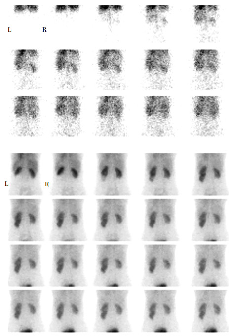
图 1 患者女性,27岁,双侧肾外型肾盂伴左肾盂重度积水99Tcm-DTPA肾动态显像图(后位)图中,前三行为血流相,每2 s采集1帧;后四行为功能相,每1 min采集1帧。血流相示:腹主动脉显影后2~4 s两侧肾动脉几乎同时显影,随后出现清晰的肾影,双肾血流灌注正常。功能相示:左肾下极在3 min时出现一卵圆形显像剂浓聚影,随时间推移可见其逐渐增浓变淡。

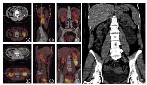
图 2 患者女性,27岁,双侧肾外型肾盂伴左肾盂重度积水99Tcm-DTPA SPECT/CT融合显像图图中,A~C:左肾99Tcm-DTPA SPECT/CT融合显像图,可见左肾盂明显扩张积液,伴肾盂内结节状致密影,其中,图A为横断位、B为矢状位、C为冠状位;D~F:右肾99Tcm-DTPA SPECT/CT融合显像图,可见右肾肾盂轻度扩张积液,其中,图D为横断位、E为矢状位、F为冠状位;G:CT定位相冠状位,双侧肾盂均位于肾门外,左肾盂明显扩张积液。

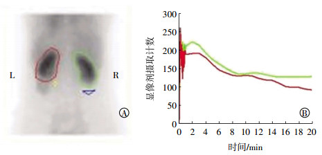
图 3 患者女性,27岁,双肾感兴趣区的勾画(后位)及时间-放射性曲线图图中,A:双肾后位感兴趣区的勾画,左肾影较右肾影体积缩小,右肾形态、大小、位置正常;B:红色曲线代表左肾时间-放射性曲线,绿色曲线代表右肾时间-放射性曲线。左肾在2~4 min可见显像剂积聚高峰,峰时、峰值正常,转移显像剂过程正常,排泄显像剂过程略延缓;右肾在2~4 min也可见显像剂积聚高峰,峰时、峰值正常,转移显像剂过程正常,排泄显像剂过程略延缓。
患者于2018年1月4日在我院行后腹腔镜下左侧肾盂成形术+肾盂切开取石术,术中发现双侧肾盂位于肾外,剪断左肾扩大的肾盂,并切除肾盂输尿管连接部,置入F6输尿管支架管(双“J”管),近端至肾盂,远端至膀胱。
-
正常人的肾盂位于肾窦内,称肾内型肾盂,少数位于肾窦外,称肾外型肾盂。组织胚胎发育至第4周以后,输尿管芽伸展方向为背后侧,随后长入中胚层的生后肾组织中,生后肾组织形态为帽状,在输尿管芽的顶端包盖,输尿管芽顶端膨大形成肾盂并分出肾大盏和肾小盏,其主干形成输尿管。肾外型肾盂可能与后肾组织、输尿管芽发育速度以及生后肾组织对输尿管芽的帽状包盖不完善有关[2-3]。
肾外型肾盂主要与肾积水、肾盂旁囊肿、尿瘘形成的尿囊肿鉴别。肾积水静脉肾盂造影表现为肾小盏的杯口形态失常,呈膨凸或饱满状,积水严重时肾皮质变薄,积水原因多由于输尿管结石或狭窄所致;肾盂旁囊肿多由先天性因素造成,行CT增强扫描时,对比剂不进入囊肿内[4-5];肾盂尿漏多由尿路梗阻、外伤所致;输尿管尿漏多继发于泌尿生殖系统、腹膜后、妇科或盆腔手术后;膀胱尿漏多为外伤所致。尿漏主要借助增强CT辅助诊断,经肘静脉注射对比剂后约30 s行皮质期扫描,60 s行髓质期扫描,50~60 min行排泄期扫描,在髓质期和排泄期可以观察肾盂、输尿管及膀胱漏口。尿漏CT间接征象为肾周积液,被包裹时形成尿囊肿,周围脂肪间隙可见条索影[6]。
本例患者“弹丸”注射99Tcm-DTPA 3 min左右,平面像可见左肾下极开始出现显像剂浓聚影。首先与尿漏鉴别,尿漏表现为随时间推移显像剂分布逐渐增浓,而本例患者随时间推移显像剂分布逐渐增浓随后减淡,所以排除尿漏的诊断;其次考虑重复肾,在上尿路通畅和肾功能正常时注射显像剂1~2 min内,肾皮质显影,随后进入髓质期和排泄期。本例患者左肾下极浓聚影在3 min左右显影,若考虑为肾组织,则可能为其功能受损而导致延迟显影,若为此种情况应注意ROI的勾画范围;若考虑为肾旋转不良,则需与背侧位型肾盂鉴别,此种情况需通过CT对肾脏进行深度校正,再去评估肾小球滤过率。从本例患者SPECT/CT冠状位可以清晰显示左肾下极浓聚影位于肾外并伴有积液,肾盂内可见液性低密度影,肾皮质未见明显受压;右肾肾盂也位于肾外。CT图像上表现为肾旁液性低密度影,还需与肾盂旁囊肿鉴别。若考虑为肾盂旁囊肿,在肾动态显像上应表现为显像剂分布稀疏或空白区,本例患者中表现为显像剂分布浓聚影,故排除肾盂旁囊肿的诊断。结合患者的实验室检查结果及临床资料,最终诊断为双侧肾外型肾盂,左肾功能轻度受损,右肾功能基本正常。
肾外型肾盂不伴感染、结石等并发症时,无需临床处理,伴有明显积水或结石时可予以手术治疗。
肾外型肾盂是一种先天性变异,双侧肾外型肾盂较为少见,但当我们在肾动态显像看到与本例患者同样特征性表现时,需考虑有肾外型肾盂合并肾盂重度积水的可能,结合SPECT/CT显像可予以进一步提示。
双侧肾外型肾盂伴左肾盂重度积水99Tcm-DTPA肾动态显像一例
A case of bilateral extrarenal pelvis accompanied with severe hydronephrosis of left pelvis of 99Tcm-DTPA renal dynamic imaging
-
摘要: 笔者报道了双侧肾外型肾盂伴左肾盂重度积水99Tcm-DTPA肾动态显像一例。肾外型肾盂临床上通常无症状,常为健康体检或其他原因行腹部超声或CT等影像学检查时偶然发现,可伴有肾结石、肾积水等。因肾盂位于肾外,肾盂积水对肾皮质一般无压迫,故患者早期血尿素、血肌酐水平正常或轻度增高。临床为评估分肾功能,将9Tcm-DTPA肾动态显像作为首选检查,肾外型肾盂伴一侧肾盂重度积水表现为患侧肾影外异常显像剂浓聚影,需与尿漏、肾旋转不良等鉴别。故笔者通过病例及文献复习加深了对肾外型肾盂伴肾盂积水9Tcm-DTPA肾动态显像特点及鉴别诊断的认识。认识这种解剖变异和提供肾功能相关信息可以帮助临床医师做出决策,并降低术中损伤肾盏肾盂的风险。Abstract: The present case describes the 99Tcm-DTPA renal dynamic imaging of bilateral extrarenal pelvis accompanied by severe hydronephrosis of the left pelvis. The extrarenal pelvis is usually asymptomatic and is often found through abdominal examination via abdominal ultrasonography or CT scan. Hydronephrosis generally does not compress the renal cortex because the renal pelvis is located outside the kidney; thus, the patient's early serum urea and creatinine levels are normal or slightly increased. 99Tcm-DTPA renal dynamic imaging is the clinicians' first choice for evaluating left or right renal functions. Bilateral extrarenal pelvis, accompanied by severe hydronephrosis of pelvis, presents an abnormal concentration of 99Tcm-DTPA outside the renal area. This abnormality needs to be identified as urinary leakage or renal dysplasia. Therefore, we explored the characteristics of 99Tcm-DTPA renal dynamic imaging and the differential diagnosis of extrarenal pelvis with severe hydronephrosis in this study. Understanding this anatomical variation and providing information about renal functions can help clinicians make appropriate clinical decisions and reduce the risk of damage in the renal pelvis during surgery.
-
Key words:
- Extrarenal pelvis /
- Hydronephrosis /
- Technetium Tc 99m pentetate /
- Radionuclicle imaging
-
图 1 患者女性,27岁,双侧肾外型肾盂伴左肾盂重度积水99Tcm-DTPA肾动态显像图(后位)图中,前三行为血流相,每2 s采集1帧;后四行为功能相,每1 min采集1帧。血流相示:腹主动脉显影后2~4 s两侧肾动脉几乎同时显影,随后出现清晰的肾影,双肾血流灌注正常。功能相示:左肾下极在3 min时出现一卵圆形显像剂浓聚影,随时间推移可见其逐渐增浓变淡。
图 2 患者女性,27岁,双侧肾外型肾盂伴左肾盂重度积水99Tcm-DTPA SPECT/CT融合显像图图中,A~C:左肾99Tcm-DTPA SPECT/CT融合显像图,可见左肾盂明显扩张积液,伴肾盂内结节状致密影,其中,图A为横断位、B为矢状位、C为冠状位;D~F:右肾99Tcm-DTPA SPECT/CT融合显像图,可见右肾肾盂轻度扩张积液,其中,图D为横断位、E为矢状位、F为冠状位;G:CT定位相冠状位,双侧肾盂均位于肾门外,左肾盂明显扩张积液。
-
[1] Zivkovi ć D, Varga J, Grebeldinger S, et al.[Ureteral triplication——a case report] [J]. Med Pregl, 2005, 58(11-12):592-595. doi: 10.2298/MPNS0512592Z [2] Nataraju G, Nandeesh BN, Gayathri MN. Extrarenal calyces:a rare anomaly of the renal collecting system[J]. Indian J Pathol Microbiol, 2009, 52(3):368-369. DOI:10.4103/0377-4929.54996. [3] Gupta T, Goyal SK, Aggarwal A, et al. Extrarenal calyces:a rare renal congenital anomaly[J]. Surg Radiol Anat, 2015, 37(4):407-410. DOI:10.1007/s00276-014-1349-8. [4] 苗新中, 段青松.多层螺旋CT诊断肾外肾盂(附47例报告)[J].医学影像学杂志, 2012, 22(11):1904-1906. doi: 10.3969/j.issn.1006-9011.2012.11.040
Miao XZ, Duan QS. Spiral CT diagnosis of extrarenal pelvis (report of 47 cases)[J]. J Med Imaging, 2012, 22(11):1904-1906. doi: 10.3969/j.issn.1006-9011.2012.11.040[5] Filimonov VB, Vasin RV.[Pelvic dystopia of the left kidney with extrarenal position of the calycopelvic system with stricture of the pelvicoureteral segment complicated with hydronephrosis] [J]. Urologiia, 2011, 2:75-76. [6] Titton RL, Gervais DA, Hahn PF, et al. Urine leaks and urinomas:diagnosis and imaging-guided intervention[J]. Radiographics, 2003, 23(5):1133-1147. DOI:10.1148/rg.235035029. -

 点击查看大图
图(3)
计量
- 文章访问数: 2780
- HTML全文浏览量: 1741
- PDF下载量: 4