


 下载:
下载:
-
急性局灶性细菌性肾炎(acute focal bacterial nephritis, AFBN)为局限于肾实质内的无液化细菌性炎症,也称“大叶性肾炎”。儿童期发病的临床症状和体征均不典型,若能早期诊断则可避免发展至肾脓肿。影像学检查可反映患肾受累程度、范围及是否存在并发症,对于病变评估、预后及随访等具有重要的临床价值[1]。对于AFBN的诊断超声为首选检查,但其诊断的灵敏度及特异度均不高;CT增强检查由于存在电离辐射并需注射碘对比剂而使其应用相对受限。MRI增强检查对于AFBN的诊断灵敏度及特异度均较高,是目前应用较为广泛的影像学检查手段[2]。扩散加权成像(diffusion weighted imaging,DWI)技术能够无创性反映活体组织或器官的功能状态,并可通过测量表观扩散系数(apparent diffusion coefficient, ADC)对患肾进行定量评估[3]。我们就儿童AFBN的DWI影像学表现进行分析总结,评判其与钆(Gadolinium,Gd)-T1加权像(T1 weighted image,T1WI)增强扫描的诊断一致性,旨在评估其诊断效能,提高对该病的早期诊断能力。
-
收集2016年2月至2017年12月在我院肾内科经临床、实验室检查而确诊的AFBN患儿共26例。其中男患儿12例,女患儿14例,年龄7个月至12岁,中位年龄7.8岁,学龄期儿童为20例(76.9%)。所有患儿均为急性发病,有26例出现发热,伴有尿频20例,肉眼血尿18例,尿痛12例,水肿尿少5例,腹痛呕吐4例,轻咳3例。查体:血压均正常,肾区叩击痛(+)12例。实验室检查显示外周血WBC增高者25例,C-反应蛋白(CRP)增高者19例。26例患儿尿常规镜检白细胞均阳性,+~ +++不等;尿培养均阳性,可检出大肠埃希菌、屎肠球菌、粪肠球菌、肺炎克雷伯菌、弗氏柠檬酸杆菌;血培养阳性者甚少,仅为3例。
入组病例在MRI检查前根据尿培养结果已基本明确泌尿系感染的临床诊断,并确认患儿肾功能指标正常,同时排除肾脓肿、肾肿瘤及肾外伤。26例患儿均行常规磁共振平扫、DWI及Gd-T1WI增强扫描,均经家属签署知情同意书。
-
患儿检查前1 h内禁止大量饮水、禁食,检查前2 h内未接受药物治疗。不能配合者需口服6.5%水合氯醛(0.8 mL/kg)予以镇静。增强检查需静脉注射0.5 mmol/mL钆双胺注射液(欧乃影,0.4 mL/kg,通用电气药业上海有限公司)。采用美国GE公司1.5 T Singa HDxt超导型MRI仪,应用腹部线圈或心脏线圈。患儿仰卧,足先进。线圈将腹部覆盖完全,以肾脏区域为中心。安放呼吸门控,清醒状态下能配合检查的患儿需于扫描前进行呼吸训练。
常规MRI序列包括:①冠状位、轴位T2WI压脂序列,施加预饱和脂肪抑制技术,呼吸触发,重复时间(repetition time,TR)/回波时间(echo time,TE):8000 ms/85 ms,激励次数(NEX)2,回波链长度(ETL)20,矩阵288×192,带宽83.33 kHz,视野(field of view,FOV)28~36 cm,层厚3.0~5.0 mm,间隔1.0 mm;②轴位T1WI序列,翻转角80°,TR/TE:255.0 ms/2.4 ms,激励次数(NEX)0.75,矩阵288×192,带宽83.33 kHz,FOV 28~36 cm,层厚3.0~5.0 mm,间隔1.0 mm。
DWI检查:单次激发平面回波(SS-EPI)序列,b值取0、500 s/mm2和0、800 s/mm2两组,同时分别在X、Y、Z轴3个方向上施加敏感梯度脉冲,TR 1000 ms,TE 39.3~68.1 ms,激励次数(NEX)5,矩阵96×128,带宽41.67 kHz,FOV 28~32 cm,层厚3.0~5.0 mm,间隔1.0 mm,在双肾范围施加自动FOV 24 cm,扫描范围覆盖双肾及肾上腺。
增强检查:采用Lava序列采集图像,扫描范围覆盖双肾及肾上腺。最低限度TR 5.9 ms,翻转角12°,FOV 28~32 cm。清醒状态下屏气10 s左右。
DWI及Lava序列均与T2WI保持同一扫描方位。
-
因AFBN可为多灶性分布,将入组病例的肾脏以肾窦区为界进一步划分为上、中、下3部,以便更精准地计算病灶的检出率。由2位放射科的主任医师采用双盲法对图像进行评估,同一患者的DWI及T1WI增强图像分开进行评估。DWI图像上肾实质内片状、楔形高信号为AFBN的阳性表现,Gd-T1WI增强图像上肾实质内低灌注区为AFBN的阳性表现。同时我们还观察肾脏大小、形态及有无肾脏畸形等异常。在病灶最大层面、健侧肾或病灶同侧正常肾组织分别勾画ROI并测量ADC,比较病灶与正常肾脏ADC的差异,测量时应尽量避开组织边缘及血管结构。
-
使用SPSS17.0软件对数据进行分析。以Gd-T1WI增强检查的阳性表现为标准评估DWI诊断AFBN的灵敏度和特异度。应用Kappa检验和McNemar检验方法计算扫描序列之间的一致性和观察者之间的可重复性。P<0.05表示差异有统计学意义。
-
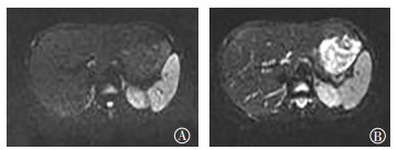
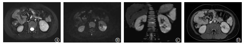
26例患儿中24例在Gd-T1WI增强图像上表现出片状或楔形低灌注区,其中21例单侧肾脏受累,3例双侧肾脏受累,共27个患肾。26例患儿于DWI图像上均发现阳性结果,其中,23例单侧肾脏受累,3例双侧肾脏受累,共29个患肾。DWI上高信号呈片状或尖端指向肾盂、底部朝向肾外的楔形表现(图 1、2)。病灶内ADC的平均值为(1.37±0.31)×10-3 mm2/s,较正常肾组织ADC的平均值[(1.92±0.38)×10-3 mm2/s]减低;且病程相对较长的患儿,其病灶内ADC与正常肾组织的差异更为明显。以肾上、中、下3部进一步细化病灶部位,Gd-T1WI增强图像上共53部检出阳性病灶(肾上、中、下部受累数目分别为15、21、17), DWI图像上共55部检出阳性病灶(肾上、中、下部受累数目分别为16、21、18)。2个患肾内单发病灶在DWI中被检出,而在Gd-T1WI增强图像上未明确显示。

图 1 急性局灶性细菌性肾炎患儿(女,8岁)的MRI图像
Figure 1. Magnetic resonance images of children with acute focal bacterial nephritis(female, 8 years old)

图 2 急性局灶性细菌性肾炎患儿(男, 6岁)的磁共振DWI图
Figure 2. Diffusion weighted images of children with acute focal bacterial nephritis (male, 6 years old)
DWI与Gd-T1WI具有很好的一致性(k=0.923),DWI对于病灶的检出率与Gd-T1WI无明显差异,且差异无统计学意义(P=0.25)。以肾脏个数计数得出DWI诊断AFBN的灵敏度为100%、特异度为92%;以肾上、中、下3部计数得出DWI诊断AFBN的灵敏度为100%、特异度为98%。各观察者应用DWI检测病变具有极好的可重复性(k=0.76)。
4例患儿(6个病灶)的常规T2WI图像仅显示出肾实质局部信号欠均匀,不能明确病灶的存在;其相应的DWI图像则可清晰显示出病灶的边界和范围(图 1)。结合常规MRI序列可发现29个患肾中共12个患肾不同程度增大,2例患儿同时合并肾脏重复畸形。
-
AFBN是急性局限性无液化的肾脏细菌感染,以往认为是急性肾盂肾炎的炎性病变过程,现多将它视为肾脓肿前期的一种状态,病灶局部充血、间质水肿、白细胞浸润,如不及时治疗可发展为肾脓肿。常见的感染途径包括血源性感染和泌尿系逆行感染[4-5]。大多数学者认为下尿路逆行感染可能为儿童AFBN主要的感染途径,本组患儿多合并尿频、尿痛、血尿,且尿白细胞、尿培养均呈阳性表现,与已有报道一致[5]。另有3例患儿合并有呼吸道感染病史,可能与体内其他部位化脓性病灶经血行感染有关。无论何种感染途径,炎性病变沿肾血管或肾小管呈放射状或扇形分布,因此AFBN病灶在MRI的任何序列上主要呈尖端指向肾盂、底部朝向肾外的楔形表现。文献报道称部分AFBN患儿存在泌尿系统畸形[6-7],本研究中有2例患肾为重复肾,故MRI在诊断AFBN的同时可明确有无合并泌尿系统畸形。
-
AFBN患儿由于肾组织炎性水肿、肾血管痉挛造成血流减少, 可导致肾脏灌注减低;肾脏病变亦影响肾脏对于水的转运功能。低b值时,ADC中主要为微循环灌注信息;高b值时,DWI主要获得的是微观水分子运动信息,接近组织真实的水分子扩散水平,因此可通过肾脏DWI来综合评价肾脏微血管灌注及水分子扩散。此外肾脏位于腹膜后,受呼吸运动的影响相对较小。这些解剖及生理功能特点使得肾脏适合行DWI研究[8]。
AFBN的DWI表现为水分子扩散受限,呈高信号改变,ADC减低。本研究b值选取了500 s/mm2、800 s/mm2两组,结果表明两个b值DWI图像上肾脏轮廓均清晰且无明显伪影,b值为500 s/mm2时的DWI图像显示解剖结构相对清晰,但b值为800 s/mm2时的病灶与正常肾组织的分界更为清楚。我们可利用高b值DWI图来判断炎性病变累及的范围,同时结合低b值DWI图像及常规MR序列来观察肾脏形态有无异常、判断有无合并泌尿系统畸形等。Faletti等[9]对52例急性肾盂肾炎及36例肾脓肿行DWI研究,证实正常肾组织、急性肾盂肾炎及肾脓肿的平均ADC之间差异有统计学意义。我们的研究结果与之相似,AFBN病灶内的ADC较正常肾组织减低,且ADC的量化减低与AFBN的病程发展相关,患儿病程越长病灶ADC越低,故我们认为ADC可作为评定AFBN病程的一个量化指标。值得注意的是,本研究中4例患儿的T2WI图像未能清晰显示出病灶,而同一层面的DWI图像可明确显示出病灶的边界及范围,故DWI较常规T2WI更为灵敏,可达到早期诊断目的。
-
Gd-T1WI增强MRI可明确诊断AFBN,以其为标准评定DWI诊断AFBN的灵敏度为100%,特异度达92%,两者具有高度的诊断一致性(k=0.923)。特别是将入组病例的MRI图像以肾窦区为界划分为上、中、下3部,可进一步细化病灶出现的范围,以便更好地明确检出率。因个别病灶出现融合的倾向,边界不甚清晰,故未以病灶数作为单位进行统计。上述结果表明DWI是诊断AFBN的可靠技术,优势在于无辐射且不使用对比剂,对于肾功能不全的患儿意义重大。De Pascale等[10]亦在研究中得出414次MRI检查中258次DWI与T1WI增强检查呈正相关,DWI诊断急性肾盂肾炎的灵敏度、特异度、准确率分别为95.2%、94.9%、94.6%。本研究中有2个病灶仅在DWI序列中被检出,回顾分析图像是因为Lava容积扫描时患儿未能屏气造成了较大的运动伪影,继而掩盖了较小的病灶,因此对于不能屏气的低龄儿童,DWI较Gd-T1WI增强MRI检查具有更广阔的应用空间。
DWI评价AFBN具有优势, 但尚存在一些问题有待阐明:b值可影响DWI图像质量及ADC的测量[11]。目前在多b值DWI的研究中,体素内不相干运动模型的临床应用价值在腹部肿瘤的相关研究中已得到认可[12],但也有报道称传统的单指数模型对于肾脏慢性疾病的评估更具有实用价值[13],因此本病的多b值模型选择仍需进一步对比研究。另外,基于Lava序列受呼吸运动影响较大,屏气扫描可更大程度地保证图像质量,本研究所纳入对象大多集中在学龄期儿童,婴幼儿所占比例较低(23.1%)。而结果表明,DWI与MRI增强检查具有很高的诊断一致性,在一定程度上也为今后低龄AFBN患儿DWI技术的应用提供了前期的理论基础。
儿童AFBN在临床并不少见,但临床症状及体征不典型,若能及时诊断可避免其发展为肾脓肿。DWI可反映患肾受累范围及程度,其诊断有效性与MRI增强扫描基本一致,在儿童群体尤其是肾功能不全、不宜使用对比剂者中具有很大的应用优势,可为临床诊断及治疗后随访观察提供准确的信息。
儿童急性局灶性细菌性肾炎的DWI诊断价值
DWI diagnosis of acute focal bacterial nephritis in children
-
摘要:
目的分析儿童急性局灶性细菌性肾炎(AFBN)的扩散加权成像(DWI)的影像学表现,并以钆-T1加权像(Gd-T1WI)增强检查为参考标准评估DWI对AFBN的诊断价值。 方法2016年2月至2017年12月经临床确诊为AFBN的患儿26例(男患儿12例、女患儿14例),均行磁共振检查,包括常规磁共振序列(T1WI、T2WI)、DWI及Gd-T1WI增强扫描,以Gd-T1WI增强表现来评定DWI诊断AFBN的灵敏度和特异度。应用Kappa检验和McNemar检验方法统计扫描序列之间的一致性和观察者之间的可重复性。 结果26例患儿中有24例在Gd-T1WI增强图像上表现出楔形低灌注区,21例单侧肾脏受累,3例双侧肾脏受累。26例患儿于DWI图像上均发现阳性结果,其中,23例单侧肾脏受累,3例双侧肾脏受累。病变在DWI上表现为高信号,表现扩散系数的平均值较正常肾组织减低。采用DWI检测病变的灵敏度和特异度分别为100%和92%。DWI与Gd-T1WI具有很好的一致性,对病灶的检测差异无统计学意义(k=0.923,P=0.25)。各观察者应用DWI检测病变具有很好的可重复性(k=0.76)。 结论DWI序列可明确诊断儿童AFBN,尤其是对肾功能不全、不宜使用对比剂者;其影像表现为楔形或片状高信号影,诊断有效性与MRI增强扫描基本一致。 Abstract:ObjectiveTo analyze the diffusion weighted imaging(DWI) manifestations of acute focal bacterial nephritis in children against the reference standard of gadolinium-enhanced T1-weighted imaging(Gd-T1WI). MethodsBetween February 2016 and December 2017, 26 cases of children (12 males, 14 females) with acute focal bacterial nephritis were examined by magnetic resonance(MR), including routine MR sequence(T1WI, T2WI), DWI, and Gd-T1WI-enhanced scan. The sensitivity and specificity of DWI in the diagnosis of acute focal bacterial nephritis were evaluated by Gd-T1WI-enhanced performance. Kappa test and McNemar test were applied for the calculation of the consistency among scanning sequences and calculation of repeatability among observers. ResultsAmong the 26 children, 24 cases showed a wedge-shaped hypoperfusion area in the enhanced Gd-T1WI images, 21 had unilateral renal involvement, and 3 had bilateral renal involvement. Twenty-six cases had positive results on DWI images, 23 had unilateral renal involvement, and 3 had bilateral renal involvement. The lesions showed high signal on DWI, and the mean apparent diffusion coefficient value is lower than that of the normal renal tissue. The sensitivity and specificity of DWI detection were 100% and 92%, respectively. DWI demonstrated excellent agreement(k=0.923) with Gd-T1WI with no significant difference(P=0.25) in detection of abnormal lesions. All the observers used DWI to detect lesions with excellent reproducibility(k=0.76). ConclusionsDWI can be used to diagnose acute focal bacterial nephritis with wedge-shaped or flaky high signal in children. The diagnostic effectiveness of DWI is basically the same as MRI enhanced scan, especially in people with renal insufficiency and inappropriate use of contrast agents. -
Key words:
- Child /
- Nephritis /
- Bacterial infections /
- Magnetic resonance imaging /
- Diffusion weighted imaging
-
图 1 急性局灶性细菌性肾炎患儿(女,8岁)的MRI图像
Figure 1. Magnetic resonance images of children with acute focal bacterial nephritis(female, 8 years old)
-
[1] Das CJ, Ahmad Z, Sharma S, et al. Multimodality imaging of renal inflammatory lesions[J]. World J Radiol, 2014, 6(11):865-873. DOI:10.4329/wjr.v6.i11.865. [2] Vivier PH, Sallem A, Beurdeley M, et al. MRI and suspected acute pyelonephritis in children:comparison of diffusion-weighted imaging with gadolinium-enhanced T1-weighted imaging[J]. Eur Radiol, 2014, 24(1):19-25. DOI:10.1007/s00330-013-2971-2. [3] Aoyagi J, Odaka J, Kuroiwa Y, et al. Utility of non-enhanced magnetic resonance imaging to detect acute pyelonephritis[J]. Pediatr Int, 2014, 56(3):e4-e6. DOI:10.1111/ped.12312. [4] 陈植, 刘小荣, 孟群, 等.儿童急性局灶性细菌性肾炎的临床特点[J].中华实用儿科临床杂志, 2017, 32(17):1343-1345. DOI:10.3760/cma.j.issn.2095-428X.2017.17.016.
Chen Z, Liu XR, Meng Q, et al. Clinical characteristics of acute focal bacterial nephritis in children[J]. Chin J Appl Clin Pediatr, 2017, 32(17):1343-1345. doi: 10.3760/cma.j.issn.2095-428X.2017.17.016[5] 王克明, 于大海, 齐士勇, 等.急性局灶性细菌性肾炎5例诊治分析[J].河北医药, 2009, 31(21):2944-2945. DOI:10.3969/j.issn.1002-7386.2009.21.053.
Wang KM, Yu DH, Qi SY, et al. Diagnosis and treatment of 5 cases of acute focal bacterial nephritis[J]. Hebei Med J, 2009, 31(21):2944-2945. doi: 10.3969/j.issn.1002-7386.2009.21.053[6] Bitsori M, Raissaki M, Maraki S, et al. Acute focal bacterial nephritis, pyonephrosis and renal abscess in children[J]. Pediatr Nephrol, 2015, 30(11):1987-1993. DOI:10.1007/s00467-015-3141-3 [7] 林俊, 张玉海.急性局灶性细菌性肾炎18例报告[J].中华泌尿外科杂志, 2003, 24(4):247-249. DOI:10.3760/j:issn:1000-6702. 2003.04.011.
Lin J, Zhang YH. A report of 18 cases of acute focal bacterial nephritis[J]. Chin J Urol, 2003, 24(4):247-249. doi: 10.3760/j:issn:1000-6702.2003.04.011[8] 邵虹, 朱铭.儿童肾脏病变的影像学诊断[J].国际放射医学核医学杂志, 2008, 32(3):188-190. DOI:10.3760/cma.j.issn.1673-4114. 2008.03.018.
Shao H, Zhu M. The imaging diagnosis of kidney diseases in children[J]. Int J Radiat Med Nucl Med, 2008, 32(3):188-190. doi: 10.3760/cma.j.issn.1673-4114.2008.03.018[9] Faletti R, Cassinis MC, Fonio P, et al. Diffusion-weighted imaging and apparent diffusion coefficient values versus contrast-enhanced MR imaging in the identification and characterisation of acute pyelonephritis[J]. Eur Radiol, 2013, 23(12):3501-3508. DOI:10.1007/s00330-013-2951-6. [10] De Pascale A, Piccoli GB, Priola SM, et al. Diffusion-weighted magnetic resonance imaging:new perspectives in the diagnostic pathway of non-complicated acute pyelonephritis[J]. Eur Radiol, 2013, 23(11):3077-3086. DOI:10.1007/s00330-013-2906-y. [11] Zhang JL, Sigmund EE, Rusinek H, et al. Optimization of b-value sampling for diffusion-weighted imaging of the kidney[J]. Magn Reson Med, 2012, 67(1):89-97. DOI:10.1002/mrm.22982. [12] Jiang L, Lu X, Hua B, et al. Intravoxel Incoherent Motion Diffusion-Weighted Imaging Versus Dynamic Contrast-Enhanced Magnetic Resonance Imaging:Comparison of the Diagnostic Performance of Perfusion-Related Parameters in Breast[J]. J Comput Assist Tomogr, 2018, 42(1):6-11. DOI:10.1097/RCT.0000000000000661. [13] 杨荷霞, 蒋振兴, 俞胜男, 等.肾脏DWI的IVIM及单指数模型预测肾小球滤过率的比较研究[J].磁共振成像, 2016, 7(9):679-682. DOI:10.12015/issn.1674-8034.2016.09.008.
Yang HX, Jiang ZX, Yu SN, et al. Assessment of glomerular filtration rate using diffusion weighted imaging:a comparative study of IVIM and mono-exponential models[J]. Chin J Magn Reson Imaging, 2016, 7(9):679-682. doi: 10.12015/issn.1674-8034.2016.09.008 -

 点击查看大图
图(2)
计量
- 文章访问数: 3162
- HTML全文浏览量: 2172
- PDF下载量: 6