


 下载:
下载:

-
骨折是临床的常见病、多发病,其愈合过程受多种因素的影响,准确判断骨折愈合情况,对骨折患者制定科学的康复锻炼计划、确定内固定物取出时间以及是否需要二次手术等具有重要临床意义。参考骨折的临床愈合标准[1]和延迟愈合或不愈合标准[2],多数骨折愈合情况的诊断较容易,但部分诊断为延迟愈合或不愈合的骨折,可能仍具有远期愈合的潜力,准确评价该类骨折的远期愈合能力可明确判断患者是否需要再次手术。笔者应用99Tcm -MDP SPECT/CT融合骨显像对76例未达到骨折临床愈合标准的患者进行骨折远期愈合能力评价,现总结报道如下。
-
收集2008年1月至2017年8月间在我院治疗的骨折患者76例,其中男性53例、女性23例,年龄8~77岁,平均年龄(41.5±6.9)岁。经手术治疗的58例骨折患者,其中31例钢板固定、15例髓内钉固定、12例外固定架固定;18例骨折采用石膏或支具固定等非手术方法治疗。骨折部位包括股骨25例、胫骨23例、肱骨11例、股骨颈7例、尺骨4例、桡骨3例和锁骨3例。病程半年至两年不等,骨折治疗时间均超过半年。76例患者均行X线及CT检查,均未达到骨折临床愈合标准。
-
76例患者中,62例采用德国Siemens Symbia-T6、14例采用德国Siemens Symbia-T16双探头SPECT/CT融合显像仪,配低能高分辨平行孔准直器。钼-锝发生器由北京原子高科股份有限公司提供,MDP由江苏省原子医学研究所江原制药厂生产。患者静脉注射99Tcm-MDP 740~925 MBq,注射后2.5 h先对骨折部位行SPECT显像,SPECT采集条件为能峰140 keV,窗宽20%,矩阵128×128,放大倍数1,断层扫描探头旋转轨迹为自动人体轨迹,360°采集,双探头各旋转180°,60°/帧,20 s/帧,连续采集30帧;扫描完毕患者保持体位不动,选择大于骨折端上下5 cm的范围行CT横断面扫描。CT扫描条件:层厚1.0 mm,螺距1,矩阵512×512。图像进行横轴位、矢状位和冠状位重建,SPECT和CT图像采用显像仪随机工作站图像融合软件进行融合。所有患者均于检查前签署了知情同意书。
-
由具有10年以上工作经验的2位放射科医师和1位核医学医师共同阅片,分析SPECT、CT及SPECT/CT融合图像中骨折端的放射性浓聚分布范围、浓聚程度和骨痂形成、骨质改变等情况,意见不一致时,共同协商确定评价结论。对骨折愈合情况的判断,依据“骨折临床愈合标准[1]”中X线检查应满足的条件:骨折线模糊,有连续性骨痂通过骨折线。对骨折不愈合和延迟愈合的判断依据“骨折不愈合和延迟愈合的定义[2]”:骨折经治疗后,若超过一般愈合所需的时间较长而仍未愈合,但又未达到骨折不愈合的程度,即属于骨折延迟愈合。骨折已半年以上,骨折断端仍有异常活动,X线上无成桥骨痂形成,骨折断端的髓腔已被浓密的硬化骨质封闭、变光滑,即为骨折不愈合。对骨折远期愈合能力的评价,参考相关文献[3-6],采用如下标准:放射性浓聚连续性通过骨折线,跨越两侧骨折端者,诊断为骨折具有远期愈合能力,否则诊断为骨折不具有远期愈合能力。76例患者的骨折远期愈合情况,经临床随访无愈合表现而再次进行手术者,经手术证实;远期愈合者经临床随访证实。
-
指导具有远期愈合能力的58例骨折病例临床应用石膏或支具加强外固定,进行对症保守治疗,每个月复查X线一次,随访3个月至1年。
-
76例患者的SPECT显像均显示骨折端区域放射性浓聚,但由于SPECT图像的空间分辨率较低,对放射性浓聚分布的部位和程度、是否连续性通过骨折线并跨越两侧骨折端无法进行准确判断。
-
76例患者中,CT检查显示45例骨折端周围可见部分、少量的骨痂形成,其中30例可见连续性通过骨折线的少量骨痂,15例未见连续性通过骨折线的骨痂,均诊断为骨折延迟愈合。31例骨折端周围未见明显骨痂形成,其中21例骨折端毛糙不整,未见明显骨质硬化和吸收,诊断为骨折延迟愈合;7例骨折端边缘骨质硬化、3例骨折端密度减低、骨质吸收,均诊断为骨折不愈合。
-
76例患者中,SPECT/CT融合骨显像示58例可见连续性通过骨折线并跨越两侧骨折端的放射性浓聚,其中包括CT检查中42例骨折端周围可见部分、少量骨痂形成的病例(图 1),16例骨折端周围未见明显骨痂形成、但无骨质硬化和吸收的病例(图 2),均诊断为骨折处于愈合过程中,具有远期愈合能力。18例骨折端未见明显连续性通过骨折线的放射性浓聚,其中包括CT检查中3例骨折端周围部分、少量骨痂形成的病例,5例骨折端毛糙不整、未见骨质硬化和吸收的病例和7例骨折端边缘骨质硬化的病例(图 3),3例骨折端密度减低、骨质吸收的病例(图 4),均诊断为骨折不愈合,且骨折端的骨代谢差,远期愈合的可能性较小。

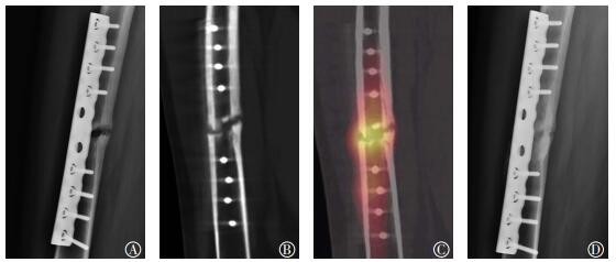
图 1 股骨骨折交锁髓内钉术后患者(男性,29岁)的DR、CT和SPECT/CT图 术后7个月股骨侧位DR片(A)及CT矢状位多平面重建图像(B),显像示骨折线清楚,边缘可见少量骨痂,但未连续性通过骨折线,诊断为骨折延迟愈合;C:SPECT/CT融合骨显像,显像示放射性浓聚连续通过骨折线并跨越两侧骨折端,提示骨折具有远期愈合能力;D:随访股骨侧位片,临床对症治疗、随访8个月后复查,骨折愈合。SPECT/CT:单光子发射计算机体层摄影术;DR:数字化X线摄影。
Figure 1. The DR, CT, SPECT/CT of patients with femoral fracture after interlocking intramedullary nail surgery

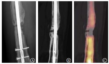
图 2 股骨骨折钢板内固定术后患者(男性,20岁)的DR、CT和SPECT/CT图 图中,A、B:术后半年股骨侧位DR片(A)及CT矢状位多平面重建图像(B),显像示骨折线清楚,骨折端未见明显骨痂形成,诊断为骨折迟延愈合;C:SPECT/CT融合骨显像,显像示放射性浓聚连续通过骨折线并跨越两侧骨折端,提示骨折具有远期愈合能力;D:随访股骨侧位片,临床对症治疗、随访半年后复查,骨折愈合。SPECT/CT:单光子发射计算机体层摄影术;DR:数字化X线摄影。
Figure 2. The DR, CT, SPECT/CT of patients with femoral fracture after plate internal fixation surgery

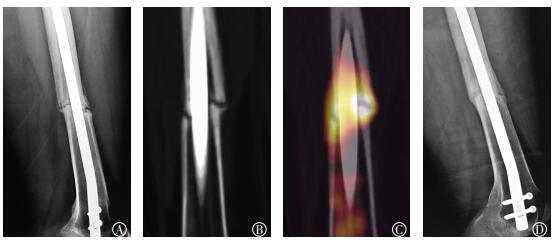
图 3 股骨骨折术后患者(男性,30岁)的CT及SPECT/CT显像图 图中,A、B:术后1年CT冠状位(A)及矢状位(B)多平面重建图像,图像示骨折线清楚,骨折端骨质硬化、封闭骨髓腔,周围未见明显骨痂形成,诊断为骨折不愈合;C:SPECT/CT融合骨显像,图像示远侧骨折端可见局限性放射性浓聚,未连续性通过骨折线,未跨越两侧骨折端,提示骨折不具有远期愈合能力,经手术治疗证实。SPECT/CT:单光子发射计算机体层摄影术;CT:计算机体层摄影术。
Figure 3. CT and SPECT/CT images of patients with femoral fracture after surgery

图 4 股骨骨折交锁髓内钉固定术后患者(男性,35岁)的DR、CT及SPECT/CT图 图中,A、B:术后15个月DR正位片(A)及CT矢状位多平面重建图像(B),图像示骨折线清楚,骨折远端骨质吸收,密度减低,骨折端周围可见骨痂形成,但未连续通过骨折线,诊断为骨折不愈合;C:SPECT/ CT融合骨显像,图像示两侧骨折端均可见放射性浓聚,但未连续性通过骨折线,未跨越两侧骨折端,提示骨折不具有远期愈合能力,经手术治疗证实。DR:数字化X线摄影;SPECT/CT:单光子发射计算机体层摄影术;CT:计算机体层摄影术。
Figure 4. DR, CT and SPECT/CT images of patients with femoral fracture after interlocking intramedullary nail surgery
-
SPECT/CT融合骨显像诊断为骨折的58例患者经治疗后:52例X线检查显示骨折端有新生的连续性骨痂通过骨折线,临床证实骨折愈合;6例骨折端仍未见明显新生骨痂形成,其中包括CT检查中2例骨折端周围可见部分、少量骨痂形成的病例,4例骨折端周围未见明显骨痂形成、但无骨质硬化和吸收的病例。6例骨折端仍未见明显新生骨痂的患者均再次进行了手术治疗,证实骨折未愈合。对58例SPECT/CT融合骨显像诊断为骨折具有远期愈合能力的病例,SPECT/CT诊断的准确率为89.66%(52/58)。18例SPECT/CT融合骨显像诊断为骨折不愈合、远期愈合可能性较小的病例,临床均进行了再次手术治疗,术中观察骨折端未见明显骨痂形成或连续性骨痂通过骨折线,其中10例骨折端可见骨质硬化或骨质吸收改变,证实为骨折不愈合,SPECT/CT对其诊断的准确率达100%(18/18)。
-
骨折是骨的连续性和完整性中断,是骨骼机械性超负荷的结果。骨折愈合是骨的原始连续性重建,是一个极其复杂的细胞组织学修复过程,其微环境受多种因素的影响[1]。对部分经过长期治疗后,骨折端仍无骨痂形成或仅有部分、少量骨痂形成的骨折,未达到骨折临床愈合标准,准确判断其是否具有远期愈合能力,选择继续等待骨折愈合还是即刻进行再次手术治疗,是临床非常棘手的难题。
CT对骨折愈合情况的判断较X线具有明显优势[7],但CT图像仍是解剖图像,观察的对象仍然是骨痂的形态、范围、骨折线是否模糊或消失等解剖结构的改变,而文献报道,X线和CT检查在骨矿含量改变少于25%时无法分辨骨痂变化,另外骨折端多有金属内固定物,检查时不可避免的会产生大量金属伪影,严重影响骨折愈合情况的判断[4]。因此,应用X线和CT检查评价骨折远期愈合能力,存在明显的时滞性和局限性。
近年来,应用放射性核素骨显像诊断骨折及判断骨折愈合成为越来越多学者探索的热点课题。放射性核素骨显像剂99Tcm-MDP参与骨代谢,可以观察骨骼病变部位显像剂的摄取,了解病变区血供、骨盐代谢和成骨细胞活跃程度等功能变化,达到诊断疾病的目的。应用放射性核素骨显像显示骨骼病变的时间远远早于X线和CT检查,当病变部位的血供、骨盐代谢和成骨过程发生改变时,即可表现为炳灶的异常摄取,具有更高的敏感性[8-12]。骨折时,骨的完整性和连续性缺失,骨小梁断裂,骨折端发生充血、水肿等血供改变,骨盐代谢及成骨细胞活跃程度加强,骨修复过程加快,骨折端即表现为明显的放射性核素高摄取,而陈旧性骨折与其不同,由于骨折端的血供、骨盐代谢和成骨细胞活跃程度低,其放射性核素呈低摄取[5]。因此,放射性核素骨显像可早期、准确地反映骨质代谢情况,敏感性较高,评价骨折远期愈合能力具有优势。但由于单纯的SPECT图像空间分辨率较低,对能引起血供及骨代谢异常变化的骨病变,如炎症、肿瘤等均可导致病变部位显像剂的异常摄取,大大降低了诊断的准确率[11-13]。
SPECT/CT融合骨显像既能显示骨折端的血供、骨盐代谢和成骨细胞的活跃程度等功能改变,又可清楚观察骨折端的解剖结构变化以及放射性分布情况,更有利于评价骨折的远期愈合能力[3-6]。应用SPECT/CT融合图像诊断骨折以及骨不连、骨折不愈合等国内外均有文献[8, 11-15]报道,但均未涉及对骨折远期愈合能力的准确评价。由于SPECT/CT融合骨显像具备对骨折远期愈合能力评价的理论基础,可根据骨折端的放射性分布情况进行远期愈合能力评价。当SPECT/CT融合骨显像显示存在部分或全部连续通过骨折线、跨越两侧骨折端的放射性浓聚时,无论此时骨折端骨痂多少、有无骨痂形成,均提示该骨折尚处于骨修复、愈合进程中,且通过临床恰当的对症治疗具有远期愈合能力;相反,无明显放射性浓聚连续通过骨折线、跨越两侧骨折端者,提示骨折端血供较差、骨盐代谢水平较低,成骨细胞活动不活跃,则骨折远期愈合的可能性就较小,无继续等待价值,需再次手术治疗。本研究76例骨折患者,其中58例通过SPECT/CT融合骨显像诊断骨折具有远期愈合能力,通过采取加强外固定等对症治疗措施,52例骨折实现远期愈合,诊断远期愈合的准确率达89.66%。18例SPECT/CT融合骨显像诊断为骨折不愈合、无远期愈合能力,均经再次手术治疗证实。远期不愈合的诊断准确率达100%。SPECT/CT融合骨显像能较准确地判断骨折是否具有远期愈合能力,既避免了部分具有远期愈合能力的骨折患者错误地进行再次手术,也可以使不具有远期愈合能力的骨折患者能得到及时科学的治疗,具有重要的临床应用价值。
由于骨折愈合受多种因素的综合影响,放射性核素骨显像也不可避免的存在假阳性和假阴性的问题[14-15],因此,应用SPECT/CT融合骨显像评价骨折远期愈合能力时,需详细分析骨折端放射性分布的程度、范围和位置,协同考虑骨折端的力学关系及解剖结构的变化,尽可能地排除引起放射性异常分布的假阳性和假阴性结果,提高诊断的特异度。需要注意的是,SPECT/CT融合骨显像评价骨折具有远期愈合能力,并不是简单地认为此骨折一定可以远期愈合,只能说明当时的骨折端血供、骨盐代谢和成骨细胞活跃程度较好,具备远期愈合的基础条件,骨折能否远期愈合,还与后期科学的临床治疗密不可分,如加强骨折端的外固定、选择合适的负重时机及强度、促进骨折愈合的药物应用等,治疗过程中应密切随访,定期复查,才能更好地促进骨折愈合[16]。
99Tcm-MDP SPECT/CT融合骨显像评价骨折远期愈合能力的应用价值
Evaluation of fracture healing by 99Tcm-MDP SPECT/CT
-
摘要:
目的探讨99Tcm-MDP SPECT/CT融合骨显像技术评价骨折远期愈合能力的应用价值。 方法回顾性分析2008年1月至2017年8月在我院治疗至少超过6个月的76例骨折患者的影像资料,其中58例经手术治疗,18例采用非手术方法治疗,所有骨折患者均未达到临床愈合标准。对76例骨折患者行SPECT显像、CT扫描和SPECT/CT融合骨显像,分析骨折端的放射性分布及CT特征,观察骨折端放射性浓聚是否连续性通过骨折线,跨越两侧骨折端,评价骨折远期愈合能力,最终骨折愈合情况经临床随访或手术证实。 结果76例骨折患者,58例SPECT/CT融合骨显像显示放射性浓聚连续性通过骨折线并跨越两侧骨折端,诊断为骨折具有远期愈合能力,经对症保守治疗,证实52例骨折远期愈合,6例未愈合,SPECT/CT对骨折远期愈合诊断的准确率达89.66%(52/58)。18例骨折端未见明显连续性通过骨折线的放射性浓聚,诊断为骨折不愈合,且不具备远期愈合能力,均进行了手术治疗并证实为骨折不愈合,SPECT/CT对骨折远期不愈合诊断的准确率达100%(18/18)。 结论SPECT/CT融合骨显像可较准确地评价骨折的远期愈合能力,具有重要的临床应用价值。 -
关键词:
- 骨折愈合 /
- 单光子发射计算机体层摄影术 /
- 亚甲基二膦酸盐
Abstract:ObjectiveTo explore the application value of SPECT/CT on fracture healing. MethodsA retrospective analysis was done among 76 patients with fracture as evidenced by image data from January 2008 to August 2017. Fifty-eight cases with fracture revealed by a SPECT/CT fusion image with surgical treatment, 18 cases with nonsurgical treatment, and 76 cases of bone fracture clinical healing that failed to meet standards were retrospectively analyzed after more than 6 months of treatment. A SPECT/CT image fusion imaging device was applied to 76 cases of fracture detected with SPECT, CT scan, and SPECT/CT fusion imaging. The characteristics of the fractures in terms of radioactive distribution and CT were examined, and the radioactive strength of the fracture was observed to determine whether radioactivity passed continuously through the fracture line or crossed both sides of the fracture. The healing ability of the fracture was evaluated, and fracture healing cases were confirmed through surgery or clinical follow-up. ResultsA total of 76 fractures were collected and examined through SPECT/CT, and 58 cases of uptake that passed through the fracture line and crossed on both sides of the fracture end were obtained. This finding indicated that the fracture was in the process of healing and that fracture could heal. Furthermore, 52 cases were confirmed after conservative treatment was administered, and 6 cases of fracture were nonunion. For 18 cases, no nuclide concentration was detected in the fracture end, and the fracture was nonunion. The final diagnosis was confirmed through surgery, and the diagnostic accuracy rates were 89.66%(52/58) for fracture healing and 100%(18/18) for nonunion. ConclusionSPECT/CT imaging could accurately evaluate fracture healing and had an important application value. -
图 1 股骨骨折交锁髓内钉术后患者(男性,29岁)的DR、CT和SPECT/CT图 术后7个月股骨侧位DR片(A)及CT矢状位多平面重建图像(B),显像示骨折线清楚,边缘可见少量骨痂,但未连续性通过骨折线,诊断为骨折延迟愈合;C:SPECT/CT融合骨显像,显像示放射性浓聚连续通过骨折线并跨越两侧骨折端,提示骨折具有远期愈合能力;D:随访股骨侧位片,临床对症治疗、随访8个月后复查,骨折愈合。SPECT/CT:单光子发射计算机体层摄影术;DR:数字化X线摄影。
Figure 1. The DR, CT, SPECT/CT of patients with femoral fracture after interlocking intramedullary nail surgery
图 2 股骨骨折钢板内固定术后患者(男性,20岁)的DR、CT和SPECT/CT图 图中,A、B:术后半年股骨侧位DR片(A)及CT矢状位多平面重建图像(B),显像示骨折线清楚,骨折端未见明显骨痂形成,诊断为骨折迟延愈合;C:SPECT/CT融合骨显像,显像示放射性浓聚连续通过骨折线并跨越两侧骨折端,提示骨折具有远期愈合能力;D:随访股骨侧位片,临床对症治疗、随访半年后复查,骨折愈合。SPECT/CT:单光子发射计算机体层摄影术;DR:数字化X线摄影。
Figure 2. The DR, CT, SPECT/CT of patients with femoral fracture after plate internal fixation surgery
图 3 股骨骨折术后患者(男性,30岁)的CT及SPECT/CT显像图 图中,A、B:术后1年CT冠状位(A)及矢状位(B)多平面重建图像,图像示骨折线清楚,骨折端骨质硬化、封闭骨髓腔,周围未见明显骨痂形成,诊断为骨折不愈合;C:SPECT/CT融合骨显像,图像示远侧骨折端可见局限性放射性浓聚,未连续性通过骨折线,未跨越两侧骨折端,提示骨折不具有远期愈合能力,经手术治疗证实。SPECT/CT:单光子发射计算机体层摄影术;CT:计算机体层摄影术。
Figure 3. CT and SPECT/CT images of patients with femoral fracture after surgery
图 4 股骨骨折交锁髓内钉固定术后患者(男性,35岁)的DR、CT及SPECT/CT图 图中,A、B:术后15个月DR正位片(A)及CT矢状位多平面重建图像(B),图像示骨折线清楚,骨折远端骨质吸收,密度减低,骨折端周围可见骨痂形成,但未连续通过骨折线,诊断为骨折不愈合;C:SPECT/ CT融合骨显像,图像示两侧骨折端均可见放射性浓聚,但未连续性通过骨折线,未跨越两侧骨折端,提示骨折不具有远期愈合能力,经手术治疗证实。DR:数字化X线摄影;SPECT/CT:单光子发射计算机体层摄影术;CT:计算机体层摄影术。
Figure 4. DR, CT and SPECT/CT images of patients with femoral fracture after interlocking intramedullary nail surgery
-
[1] 吴作培, 孙贵新.骨不连治疗的研究与技术应用进展[J].中国组织工程研究, 2013, 17(35):6333-6338. DOI:10.3969/j.issn.2095-4344.2013.35.018.
Wu ZP, Sun GX. Progress of research and technology application of the treatment of fracture nonunion[J]. J Clin Rehabil Tis Eng Res, 2013, 17(35):6333-6338. doi: 10.3969/j.issn.2095-4344.2013.35.018[2] 韩萍, 于春水.医学影像诊断学[M].北京:人民卫生出版社, 2017:560-561.
Han P, Yu CS. Diagnosic Medical Imaging[M]. Beijing:People's Medical Publishing House, 2017:560-561.[3] 张建媛, 尤立强.临床愈合期后骨折部位核素骨显像表现分析[J].医学信息, 2014, 27(3):66-67. DOI:10.3969/j.issn.1006-1959. 2014.03.076.
Zhang JY, You LQ. Analysis of radionuclide bone imaging performance of fracture after the clinical healing period[J]. Med Inf, 2014, 27(3):66-67. doi: 10.3969/j.issn.1006-1959.2014.03.076[4] 张敏, 刘玉珂, 陈亚玲, 等.应用图像融合技术诊断骨折愈合——图像融合技术在骨与关节疾病诊断中的应用(二)[J].中医正骨, 2010, 22(10):22-24. DOI:10.3969/j.issn.1001-6015. 2010.10.007.
Zhang M, Liu YK, Chen YL, et al. Diagnosis of fracture healing by image fusion-application of image fusion in the diagnosis of bone and joint diseases[J]. J Tradit Clin Orthop Traumatol, 2010, 22(10):22-24. doi: 10.3969/j.issn.1001-6015.2010.10.007[5] 张斌青, 宋青凤, 王军辉, 等. SPECT/CT显像对骨折延迟愈合的诊断价值[J].中华核医学与分子影像杂志, 2014, 34(4):305-307. DOI:10.3760/cma.j.issn.2095-2848.2014.04.009.
Zhang BQ, Song QF, Wang JH, et al. Diagnostic value of SPECT/CT for fracture delayed union[J]. Chin J Nucl Med Mol Imaging, 2014, 34(4):305-307. doi: 10.3760/cma.j.issn.2095-2848.2014.04.009[6] 贠新泉, 刘学军, 葛丽红, 等. 放射性核素骨显像和SPECT/CT断层融合显像技术在骨折愈合过程中的应用及预后研究[J/OL]. 临床医药文献电子杂志, 2017, 4(22): 4297[2018-03-11]. hppt://www.wanfangdata.com.cn/details/detail.do?_type=perio&id=lcyydzzz201722109. DOI:10.3877/j.issn.2095-8242.2017.22.109.
Yun XQ, Liu XJ, Ge LH, et al. Application of SPECT and SPECT/CT in fracture healing and prognosis[J/OL]. J Clin Med Lit(Electronic Edition), 2017, 4(22): 4297[2018-03-11]. http://www.wanfangdata.com.cn/details/detail.do?_type=perio&id=lcyydzzz201722109.[7] 刘鸿麒, 丁真奇.临床研究中骨折愈合过程的评价方法进展[J].中国现代医生, 2010, 48(4):43-44, 59. DOI:10.3969/j.issn.1673-9701.2010.04.018.
Liu HQ, Ding ZQ. Progress in evaluation of fracture healing in clinical studies[J]. Chin Modern Doctor, 2010, 48(4):43-44, 59. doi: 10.3969/j.issn.1673-9701.2010.04.018[8] 张斌青, 宋青凤, 刘玉珂, 等. 99Tcm-MDP SPECT/CT图像融合诊断隐匿性骨折[J].中国医学影像技术, 2014, 30(4):587-590. DOI:10.13929/j.1003-3289.2014.04.052.
Zhang BQ, Song QF, Liu YK, et al. 99Tcm-MDP SPECT/CT fusion in diagnosis of occult bone fractures[J]. Chin J Med Imaging Technol, 2014, 30(4):587-590. doi: 10.13929/j.1003-3289.2014.04.052[9] Hamann M, Aldridge M, Dickson J, et al. Evaluation of a low-dose/slow-rotating SPECT-CT system[J]. Phys Med Biol, 2008, 53(10):2495-2508. DOI:10.1088/0031-9155/53/10/003. [10] Zhao Z, Li L, Li F, et al. Single photon emission computed tomography/spiral computed tomography fusion imaging for the diagnosis of bone metastasis in patients with known cancer[J]. Skeletal Radiol, 2010, 39(2):147-153. DOI:10.1007/s00256-009-0764-0. [11] 叶谦, 董海锋, 王卫伟, 等.放射性核素骨显像在骨折诊断中的应用[J].创伤外科杂志, 2007, 9(6):530-533. DOI:10.3969/j.issn.1009-4237.2007.06.016.
Ye Q, Dong HF, Wang WW, et al. Application of radionuclide bone imaging in the diagnosis of fracture[J]. J Traum Surg, 2007, 9(6):530-533. doi: 10.3969/j.issn.1009-4237.2007.06.016[12] 张斌青, 刘玉珂, 郭会利, 等. 99Tcm-MDP SPECT/CT诊断骨骺早闭[J].中国医学影像技术, 2014, 30(10):1557-1560. DOI:10.13929/j.1003-3289.2014.10.032.
Zhang BQ, Liu YK, Guo HL, et al. 99Tcm-MDP SPECT/CT in diagnosing early closure of epiphyseal[J]. Chin J Med Imaging Technol, 2014, 30(10):1557-1560. doi: 10.13929/j.1003-3289.2014.10.032[13] 高建青, 魏玲格, 傅鹏, 等.放射性核素骨显像对骨肿瘤样病变的影像学研究价值[J].国际放射医学核医学杂志, 2014, 38(4):266-270. DOI:10.3760/cma.j.issn.1673-4114.2014.04.014.
Gao JQ, Wei LG, Fu P, et al. Value of imaging research in tumor-like disorders by radionuclide bone imaging[J]. Int J Radiat Med Nucl Med, 2014, 38(4):266-270. doi: 10.3760/cma.j.issn.1673-4114.2014.04.014[14] Liodakis E, Liodaki E, Krettek C, et al. Can the viability of a nonunion be evaluated using SPECT/CT? A preliminary retrospective study[J]. Technol Health Care, 2011, 19(2):103-108. DOI:10.3233/THC-2011-0617. [15] Rager O, Schaller K, Payer M, et al. SPECT/CT in differentiation of pseudarthrosis from other causes of back pain in lumbar spinal fusion:report on 10 consecutive cases[J]. Clin Nucl Med, 2012, 37(4):339-343. DOI:10.1097/RLU.0b013e318239248b. [16] 曹克勇, 方彩莲, 叶积飞.不同强度的早期负重对股骨干骨折愈合的影响[J].中国康复, 2014, 29(6):458-459. DOI:10.3870/zgkf.2014.06.021.
Cao KY, Fang CL, Ye JF. Effect of early loading of different strength on femoral shaft fracture healing[J]. Chin J Rehabil, 2014, 29(6):458-459. doi: 10.3870/zgkf.2014.06.021 -

 点击查看大图
计量
- 文章访问数: 2955
- HTML全文浏览量: 2052
- PDF下载量: 3