


 下载:
下载:
-
腰椎横突骨折(lumbar vertebral transverse process fracture,LTF)属于腰椎附件骨折,在腰椎骨折中的发病率仅次于椎体骨折[1]。腰椎横突较薄,呈带状,为由椎弓根与椎弓板会合处向外突出的骨性结构,是腰大肌、腰方肌、棘肌及腰部筋膜的附着点[2]。腰椎横突可直接受损伤或因附着其上肌肉猛烈收缩而发生骨折,以第2及第3腰椎(L2、L3)受伤较多,患者多以腰痛、腰部活动障碍为常见症状。SPECT/CT同机融合显像集合了SPECT的高灵敏度与CT的高特异度的优点,大大提高了LTF诊断的准确率。本研究通过回顾性分析我院LTF患者的SPECT/CT同机融合影像学特征,探讨SPECT/CT同机融合显像在LTF诊断中的应用价值。
-
收集本院2013年6月至2015年6月腰部不适并经X射线检查排除腰椎退变及转移的患者共203例,根据病理结果及6个月以上随访结果,最终入选42例患者,共49个病灶。其中男性31例、女性11例,年龄39~75岁,平均(52 ± 8)岁。纳入标准:(1)腰部不适;(2)经SPECT/CT同机融合检查。排除标准:(1)年龄在18岁以下或75岁以上患者;(2)妊娠或哺乳期妇女;(3)合并严重心血管、脑血管、肝肾造血系统或内分泌等疾病和精神病患者。本研究经本院医学伦理委员会批准。所有患者或其家属均于检查前签署了知情同意书。
-
(1)患者有明确的外伤史;(2)腰部困痛;(3)局部压痛、疼痛、活动受限或伴有臀部放射痛;(4)腰椎正侧位片或CT扫描见腰椎横突骨质连续性破坏。
-
采用美国GE公司Discovery NM-CT D670型SPECT/CT仪,同机CT为16层螺旋CT。患者静脉注射740~925 MBq 99Tcm-MDP 3 h后行常规全身骨显像,再对腰椎阳性病灶进行融合断层显像,SPECT采集矩阵128×128,放大倍数1.28,连续采集360°;随后行同机CT扫描,电压120 kV,电流80 mA,层厚3.75 mm,矩阵512 × 512,利用自带软件进行SPECT/CT同机融合图像处理。
-
由2位工作10年以上的高年资核医学科医师通过双盲法分别进行CT及SPECT/CT阅片。判断标准:以相邻的上下正常椎体为对照,对CT图像或SPECT/CT融合图像难以确诊的病灶进一步行CT冠状位及矢状位重建,排除正常变异及其他病变后确定为骨折。
-
采用SPSS 17.0软件进行统计学分析,CT与SPECT/CT融合显像的病灶检出率比较采用χ2检验,P < 0.05表示差异有统计学意义。
-
SPECT/CT同机融合显像发现LTF病灶共45个,检出率为91.8%(45/49),其中第1至第5腰椎(L1-L5)检出病灶数分别为4、12、21、8、0个,以L2(26%)和L3(46%)(图 1)的发病率较高;而CT扫描发现病灶共38个,检出率为77.6%(38/49),其中L1-L5检出病灶数分别为2、11、18、7、0个,对于斜行骨折及隐匿骨折未能发现。两种检查方法对LTF的检出率差异具有统计学意义( χ2 = 4.0,P < 0.05)。

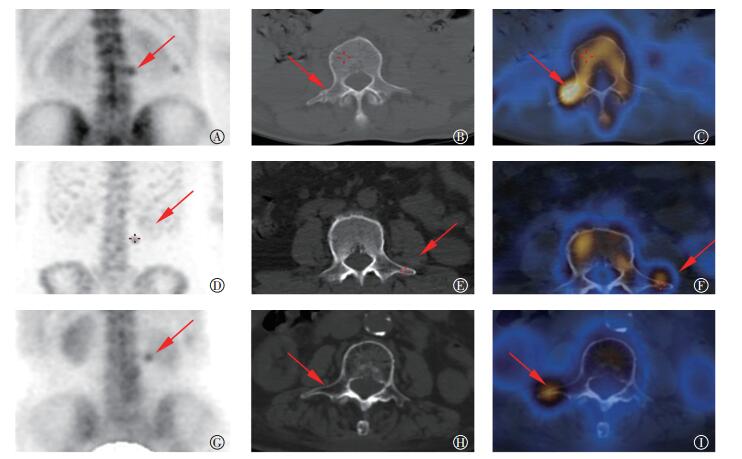
图 1 腰椎横突骨折患者的 SPECT/CT 图像 图中, A~C: 患者男性, 63 岁。 A: 全身骨扫描(后位)示 L3 异常核素浓聚(箭头示);B: CT 示 L3 右侧横突骨皮质不连续, 见线样低密度影, 骨痂形成(箭头示); C: SPECT/CT 示 L3 右侧横突核素异常浓聚处骨皮质不连续, 可见骨痂形成(箭头示)。 D~F: 患者女性, 58 岁。 D: 全身骨扫描(前位)示 L3 异常核素浓聚(箭头示); E: CT 示 L3 左侧横突见线性密度影, 未见骨痂形成(箭头示); F: SPECT/CT 示 L3 左侧横突核素异常浓聚处骨皮质不光整, 髓腔内见线样密度影, 未见骨痂形成(箭头示)。 G~I: 患者男性, 73 岁。 G: 全身骨扫描(后位)示 L3 异常核素浓聚(箭头示); H: CT 示 L3 右侧横突骨皮质连续, 髓腔内未见异常密度影(箭头示); I: SPECT/CT 示 L3 右侧横突核素异常浓聚处骨皮质连续, 髓腔内未见异常密度影(箭头示)。
Figure 1. SPECT/CT images of lumbar vertebral transverse process fracture
-
LTF主要分为3种类型:Ⅰ型为横形骨折,骨折线呈前后走向,与正中矢状面平行,可有或无断端分离或错位;Ⅱ型为斜形骨折,骨折线走向与正中矢状面呈不同程度夹角;Ⅲ型为横突尖的撕脱性骨折,撕脱小碎骨片位于横突尖旁肌组织内[3]。Ⅱ型及Ⅲ型骨折在影像学检查中常易漏诊。
LTF的诊断可依据:①患者局部疼痛、压痛、活动受限等;②X射线腰椎正侧位片或CT扫描发现横突骨质连续性破坏。但常规X线片的漏诊、误诊率较高[4-6],文献报道其假阴性率可达70%[7]。CT扫描在发现LTF及骨折类型方面的价值明显优于X线片[3, 8]。常规CT扫描虽然能够明确诊断LTF,但对于一些有临床症状而骨质改变轻微隐匿的患者不易作出早期诊断,本研究中49个病灶通过CT扫描漏诊 11个,分析原因可能为骨折方向与轴位图像相近或一致,或患者骨折较为轻微隐匿所造成。
近年来,SPECT/CT同机融合技术的应用大大提高了LTF的诊断检出率。由于一些骨折部位隐蔽或太小导致X射线或CT检查难以发现[9]。Cho等[10]分析130例胸外伤患者发现单纯轴位CT扫描中有58处肋骨骨折被漏诊。而SPECT在早期诊断应力性骨折、隐性骨折中有着重要的诊断价值[11]。SPECT能显示骨代谢异常活跃的部位,对骨折的诊断灵敏度几乎达100%[12],但特异度较低。常规SPECT显像很难对骨代谢活跃部位进行定性诊断,同机CT可进一步观察骨质、髓腔及周边软组织情况,两者结合对骨折的诊断效能较好[13-14]。本研究中49个病灶通过SPECT/CT融合显像诊断45个,漏诊4个,可能由于骨折修复时间较长对放射性摄取不明显,阅片医师疏忽未进一步分析其CT图像而导致漏诊。
综上所述,腰椎横突由于其解剖生理的特点,患者若有腰部外伤史,疼痛明显,应考虑横突骨折,CT图像尤其是多层螺旋CT后处理技术的应用,明显提高了骨折的检出率[15]。MRI对该病的早期诊断也具有较高的灵敏度及特异度[6],但检查费用高,不宜作为临床首选。SPECT/CT融合显像充分发挥了SPECT的高灵敏度与CT的高特异度的优点,大大提高了微小隐匿骨折诊断的准确率。
SPECT/CT融合显像在腰椎横突骨折诊断中的应用价值
Application value analysis on SPECT/CT in the diagnosis of lumbar vertebral transverse process fracture
-
摘要:
目的探讨SPECT/CT同机融合显像在腰椎横突骨折(LTF)诊断中的应用价值。 方法收集2013年6月至2015年6月腰部不适并经X射线检查排除腰椎退变及转移的患者共203例,根据病理结果及6个月以上随访结果,最终42例患者共49个病灶确诊为LTF,阳性病灶均行SPECT/CT同机融合显像,对病灶进行CT及SPECT/CT阅片。病灶检出率比较采用χ2检验。 结果 SPECT/CT同机融合显像发现LTF病灶共45个,其中,第1至第5腰椎(L1-L5)分别为4、12、21、8、0个,以L2(26%)和L3(46%)发病率较高。CT扫描发现LTF病灶共38个。两种方法对LTF的检出率差异具有统计学意义(χ2=4.0,P < 0.05)。 结论 SPECT/CT同机融合显像在诊断LTF方面具有良好的临床应用价值。 -
关键词:
- 骨折 /
- 体层摄影术, 发射型计算机, 单光子 /
- 体层摄影术, X线计算机 /
- 腰椎横突
Abstract:Objective To investigate the diagnostic value of SPECT/CT on lumbar vertebral transverse process fracture (LTF). Methods From June 2013 to June 2015, 203 patients with flank pain were collected by X-ray eliminating lumbar vertebral degeneration or bone metastasis. A total of 42 patients with 49 LTF lesions confirmed by pathology or follow-up over six months of imaging underwent both SPECT/CT and CT examinations. Imaging findings in all patients were analyzed independently. The prevalence of lesion was statistically analyzed using χ2 test. Results SPECT/CT was able to detect 45 lesions, wherein 4, 12, 21, 8, 0 lesions were observed from L1 (the first lumbar) to L5 (the fifth lumbar), respectively. Meanwhile, the CT images showed 38 lesions. Significant differences were observed between SPECT/CT and CT (χ2=4.0, P < 0.05). Conclusion SPECT/CT imaging was significantly superior to CT in diagnosis of LTF. -
图 1 腰椎横突骨折患者的 SPECT/CT 图像 图中, A~C: 患者男性, 63 岁。 A: 全身骨扫描(后位)示 L3 异常核素浓聚(箭头示);B: CT 示 L3 右侧横突骨皮质不连续, 见线样低密度影, 骨痂形成(箭头示); C: SPECT/CT 示 L3 右侧横突核素异常浓聚处骨皮质不连续, 可见骨痂形成(箭头示)。 D~F: 患者女性, 58 岁。 D: 全身骨扫描(前位)示 L3 异常核素浓聚(箭头示); E: CT 示 L3 左侧横突见线性密度影, 未见骨痂形成(箭头示); F: SPECT/CT 示 L3 左侧横突核素异常浓聚处骨皮质不光整, 髓腔内见线样密度影, 未见骨痂形成(箭头示)。 G~I: 患者男性, 73 岁。 G: 全身骨扫描(后位)示 L3 异常核素浓聚(箭头示); H: CT 示 L3 右侧横突骨皮质连续, 髓腔内未见异常密度影(箭头示); I: SPECT/CT 示 L3 右侧横突核素异常浓聚处骨皮质连续, 髓腔内未见异常密度影(箭头示)。
Figure 1. SPECT/CT images of lumbar vertebral transverse process fracture
-
[1] 鲁光钱.腰椎横突骨折的影像学分析及临床意义[J].中国中医骨伤科杂志, 2001, 9(3):41-42. DOI:10.3969/j.issn.1005-0205. 2001. 03.014.
Lu GQ. Imaging analysis and clinical significance of lumbar vertebral transverse process fracture[J]. Chinese J Trad Med Traum & Orthop, 2001, 9(3):41-42. doi: 10.3969/j.issn.1005-0205.2001.03.014[2] Gilsanz V, Miranda J, Cleveland R, et al. Scoliosis secondary to fractures of the transverse processes of lumbar vertebrae[J]. Radiology, 1980, 134(3):627-629. DOI:10.1148/radiology. 134.3. 7355208. [3] 刘三军, 钱新初, 唐肇普, 等.腰椎横突骨折及其并发伤的CT分析[J].放射学实践, 2005, 20(11):993-995. DOI:10.3969/j.issn.1000-0313.2005.11.016.
Liu SJ, Qian XC, Tang ZP, et al. The CT analysis of lumbar transverse process fracture and its concurrent injury[J]. Radiol Pract, 2005, 20(11):993-995. doi: 10.3969/j.issn.1000-0313.2005.11.016[4] Patten RM, Gunberg SR, Brandenburger DK. Frequency and importance of transverse process fractures in the lumbar vertebrae at helical abdominal CT in patients with trauma[J]. Radiology, 2000, 215(3):831-834. DOI:10.1148/radiology.215.3.r00jn27831. [5] 李晓松, 卜德忠, 李健, 等. CR平片对62例腰椎横突骨折的漏诊分析[J].中外健康文摘, 2012, 9(45):84. DOI:10.3969/j.issn.1672-5085.2012.45.077.
Li XS, Bu DZ, Li J, et al. Missed diagnosis analysis of 62 cases of lumbar vertebral transverse process fracture by CR plain film[J]. World Health Digest, 2012, 9(45):84. doi: 10.3969/j.issn.1672-5085.2012.45.077[6] 石冬, 刘志, 杜天会, 等.磁共振STIR和DWI在疲劳骨折诊断中的应用价值[J].医学影像学杂志, 2014, 24(10):1824-1826.
Shi D, Liu Z, Du TH, et al. The application value of STIR and DWI of MRI in the stress fracture[J]. J Med Imaging, 2014, 24(10):1824-1826.[7] 蔡振颖. 3种影像学检查方法在胫骨疲劳骨折的诊断价值[J].检验医学与临床, 2014, 11(16):2260, 2262. DOI:10.3969/j.issn.1672-9455.2014.16.029.
Cai ZY. Values of 3 imaging methods in the diagnosis of tibial fatigue fracture[J]. Lab Med Clin, 2014, 11(16):2260, 2262. doi: 10.3969/j.issn.1672-9455.2014.16.029[8] 李莉, 吕晓艳.腰椎横突骨折的影像学分析及临床意义[J].齐齐哈尔医学院学报, 2015, 36(27):4107-4108.
Li L, Lyu XY. Imaging analysis and clinical significance of lumbar vertebral transverse process fracture[J]. J Qiqihar Univ Med, 2015, 36(27):4107-4108.[9] 周前.中华影像医学:影像核医学卷[M].北京:人民卫生出版社, 2002:176.
Zhou Q. Chinese Medical Imaging (Nuclear Medicine Volume)[M]. Beijing:People's Medical Publishing House, 2002:176.[10] Cho SH, Sung YM, Kim MS. Missed rib fractures on evaluation of initial chest CT for trauma patients:pattern analysis and diagnostic value of coronal multiplanar reconstruction images with multidetector row CT[J/OL]. Br J Radiol, 2012, 85(1018):E845-E850[2017-03-03]. https://www.ncbi.nlm.nih.gov/pmc/articles/PMC3474015.DOI:10.1259/bjr/28575455. [11] 应红.核素骨显像在应力性骨折、隐性骨折早期诊断中的价值[J].重庆医学, 2007, 36(14):1413, 1447. DOI:10.3969/j.issn.1671-8348.2007.14.033.
Ying H. Value of bone scintigraphy in early diagnosis of stress and occult bone fracture[J]. Chongqing Med, 2007, 36(14):1413, 1447. doi: 10.3969/j.issn.1671-8348.2007.14.033[12] Giladi M, Nili E, Ziv Y, et al. Comparison between radiography, bone scan, and ultrasound in the diagnosis of stress fractures[J]. Mil Med, 1984, 149(8):459-461. [13] 蔡良, 曾贵刚, 陈曙光, 等. SPECT/CT融合图像三维半定量分析诊断应力性骨折的实验研究[J].中华核医学与分子影像杂志, 2014, 34(1):53-56. DOI:10.3760/cma.j.issn.2095-2848. 2014. 01.015.
Cai L, Zeng GG, Chen SG, et al. Three dimensional semi-quantitative analysis of SPECT/CT in the diagnosis of stress fracture[J]. Chin J Nucl Med Mol Imaging, 2014, 34(1):53-56. doi: 10.3760/cma.j.issn.2095-2848.2014.01.015[14] 王妮, 王喆, 李成, 等.全身骨显像及局部SPECT/CT断层显像在疲劳性骨折诊断中的临床价值[J].中华核医学与分子影像杂志, 2016, 36(3):261-262. DOI:10.3760/cma.j.issn. 2095-2848. 2016. 03.013.
Wang N, Wang Z, Li C, et al. Diagnostic value of whole-body bone scan and SPECT/CT for stress fracture[J]. Chin J Nucl Med Mol Imaging, 2016, 36(3):261-262. doi: 10.3760/cma.j.issn.2095-2848.2016.03.013[15] 张鹏, 孙百胜, 都基权, 等. 64层螺旋CT后处理技术对肋骨骨折的诊断价值[J].医学影像学杂志, 2014, 24(7):1211-1213.
Zhang P, Sun BS, Du JQ, et al. Diagnotic value of post-processing techniques in rib fracture using 64-slice spiral CT[J]. J Med Imaging, 2014, 24(7):1211-1213. -

 点击查看大图
图(1)
计量
- 文章访问数: 2886
- HTML全文浏览量: 1962
- PDF下载量: 6