


-
核医学诊断技术可以提供身体各组织的功能性变化,可以更早地发现疾病,判断疾病的发展程度和性质,因此被广泛应用于临床诊断、医学研究等方面,在临床医学中占有重要的地位[1-2]。随着放射性核素广泛应用于核医学,其释放的射线导致许多生物不良反应,因此与核医学辐射防护相关的研究受到广泛关注[3]。本文介绍了国内外近年来核医学的发展状况,对核医学工作人员和受检者在不同检查项目中的有效剂量和辐射防护现状进行综述,并对核医学的辐射防护方法做了针对性介绍。
-
从1950年开始,美国食品药品管理局和公共卫生服务机构多次对美国核医学诊断状况进行调查[4],旨在评估核医学诊疗给公众以及专业从业人员带来的辐射安全问题。美国不同年份进行核医学诊疗的人次见表 1,表中数据显示核医学诊疗人数有明显的增长趋势。
检查项目 1973年 1982年 2005年 骨扫描 12.5 181.1 345.0 甲状腺 46.0 67.7 - 肾脏 12.2 23.6 47.0 肿瘤 1.4 12.1 34.0 心脏 3.3 95.0 980.0 注:表中“-”表示未做详细统计。 表 1 美国不同年份进行核医学诊疗人次(万次)
Table 1. The number of patients for nuclear medicine diagnosis and treatment in United States in different years (ten thousand)
从1982年开始,日本同位素协会每隔5年对日本核医学的诊疗状况进行普查[5],最近一次2012年的普查结果显示,SPECT检查的人数逐渐增多,PET/CT检查的人数亦呈上升趋势,用于肿瘤检查方面的增长尤为显著。
2014年中华医学会核医学分会进行了全国核医学现状普查[6],统计结果显示,行SPECT检查的人数明显增多;行PET/CT检查的总人数较2011年增加45.3%。
-
核医学工作人员的职业照射主要来自工作中接触的各种放射性核素释放的射线,这种照射与工作人员的工作性质以及工作中所使用的放射性核素种类有关,简单来说就是与工作人员的受照剂量有关[7]。为了评估放射性药物对工作人员所致的辐射剂量,美国食品药品管理局和公共卫生服务机构对美国核医学工作人员是否采取铅屏蔽防护受到不同放射性药物辐射的有效剂量进行调查[4],结果如图 1所示。从图 1中可以看出,采取铅屏蔽防护的工作人员的受照剂量低于无铅屏蔽防护的工作人员,将两者的受照剂量进行单样本t检验,差异有统计学意义(t=-3.885,P < 0.05),说明工作人员进行科学的辐射防护非常必要。张庆召等[8]研究发现,患者行SPECT检查注射99Tcm药物后,剂量当量率随距离的增加而下降,注射药物5.5 h后剂量当量率降到最初的10%~15%,说明在辐射防护中距离和时间要素的重要性。刘斌等[9]的研究结果显示,核医学技师接触1例18F-FDG显像患者的平均辐射剂量为(5.83±0.69)μSv,按照国际原子能机构和国际辐射防护委员会提出的放射性工作人员的年平均有效剂量限值20 mSv计算[10],1年内接触3430例才达到该剂量限值,说明18F-FDG PET/CT显像患者对核医学技师的辐射剂量低于其剂量限值。

图 1 核医学工作人员是否采取铅屏蔽防护受到不同放射性药物辐射的有效剂量抗图中,99TcmO4-:99Tcm-高锝酸盐。
Figure 1. In the case of lead and lead-free shielding protection, the exposed dose to the nuclear medicine workers in different examination items
-
核医学检查是将标记有放射性核素的显像剂通过注射、吸入或口服的方式引入体内,显像剂与靶器官或组织特异性结合,用探测器对核素发射的γ射线进行探测,获得显像剂在体内的位置和分布图像,从而对疾病进行诊断。由于核医学检查直接将放射性药物引入体内,受检者会受到一定的辐射影响。
根据检查仪器类型不同,可将核医学检查分为SPECT检查和PET/CT检查,SPECT检查主要使用99Tcm标记的显像剂,由核素发生器(99Mo-99Tcm)生产,99Tcm核素的半衰期为6.02 h,衰变产生的γ射线能量为140 keV。PET/CT检查使用的显像剂主要采用18F和11C标记的正电子药物,它们都是通过加速器生产的,半衰期分别为109.7 min和20.4 min,它们衰变出来的正电子和电子发生湮灭辐射,转变成两个方向相反,能量皆为511 keV的γ光子。
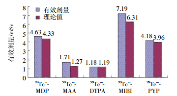
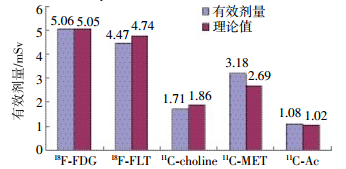
王风等[11]研究了受检者在不同检查项目中的受照剂量,采用标准人体模型,SPECT受检者的接受剂量根据美国核医学会医用内照射剂量学委员会提出的内照射剂量计算方法和国际辐射防护委员会106号报告的计算公式计算受检者的受照剂量。PET/CT受检者接受的CT扫描剂量参考欧盟委员会16262号报告的计算方法。SPECT受检者注射单光子药物99Tcm-MDP、99Tcm-大颗粒聚合人血清白蛋白、99Tcm-DTPA、99Tcm-MIBI和锝[99Tcm]焦磷酸纳注射液的活度分别为(760±64)、(155±37)、(242±80)、(799±128)、(733±92)MBq,有效剂量计算结果与利用国际辐射防护委员会53、80和106号报告计算的理论值如图 2所示。两组剂量进行单样本t检验,差异无统计学意义(t=2.477,P > 0.05)。赵海敏等[12]研究40位骨扫描加局部CT扫描受检者的受照剂量,受检者注射99Tcm-MDP的活度为(839.9±51.8)MBq,根据医学内照射剂量的程序计算受检者的受照剂量。通过记录每位受检者局部CT的扫描长度和容积CT剂量加权指数计算CT扫描所致的辐射剂量,结果得出40位受检者单纯骨扫描所致的辐射剂量为(4.73±0.97)mSv,骨扫描加局部CT扫描总的辐射剂量为(7.2±1.3)mSv,两者进行单样本t检验,差异无统计学意义(t=0.796,P > 0.05),说明在SPECT检查时局部CT扫描所致的辐射剂量可忽略不计。受检者行PET/CT检查使用低剂量CT参数,正电子放射性药物18F-FDG、3’-脱氧-3’-(18)F-氟胸腺嘧啶核苷、11C-胆碱、11C-甲基-L-蛋氨酸和11C-乙酸盐的注射活度分别为(266±38)、(316±87)、(388±50)、(379±83)、(309±56)MBq,受检者接受正电子放射性药物的辐射剂量和理论值比较如图 3所示。两组剂量进行单样本t检验,差异无统计学意义(t=0.216,P > 0.05)。通过体模测量并由扫描计算机读取头部和体部的容积CT剂量加权指数,计算得到CT扫描所致的辐射剂量为(8.80±0.85)mSv,PET/CT检查中CT扫描所致的辐射剂量占很大比重。Huang等[13]研究全身PET/CT的辐射剂量及其带来的潜在致癌危险,采用奥尔德森仿真模型热释光剂量计测量CT的辐射剂量,根据医学内照射剂量的程序进行理论计算得到PET的辐射剂量,假设注射18F-FDG的剂量为370 MBq,计算得到PET单独的辐射剂量为6.23 mSv。实验中模拟3种不同的CT采集条件,分别计算出不同采集条件下的辐射剂量,将PET的辐射剂量与CT的辐射剂量相加得到PET/CT总的辐射剂量,计算结果显示,CT所致的辐射剂量占总辐射剂量的54%~81%,他们的研究结果与王风等[11]的实验结果类似,所以在接受PET/CT检查时应选择适当的放射性药物活度和CT采集条件来降低受检者的辐射剂量。

图 2 受检者SPECT检查受到不同单光子药物辐射的有效剂量图中,99Tcm-MAA:99Tcm-大颗粒聚合人血清白蛋白;99Tcm-PYP:锝[99Tcm]焦磷酸纳。
Figure 2. The radiation dose to patients from single photon radiopharmaceutical was compared with the theoretical value

图 3 受检者PET/CT检查受到不同单光子药物辐射的有效剂量图中,18F-FLT:3’-脱氧-3’-(18)F-氟代胸腺嘧啶核苷;11C-choline:11C-胆碱;11C-MET:11C-甲基-L-蛋氨酸;11C-Ac:11C-乙酸盐。
Figure 3. The radiation dose to patients from positron radiopharmaceutical was compared with the theoretical value
受检者在核医学检查中接受的辐射剂量与注射的放射性药物活度、类型、停留时间和体内分布等因素有关[14],因此,在保证检查图像质量的前提下应采取适当的防护措施,合理降低辐射剂量,贯彻放射防护的最优化原则。
-
辐射防护的目的是防止确定性效应的发生,降低随机性效应发生的概率,使防护的结果达到可以接受的水平。辐射防护的原则是防护的最优化和实践的正当性。核医学辐射具有两大特点:① 对工作人员主要是外照射,即射线从人体外对人体产生的照射,对受检者则主要是内照射,即放射性药物进入人体产生的照射。② 由于放射性药物的特异性结合,受检者全身的受照剂量低而靶器官的受照剂量高。
核医学工作人员、受检者要做好内照射和外照射的防护,在实际的防护工作中,内照射防护的原则是采取有效措施切断放射性核素进入体内的各种途径;外照射防护的3个基本原则是距离防护、时间防护和屏蔽防护[15]。① 距离防护:对于γ射线点源,某一点所受到的辐射剂量与距源的距离的平方成反比,离放射源的距离越远,人体受到的辐射越少,工作人员应在保证完成工作的前提下,尽可能使用远距离操作器材来增加操作距离。② 时间防护:辐射剂量是一个积累的过程,基本与放射源的接触时间成正比。因此工作人员一定要做好准备工作再开始操作,尽可能缩短与放射源的接触时间。③ 屏蔽防护:针对不同的射线采用不同的屏蔽防护方法,α射线的穿透能力很弱,无法穿透皮肤或面巾纸,所以一般不需要屏蔽防护。对于α射线主要是防止放射性核素进入体内,因为α射线的电离能力很强,可引起细胞大量损伤。β射线比α射线具有更强的穿透能力,一般采用塑料、玻璃和树脂玻璃等防护。γ射线的穿透能力很强,一般采用高原子序数材料防护,在日常工作中主要用铅进行防护。
总之,在核医学的诊疗中,工作人员要严格遵守外照射防护的3个基本原则,在保证完成工作的前提下,采用远距离操作器材、轮流操作和穿戴防护工具等防护措施,保护身体健康。对于受检者,应鼓励他们多喝水多排尿,将放射性药物尽快排出体外,减少内照射。
核医学工作人员和受检者辐射防护现状
The status of radiation protection and control strategy for nuclear medicine workers and patients
-
摘要: 诊断核医学使用的放射性药物具有电离辐射效应,有可能损害核医学工作人员和受检者的健康或危及生命。笔者介绍了国内外诊断核医学近年来的发展趋势,并对核医学工作人员和受检者在不同检查项目中的辐射剂量进行重点分析,评估他们的辐射防护状况。笔者分析了近年来核医学辐射防护领域的研究,结果表明在采取有效防护措施的情况下,核医学工作人员的年有效剂量低于放射性工作人员的年剂量限值。受检者接受SPECT检查所致的辐射剂量主要来自99Tcm标记的单光子放射性药物,接受PET/CT检查所致的辐射剂量绝大部分来自CT扫描,所以选择合适的放射性药物活度和CT采集条件可以显著降低受检者的辐射剂量。
-
关键词:
- 体层摄影术,发射型计算机,单光子 /
- 正电子发射断层显像术 /
- 体层摄影术,X线计算机 /
- 核医学 /
- 辐射防护 /
- 吸收剂量
Abstract: In diagnostic nuclear medicine, the used of radioactive drugs with ionizing radiation effect and may place nuclear medicine workers and patients at risk of infection in terms of health.In this review the global trends in diagnostic nuclear medicine modalities applied in medical procedure were introduced at first.And then the exposure dose of nuclear medicine workers and patients in different examination items were analyzed emphatically.The radiation protection status was evaluated based on the analysis.In recent years, the researches in nuclear medicine radiation protection showed that the annual effective dose of workers was less than yearly dose limit of occupational exposure.The effective dose was mainly from single photon radiopharmaceuticals labeled by 99Tcm for patients undergoing SPECT examinations.However for PET/CT examinations, the effective dose was mainly from CT procedure.It could significantly reduce the effective dose to the patients when the activity of radioactive drugs and CT acquisition conditions were selected appropriately. -
图 1 核医学工作人员是否采取铅屏蔽防护受到不同放射性药物辐射的有效剂量抗图中,99TcmO4-:99Tcm-高锝酸盐。
Figure 1. In the case of lead and lead-free shielding protection, the exposed dose to the nuclear medicine workers in different examination items
图 2 受检者SPECT检查受到不同单光子药物辐射的有效剂量图中,99Tcm-MAA:99Tcm-大颗粒聚合人血清白蛋白;99Tcm-PYP:锝[99Tcm]焦磷酸纳。
Figure 2. The radiation dose to patients from single photon radiopharmaceutical was compared with the theoretical value
图 3 受检者PET/CT检查受到不同单光子药物辐射的有效剂量图中,18F-FLT:3’-脱氧-3’-(18)F-氟代胸腺嘧啶核苷;11C-choline:11C-胆碱;11C-MET:11C-甲基-L-蛋氨酸;11C-Ac:11C-乙酸盐。
Figure 3. The radiation dose to patients from positron radiopharmaceutical was compared with the theoretical value
表 1 美国不同年份进行核医学诊疗人次(万次)
Table 1. The number of patients for nuclear medicine diagnosis and treatment in United States in different years (ten thousand)
检查项目 1973年 1982年 2005年 骨扫描 12.5 181.1 345.0 甲状腺 46.0 67.7 - 肾脏 12.2 23.6 47.0 肿瘤 1.4 12.1 34.0 心脏 3.3 95.0 980.0 注:表中“-”表示未做详细统计。  下载: 导出CSV
-
[1] Dzhilavyan LZ, Karev AI, Raevsky VG. Possibilities for the production of radioisotopes for nuclear-medicine problems by means of photonuclear reactions[J]. Phys Atom Nucl, 2011, 74(12):1690-1696. DOI:10. 1134/S1063778811120040. [2] 郝大为, 孙俊杰.分子核医学用于肿瘤显像的研究进展[J].中华全科医学, 2011, 9(5):788-789.
Hao DW, Sun JJ. The research progress of molecular nuclear medicine for tumor imaging[J]. Chin J Gen Pract, 2011, 9(5):788-789.[3] 袁志斌, 周志俊.核医学从业人员的职业暴露与放射防护[J].国际放射医学核医学杂志, 2011, 35(1):49-52. DOI:10. 3760/cma. j. issn.1673-4114. 2011. 01. 014.
Yuan ZB, Zhou ZJ. Occupational exposure and radiation protection of nuclear medicine professional staffs[J]. Int J Radiat Med Nucl Med, 2011, 35(1):49-52. doi: 10.3760/cma.j.issn.1673-4114.2011.01.014[4] Mettler FA Jr, Bhargavan M, Faulkner K, et al. Radiologic and nuclear medicine studies in the United States and worldwide:frequency, radiation dose, and comparison with other radiation sources——1950-2007[J]. Radiology, 2009, 253(2):520-531. DOI:10. 1148/radiol. 2532082010. [5] Kinuya S, Kuwabara Y, Inoue K, et al. Nuclear medicine practice in Japan:a report of the seventh nationwide survey in 2012[J]. Ann Nucl Med, 2014, 28(10):1032-1038. DOI:10.1007/s12149-014-0893-y. [6] 中华医学会核医学分会. 2014年全国核医学现状普查简报[J].中华核医学与分子影像杂志, 2014, 34(5):389-390. DOI:10. 3760/cma.j. issn. 2095-2848. 2014. 05. 012.
Chinese Society of Nuclear Medicine. National nuclear medicine situation census bulletin in 2014[J]. Chin J Nucl Med Mol Imaging, 2014, 34(5):389-390. doi: 10.3760/cma.j.issn.2095-2848.2014.05.012[7] Salvatori M, Lucignani G. Radiation exposure, protection and risk from nuclear medicine procedures[J]. Eur J Nucl Med Mol Imaging, 2010, 37(6):1225-1231. DOI:10. 1007/s00259-010-1474-5. [8] 张庆召, 刘梅, 梁婧, 等. SPECT检查过程中放射性核素99Tcm的剂量评价[J].中国医学装备, 2015, 12(5):65-67. DOI:10. 3969/J.ISSN. 1672-8270. 2015. 05. 020.
Zhang QZ, Liu M, Liang J, et al. The evaluation of radiation dose to people after administered 99Tcm radionuclide during SPECT examinations[J]. Chin Med Equipment, 2015, 12(5):65-67. doi: 10.3969/J.ISSN.1672-8270.2015.05.020[9] 刘斌, 蒋丽莎, 杨晓川, 等. 18F-FDG显像患者对公众人群及核医学技师辐射剂量的估算[J].中国医学影像技术, 2016, 32(1):134-137. DOI:10. 13929/j. 1003-3289. 2016. 01. 035.
Liu B, Jiang LS, Yang XC, et al. Radiation dose estimation from patients undergoing 18F-FDG scanning:Implications for general public and nuclear medicine technicians[J]. Chin J Med Imaging Technol, 2016, 32(1):134-137. doi: 10.13929/j.1003-3289.2016.01.035[10] Chen MY. Radiation protection and regulations for the nuclear medicine physician[J]. Semin Nucl Med, 2014, 44(3):215-228. DOI:10. 1053/j.semnuclmed. 2014. 03. 005. [11] 王风, 赵伟, 赵起超, 等.核医学检查受检者所受辐射剂量分析[J].中华放射医学与防护杂志, 2015, 35(9):705-708. DOI:10. 3760/cma.j. issn. 0254-5098.
2015. 09. 017. Wang F, Zhao W, Zhao QC, et al. Analysis of patient radiation dose from nuclear medicine examinations[J]. Chin J Radiol Med Prot, 2015, 35(9):705-708. doi: 10.3760/cma.j.issn.0254-5098[12] 赵海敏. 99Tcm-MDP SPECT/CT扫描对骨转移瘤的诊断价值及所致辐射剂量的研究[D]. 济南: 山东大学, 2014.
Zhao HM. Diagnostic value for bone metastasis on 99Tcm-MDP SPECT/CT scanning and studies of related radiation dose[D]. Jinan:Shandong Universty, 2014.[13] Huang B, Law MW, Khong PL. Whole-body PET/CT scanning:estimation of radiation dose and cancer risk[J]. Radiology, 2009, 251(1):166-174. DOI:10. 1148/radiol. 2511081300. [14] 王风, 杨志, 张岩, 等. 18F-氟代脱氧葡萄糖PET/CT检查患者所受辐射剂量的测量分析[J].中华放射医学与防护杂志, 2013, 33(6):662-663. DOI:10. 3760/cma.j. issn. 0254-5098. 2013. 06. 026.
Wang F, Yang Z, Zhang Y, et al. Analysis of patient radiation dose from 18F-FDG PCT/CT scanning[J]. Chin J Radiol Med Prot, 2013, 33(6):662-663. doi: 10.3760/cma.j.issn.0254-5098.2013.06.026[15] Bolus NE. Review of common occupational hazards and safety concerns for nuclear medicine technologists[J]. J Nucl Med Technol, 2008, 36(1):11-17. DOI:10. 2967/jnmt. 107. 043869. -

 点击查看大图

计量
- 文章访问数: 3611
- HTML全文浏览量: 2641
- PDF下载量: 13