


 下载:
下载:


-
细胞凋亡是机体通过特定机制密切调控的细胞自发的、有序的死亡现象,它发生于胚胎发育、免疫防御及细胞稳态等阶段,具有广泛的生物学意义。而凋亡异常可引起多种疾病,例如,当机体凋亡过度增强时,往往可引起自身免疫性疾病、神经退行性病变等。与之相反,当机体凋亡缺陷时,突变的细胞增殖失控,最终形成恶性肿瘤[1]。许多抗肿瘤治疗手段(如化疗、放疗)均通过促进肿瘤细胞凋亡来达到治疗肿瘤的目的。
临床实践中,病理学检查常被用来评价肿瘤细胞凋亡情况[2]。但是,病理学检查为有创性检查,需考虑取材的可行性及患者的耐受性。同时,肿瘤具有空间异质性及时间异质性,病理学检查不能评价全身治疗效应,也不能动态地监测凋亡在人体的发生及发展过程。因此,针对细胞凋亡的影像学检查已成为研究热点。目前,用于检测细胞凋亡的影像学检查共有4类:① MRI。细胞出现凋亡时,细胞内水份减少,同时还可以产生脂质体等代谢物,磁共振设备可检测到凋亡细胞弛豫时间和弥散时间改变,而脂质磁共振波谱也可监测代谢物的产生[3]。虽然磁共振成像时间分辨率和空间分辨率较高,但它只能用于凋亡晚期的监测。② 超声检查。凋亡细胞通常可出现DNA断裂、染色体浓集等超微结构表现,超声检查结果会呈现回声增强[4]。但是,超声监测深度较浅,只能用于浅表部位的检查。③ 光学成像。研究者研发出一系列光学成像手段,如Cy5.5-膜联蛋白V(其中Cy5.5是一种荧光团)[5]、双报告基因(Rluc/Flue)生物发光成像[6]等都可用来评价凋亡。虽然光学成像具有无创性,可反复进行动态监测,但荧光染料价格较昂贵,且探测深度较浅,因此未能广泛进入临床。④ 放射性核素标记的凋亡显像剂。它可与凋亡细胞特异性结合,不受探测深度制约,同时又可应用于凋亡的早期检测,因此具有最广泛的应用前景。放射性核素标记的凋亡显像剂分为4类:靶向外翻细胞膜磷脂的显像剂、靶向凋亡细胞膜印迹显像剂、靶向半胱天冬酶(caspases)显像剂、线粒体膜势能改变显像剂(图 1)。理想的肿瘤凋亡放射性显像剂需具备以下特点:① 对凋亡细胞有高度特异性;② 在体内外性状稳定;③ 适宜的药理学特性;④ 便于放化合成及量化分析;⑤ 免疫原性低、毒性小;⑥ 经济上可行[3]。现在已有靶向外翻细胞膜磷脂显像剂、靶向凋亡细胞膜印迹显像剂和靶向半胱天冬酶显像剂进入临床研究阶段,本文对此类显像剂的作用机制及其临床研究应用进行综述。

图 1 凋亡显像剂的成像机制图中,ML-10:2-(5-氟代戊基)-2-甲基丙二酸。
Figure 1. Mechanism for nuclear medicine traces to image apoptotic cell
-
正常细胞膜为不对称性分布,磷脂酰丝氨酸(phosphatidylserine,PS)与磷脂酰乙醇胺(phos-phatidylethanol,PE)位于胞膜内侧。当细胞出现凋亡时,胞膜内的PS和PE外翻至细胞膜外表面,出现大量结合位点。目前科研人员已发现膜联蛋白V(Annexin V)和二(2,2′-二吡啶甲基胺)-Zn2+类配合物[8-9]等可与细胞外膜表面PS特异性结合,而耐久霉素和肉桂霉素等可与PE特异性结合[10],进而标记这些特异性小分子化合物、多肽和蛋白质,可应用在凋亡成像研究中。目前,99Tcm-Annexin V和123I-Annexin V[11]已应用于临床研究(图 2),但后者合成困难、易出现卤化反应,制约了其进一步临床转化,因此,本节我们主要介绍99Tcm-Annexin V。

图 2 凋亡细胞摄取膜联蛋白V的机制
Figure 2. Mechanism for Annexin V uptake by apoptotic cells
99Tcm-4,5-2(硫代乙酰胺)戊酰-膜联蛋白V (99Tcm-BTAP-Annexin V)是最早应用于动物实验和人体试验的分子探针[12-13],但由于其合成步骤复杂、耗时长,且放射性化学产率较低,因此研究者合成了另外一种Annexin V显像剂——99Tcm-联肼尼克酰胺-膜联蛋白V(99Tcm-HYNIC-Annexin V)[14]。相对于99Tcm-BTAP-Annexin V,99Tcm-HYNIC-Annexin V合成较快、步骤简单。作为烟碱酸类似物,可以与氨基末端和赖氨酸残基结合,同时与99Tcm结合。目前已广泛用于Ⅰ、Ⅱ期临床试验。
2003年,Kemerink等[15]募集了6名健康志愿者,进行了Ⅰ期临床研究,结果发现,注射99Tcm-HYNIC-Annexin V后,受试者主要是通过泌尿系统将其排泄,肾、肝、膀胱的摄取值较高。肾、肝、膀胱对99Tcm-BTAP-Anenxin V的摄取量也较多,但99Tcm-BTAP-Anenxin V可快速、大量从肠道排泄,形成肠道放射性浓聚影,制约了它在临床上的进一步推广[13]。
从2003年开始,van de Wiele等[16]开始募集头颈癌患者,进行一系列Ⅱ期临床试验,发现治疗前99Tcm-HYNIC-Annexin A5的摄取值与死亡细胞数成正比,而且定量检查具有可重复性[17]。虽然99Tcm-HYNIC-Annexin A5对头颈癌原发灶检查的灵敏度高,但它对转移淋巴结的检出率较低,考虑为SPECT的低分辨率及淋巴结直径较小所致[18]。治疗前99Tcm-HYNIC-Annexin A5的摄取值还可以预测预后,2008年,Loose等[19]对29例头颈癌患者进行随访发现,99Tcm-HYNIC-Annexin A5的T/N值与无病生存率及总生存率成负相关。
99Tcm-HYNIC-Annexin A5还可对肿瘤治疗进行早期监测。目前已证实它可对淋巴瘤、头颈癌和非小细胞肺癌的抗肿瘤治疗监测提供参考[20-23]。Kartachova等[24]募集16例晚期非小细胞肺癌患者,在化疗前和化疗后48 h内分别行99Tcm-HYNIC-An-nexin A5检查,发现治疗后显像剂摄取值升高的患者得到完全或部分缓解,而摄取值不变或降低的患者病情未能缓解,甚至出现进展。
虽然99Tcm-Annexin V标记快捷,技术最成熟,目前已应用于多个Ⅱ期临床试验,但仍存在以下局限性:① 特异性较差,当细胞出现坏死、自噬性坏死时,PS暴露,可被Annexin V识别。同时肿瘤血管形成、炎症、血小板活化均可造成假阳性[25]。② 肝肾摄取值高[13, 15],阻碍它在上腹部肿瘤的临床应用。③99Tcm半衰期较长,未能动态反映细胞凋亡过程。有研究者合成了18F-Annexin V[26]行PET扫描,但由于18F-Annexin V合成步骤复杂,放射性化学产率低,限制了其在临床上的广泛应用。因此研究者开始把研究重心转移至研发其他凋亡探针上。
-
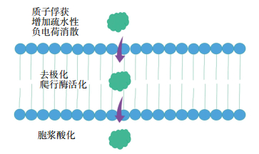
在细胞凋亡早期,细胞膜印迹会发生以下改变:爬行酶系统活化、外膜小叶和细胞内液酸化[27]。针对这一机制,研究人员已研发出一类小分子探针——Aposense家族,包括丹磺酰半胱氨酸(DDC)、2-(5-氟代戊基)-2-甲基丙二酸(ML-10)、2-(5-氟代丙基)-2-甲基丙二酸(ML-8)[28]等。在对Aposense家族研发的早期阶段,常将其作为光学成像显像剂来进行体内凋亡成像[29-30]。随着PET技术的发展,放射性核素标记的靶向细胞膜印迹显像剂应运而生。其中,18F-ML-10已进入临床研究阶段。18F-ML-10以丙二酸模体为核心,当细胞凋亡时,2个羧基通过氢键共用质子,使负离子分散在环状结构中,从而增加疏水性。酸化的探针通过疏水的羟基转移至胞质,与带负离子的蛋白质结合,从而在胞质集聚。戊烷基链进一步增加了18F-ML-10的疏水性,使其更易穿透细胞膜[31](图 3)。

图 3 凋亡细胞摄取2-(5-氟代戊基)-2-甲基丙二酸(18F-ML-10)的机制
Figure 3. Mechanism for 18F-ML-10 uptake by apoptotic cells
健康志愿者的Ⅰ期临床研究表明,18F-ML-10安全性高,性状稳定,可在体内快速分布和清除,主要通过尿路排泄。同时在男性睾丸中的摄取值高。膀胱接受的辐射剂量最高,是剂量限制器官[32]。
2012年,Allen等[33]对10例接受全脑放疗的脑转移患者进行Ⅱ期临床研究,使用影像学技术进行随访,在放疗开始前和结束时行18F-ML-10检查,在治疗结束6~8周时行MRI,发现18F-ML-10摄取值的早期改变与MRI的解剖学改变高度相关。2014年,Oborski等[34]在治疗1例初治多形性成胶质细胞瘤患者的同时,使用18F-ML-10进行监测,结果发现,18F-ML-10摄取值的改变与肿瘤细胞凋亡有关。
虽然18F-ML-10已成功进入临床转化阶段,但它的作用机制尚未完全明确。与此同时,一系列体内外试验发现,18F-ML-10在肿瘤细胞的集聚具有pH依赖性[35],制约了它的广泛应用。因此仍需行大量研究以明确和改善其作用机制。
-
靶向caspases显像剂分为Caspase-3底物衍生物类和Caspase-3直接拮抗剂类两类,18F-CP18属于Caspase-3底物衍生物类,而18F-(S)-1-((1-(2-氟乙基)-1H-[1, 2, 3]-三氮唑-4-基)甲基)-5-(2(2,4-二氟苯氧甲基)-四氢化吡咯-1-磺酰基)靛红(18F-ICMT-11)属于Caspase-3直接拮抗剂类,这两者皆已用于临床。
-
各种凋亡信号转导通路均涉及到caspase-3的活化,因此caspase-3可成为显像剂的识别靶点。目前已研发出多种靶向caspase酶的凋亡显像剂。其中(18S,21S,24S,27S,30S)-27-(2-羧乙基)-21-(羧甲基)-30-((2S,3R,4R,5R,6S)-6-((2-(4-(3-18F-氟代丙基)-1H-1,2,3-三氮唑-1-基)乙酰氨基)甲基)-3,4,5-三羟基四氢-2H-吡喃-2-羧基氨基)-24-异丙基-18-甲基-17,20,23,26,29-五氧杂-4,7,10,13-四氧杂-16,19,22,25,28-五氮杂三十烷-1,32-二酸(18F-CP18)含四肽类Caspase-3蛋白的识别序列是DEVD(Asp-Glu-Val-Asp),该序列与Caspase-3的结合力高,同时含有聚乙二醇(Polyethylene glycol,PEG)链协助显像剂跨膜转运。在凋亡细胞中,Caspase-3活性增高,可剪切PEG链,从而使造影剂滞留在细胞内。而在正常细胞中,PEG链不被剪切,显像剂可自由进出细胞(图 4)[36]。

图 4 凋亡细胞摄取18F-CP-18的机制图中,PEG:聚乙二醇;DEVD:含四肽类Caspase-3蛋白的识别序列。
Figure 4. Mechanism for 18F-CP-18 uptake by apoptotic cells
Xia等[37]选用多种细胞株(U87、A427、LNCaP、PC3)建立裸鼠肿瘤模型,予18F-CP18显像发现肿瘤摄取值升高,提示18F-CP18可用于肿瘤诊断。2016年Rapic等[38]建立Colo205移植瘤小鼠模型,分别用氟尿嘧啶(fluorouracil,5-FU)、伊立替康和5-FU+伊立替康处理,发现肿瘤造影剂摄取值与凋亡水平有关,而与坏死水平无关,提示18F-CP18可应用于肿瘤治疗的疗效监测。国内也对18F-CP18进行了一系列的临床前研究,研究者选用A549细胞株建立裸鼠肿瘤模型,予化疗及放疗后,在荷瘤鼠体内注入18F-CP18进行显像分析,发现治疗后肿瘤部位出现放射性浓聚影,提示18F-CP18可用于肿瘤治疗早期凋亡检测[39-40]。
Doss等[41]将7名健康志愿者纳入Ⅰ期临床研究后发现,18F-CP18可迅速通过泌尿系统排出,膀胱壁和肾脏的吸收剂量过高。因此,在检查过程中,有必要予受检者利尿处理,来降低膀胱和肾脏的照射剂量。
-
18F-ICMT-11为靛红类小分子化合物抑制剂,可通过二羰基与Caspase-3活性中心的半胱氨酸残基共价结合,从而用于凋亡显像[42]。
2009年,研究者行动物体外实验和体内实验时发现,18F-ICMT-11可以与抗肿瘤治疗诱导的凋亡细胞结合,随着凋亡细胞的增加,其摄取值也随之增加[43]。2013年,Nguyen等[44]制作38C13B淋巴细胞荷瘤鼠、HCT116结肠癌荷瘤鼠、MDA-MB-231乳腺癌荷瘤鼠模型,使用单剂环磷酰胺和birinapant对上述荷瘤鼠进行治疗,治疗过程中使用18F-ICMT-11进行监测发现,18F-ICMT-11可反映肿瘤凋亡的时空演变过程,进而可指导药物治疗和新药研发。
Challapalli等[45]纳入8名健康志愿者进行Ⅰ期临床试验。试验过程中,受试者耐受性好,未见明显不良反应。胆囊壁、肠道、膀胱和肝脏接受的剂量较高,提示18F-ICMT-11主要通过肝肾代谢,从而限制上腹部成像。进食后的受试者相对空腹受试者的肝脏及肠道摄取有所减少,提示可通过进食的方法,使上腹部成像成为可能。该试验同时还发现骨髓生理性摄取。
但18F-ICMT-11仍有其局限性。首先,它可在体内非特异性地与表达半胱氨酸基团的组织和蛋白结合,因此它作为凋亡显像剂在体内显像缺乏特异性。此外,它在活体内对Caspase-3的亲和力下降,活体显像时需增加给药剂量,因此有必要优化该探针的结构[46]。
-
在常规临床实践中,CT、MRI等传统解剖学成像检查通过实体瘤疗效评价标准来评估治疗效果。但是,肿瘤治疗后形态学改变存在滞后性,同时肿瘤在治疗后常出现炎症、纤维化等病理学改变,难以与肿瘤残余病灶相鉴别。PET/CT因其能成功将功能性成像和解剖性成像结合,可多维度提供病灶信息,已广泛应用于临床实践中。其中,应用最广泛的显像剂是18F-FDG。它可以先于肿瘤形态学改变前提供肿瘤代谢信息,通常摄取值减少提示治疗有效。但是18F-FDG在临床应用中仍有局限性,首先,炎症细胞也可显示摄取值升高,难以与肿瘤细胞相鉴别[47];其次,18F-FDG对部分肿瘤(如胃印戒细胞癌、前列腺癌)显示欠佳。
凋亡显像不受炎症、纤维化及糖代谢影响,在临床转换中具有广泛前景。首先,凋亡显像可早期检测到肿瘤对治疗的反应,有利于筛选肿瘤患者接受个体化治疗,避免不必要的治疗毒性,提高患者生存率。与此同时,凋亡显像也可以预测患者预后。
目前,随着分子生物学、影像学及应用化学领域知识的不断更新,科技人员开发出一系列肿瘤凋亡核医学显像剂。目前,已有99Tcm-Annexin V、18F-CP18、18F-ICMT-11、18F-ML-10进入临床试验阶段,但它们仍都存在一定的局限性。我们相信,随着人们对凋亡的生化特性的理解的进一步加深,借用不断完善的化学合成策略及转化技术,理想的肿瘤凋亡显像剂的问世指日可待。
临床用肿瘤细胞凋亡核医学显像剂研究进展
Progress of nuclear medicine imaging agents for the clinical apoptosis imaging of tumors
-
摘要: 凋亡在肿瘤的发生、发展和治疗中发挥重要的作用,因此无创、动态监测凋亡已成为目前抗肿瘤治疗的研究热点。目前已有靶向外翻细胞膜磷脂显像剂99Tcm-4,5-2(硫代乙酰胺)戊酰-膜联蛋白V和99Tcm-联肼尼克酰胺-膜联蛋白V、靶向半胱天冬酶(caspases)显像剂18F-ICMT-11和18F-CP18、靶向凋亡细胞膜印迹显像剂18F-ML-10进入临床试验阶段,虽然它们存在局限性,但当与传统的成像手段比较时,它们在肿瘤的诊断、治疗监测上具备一定的优越性。因此,肿瘤凋亡显像剂具有广泛的应用前景。笔者就目前核医学肿瘤细胞凋亡显像剂在肿瘤诊疗中的研究进展进行综述。Abstract: Apoptosis plays a key role in the development and treatment of tumors; thus, the invasive dynamic monitoring of apoptosis is of considerable interest in the management of cancer.At present, 99Tcm-BTAP-Annexin V, 99Tcm-HYNIC-Annexin V, 18F-ICMT-11, 18F-CP18, and 18F-ML-10 have achieved clinical application.Despite their limitations, these agents are superior to conventional imaging technique in the diagnosis and therapeutic monitoring of cancer and can exhibit potential in clinical applications.This review provides an overview of the recent development in apoptosis imaging by using radionuclide-labeled tracers in the management of cancer.
-
Key words:
- Apoptosis /
- Neoplasms /
- Molecular probes /
- In vivo imaging
-
图 1 凋亡显像剂的成像机制图中,ML-10:2-(5-氟代戊基)-2-甲基丙二酸。
Figure 1. Mechanism for nuclear medicine traces to image apoptotic cell
图 3 凋亡细胞摄取2-(5-氟代戊基)-2-甲基丙二酸(18F-ML-10)的机制
Figure 3. Mechanism for 18F-ML-10 uptake by apoptotic cells
-
[1] Green DR, Kroemer G. The pathophysiology of mitochondrial cell death[J]. Science, 2004, 305(5684):626-629. DOI:10.1126/science.1099320. [2] Martinez MM, Reif RD, Pappas D. Detection of apoptosis:A review of conventional and novel techniques[J]. Anal Methods, 2010, 2(8):996-1004. DOI:10.1039/c0ay00247j. [3] Liimatainen T, Hakumaki JM, Kauppinen RA, et al. Monitoring of gliomas in vivo by diffusion MRI and 1H MRS during gene therapy-induced apoptosis:interrelationships between water diffusion and mobile lipids. NMR Biomed, 2009, 22(3):272-279. DOI:10.1002/nbm.1320 [4] Czarnota GJ, Kolios MC, Abraham J, et al. Ultrasound imaging of apoptosis:high-resolution non-invasive monitoring of programmed cell death in vitro, in situ and in vivo[J]. Br J Cancer, 1999, 81(3):520-527. DOI:10.1038/sj.bjc.6690724. [5] Petrovsky A, Schellenberger E, Josephson L, et al. Near-infrared fluorescent imaging of tumor apoptosis[J]. Cancer Res, 2003, 63(8):1936-1942. [6] Shah K, Tang Y, Breakefield X, et al. Real-time imaging of TRAIL-induced apoptosis of glioma tumors in vivo[J]. Oncogene, 2003, 22(44):6865-6872. DOI:10.1038/sj.onc.1206748. [7] Zeng W, Wang X, Xu P, et al. Molecular imaging of apoptosis:from micro to macro[J]. Theranostics, 2015, 5(6):559-582. DOI:10.7150/thno.11548. [8] Wang H, Wu Z, Li S, et al. Synthesis and evaluation of a radiolabeled bis-zinc(Ⅱ)-cyclen complex as a potential probe for in vivo imaging of cell death[J]. Apoptosis, 2017, 22(4):585-595. DOI:10.1007/s10495-017-1344-8. [9] Sun T, Tang G, Tian H, et al. Positron emission tomography imaging of cardiomyocyte apoptosis with a novel molecule probe 18F FP-DPAZn2[J/OL]. Oncotarget, 2015, 6(31):30579-30591[2017-04-20]. http://www.ncbi.nlm.nih.gov/pubmed/26416423. DOI:10. 18632/oncotarget.5769. [10] Yao S, Hu K, Tang G, et al. Positron emission tomography imaging of cell death with 18F FPDuramycin[J]. Apoptosis, 2014, 19(5):841-850. DOI:10.1007/s10495-013-0964-x. [11] Lahorte CM, van de Wiele C, Bacher K, et al. Biodistribution and dosimetry study of 123I-rh-annexin V in mice and humans[J]. Nucl Med Commun, 2003, 24(8):871-880. DOI:10.1097/01.mnm.0000084585.29433.58. [12] Stratton JR, Dewhurst TA, Kasina S, et al. Selective uptake of radiolabeled annexin V on acute porcine left atrial thrombi[J]. Circulation, 1995, 92(10):3113-3121. doi: 10.1161/01.CIR.92.10.3113 [13] Kemerink GJ, Boersma HH, Thimister PW, et al. Biodistribution and dosimetry of 99mTc-BTAP-annexin-V in humans[J]. Eur J Nucl Med, 2001, 28(9):1373-1378. doi: 10.1007/s002590100578 [14] Blankenberg FG, Katsikis PD, Tait JF, et al. In vivo detection and imaging of phosphatidylserine expression during programmed cell death[J]. Proc Natl Acad Sci U S A, 1998, 95(11):6349-6354. doi: 10.1073/pnas.95.11.6349 [15] Kemerink GJ, Liu X, Kieffer D, et al. Safety, biodistribution, and dosimetry of 99mTc-HYNIC-annexin V, a novel human recombinant annexin V for human application[J]. J Nucl Med, 2003, 44(6):947-952. [16] van de Wiele C, Lahorte C, Vermeersch H, et al. Quantitative tumor apoptosis imaging using technetium-99m-HYNIC annexin V single photon emission computed tomography[J]. J Clin Oncol, 2003, 21(18):3483-3487. DOI:10.1200/JCO.2003.12.096. [17] Vermeersch H, Ham H, Rottey S, et al. Intraobserver, interobserver, and day-to-day reproducibility of quantitative 99mTc-HYNIC annexin-V imaging in head and neck carcinoma[J]. Cancer Biother Radiopharm, 2004, 19(2):205-210. DOI:10.1089/108497804323071986. [18] Vermeersch H, Loose D, Lahorte C, et al. 99mTc-HYNIC Annexin-V imaging of primary head and neck carcinoma[J]. Nucl Med Commun, 2004, 25(3):259-263. doi: 10.1097/00006231-200403000-00008 [19] Loose D, Vermeersch H, de Vos F, et al. Prognostic value of 99mTc-HYNIC annexin-V imaging in squamous cell carcinoma of the head and neck[J]. Eur J Nucl Med Mol Imaging, 2008, 35(1):47-52. DOI:10.1007/s00259-007-0577-0. [20] Rottey S, Slegers G, Van Belle S, et al. Sequential 99mTc-hydrazinonicotinamide-annexin V imaging for predicting response to che-motherapy[J]. J Nucl Med, 2006, 47(11):1813-1818. [21] Rottey S, van den Bossche B, Slegers G, et al. Influence of chemo-therapy on the biodistribution of[99mTc] hydrazinonicotinamide annexin V in cancer patients[J]. Q J Nucl Med Mol Imaging, 2009, 53(2):127-132. [22] Haas RL, de Jong D, Valdés Olmos RA, et al. In vivo imaging of radiation-induced apoptosis in follicular lymphoma patients[J]. Int J Radiat Oncol Biol Phys, 2004, 59(3):782-787. DOI:10.1016/j.ijrobp.2003.11.017. [23] Hoebers FJ, Kartachova M, de Bois J, et al. 99mTc Hynic-rh-Annexin V scintigraphy for in vivo imaging of apoptosis in patients with head and neck cancer treated with chemoradiotherapy[J]. Eur J Nucl Med Mol Imaging, 2008, 35(3):509-518. DOI:10.1007/s00259-007-0624-x. [24] Kartachova M, van Zandwijk N, Burgers S, et al. Prognostic significance of 99mTc Hynic-rh-annexin V scintigraphy during platinum-based chemotherapy in advanced lung cancer[J]. J Clin Oncol, 2007, 25(18):2534-2539. DOI:10.1200/JCO.2006.10.1337. [25] Corsten MF, Hofstra L, Narula J, et al. Counting heads in the war against cancer:defining the role of annexin A5 imaging in cancer treatment and surveillance[J]. Cancer Res, 2006, 66(3):1255-1260. DOI:10.1158/0008-5472.CAN-05-3000. [26] Murakami Y, Takamatsu H, Taki J, et al. 18F-labelled annexin V:a PET tracer for apoptosis imaging[J]. Eur J Nucl Med Mol Imaging, 2004, 31(4):469-474. DOI:10.1007/s00259-003-1378-8. [27] Cohen A, Shirvan A, Levin G, et al. From the Gla domain to a novel small-molecule detector of apoptosis[J]. Cell Res, 2009, 19(5):625-637. DOI:10.1038/cr.2009.17. [28] Yao S, Hu K, Tang G, et al. Molecular PET imaging of cyclophos-phamide induced apoptosis with 18F-ML-8[J/OL]. Biomed Res Int, 2015:317403[2017-04-20]. http://www.ncbi.nlm.nih.gov/pubmed/25977920. DOI:10.1155/2015/317403. [29] Damianovich M, Ziv I, Heyman SN, et al. ApoSense:a novel technology for functional molecular imaging of cell death in models of acute renal tubular necrosis[J]. Eur J Nucl Med Mol Imaging, 2006, 33(3):281-291. DOI:10.1007/s00259-005-1905-x. [30] Aloya R, Shirvan A, Grimberg H, et al. Molecular imaging of cell death in vivo by a novel small molecule probe[J]. Apoptosis, 2006, 11(12):2089-2101. DOI:10.1007/s10495-006-0282-7. [31] 张晓军, 李云钢, 刘健, 等. 18F-ML-10的制备、生物分布和临床应用[J].中华核医学与分子影像杂志, 2016, 36(2):131-136. DOI:10.3760/cma.j.issn. 2095-2848.2016.02.008.
Zhang XJ, Li YG, Liu J, et al. Preparation and biodistribution of 2-(5-[18F] fluoro-pentyl)-2-methyl-malonic acid and its clinical application[J]. Chin J Nucl Med Mol Imaging, 2016, 36(2):131-136. doi: 10.3760/cma.j.issn.2095-2848.2016.02.008[32] Höglund J, Shirvan A, Antoni G, et al. 18F-ML-10, a PET tracer for apoptosis:first human study[J]. J Nucl Med, 2011, 52(5):720-725.DOI:10.2967/jnumed.110.081786. [33] Allen AM, Ben-Ami M, Reshef A, et al. Assessment of response of brain metastases to radiotherapy by PET imaging of apoptosis with 1F-ML-10[J]. Eur J Nucl Med Mol Imaging, 2012, 39(9):1400-1408. DOI:10.1007/s00259-012-2150-8. [34] Oborski MJ, Laymon CM, Lieberman FS, et al. First use of 18F-labeled ML-10 PET to assess apoptosis change in a newly diagnosed glioblastoma multiforme patient before and early after therapy[J].Brain Behav, 2014, 4(2):312-315. DOI:10.1002/brb3.217. [35] Bauwens M, de Saint-Hubert M, Cleynhens J, et al. Radioiodinated phenylalkyl malonic acid derivatives as pH-sensitive SPECT tracers[J/OL]. PLoS One, 2012, 7(6):e38428[2017-04-20]. http://www.ncbi.nlm.nih.gov/pubmed/22719886. DOI:10.1371/journal.pone.0038428. [36] Su H, Chen G, Gangadharmath U, et al. Evaluation of 18F-CP18 as a PET imaging tracer for apoptosis[J]. Mol Imaging Biol, 2013, 15(6):739-747. DOI:10.1007/s11307-013-0644-9. [37] Xia CF, Chen G, Gangadharmath U, et al. In vitro and in vivo evaluation of the caspase-3 substrate-based radiotracer 18F-CP18 for PET imaging of apoptosis in tumors[J]. Mol Imaging Biol, 2013, 15(6):748-757. DOI:10.1007/s11307-013-0646-7. [38] Rapic S, Vangestel C, Elvas F, et al. Evaluation of 18F CP18 as a Substrate-Based apoptosis imaging agent for the assessment of early treatment response in oncology[J/OL]. Mol Imaging Biol, 2017[2017-04-20]. http://www.ncbi.nlm.nih.gov/pubmed/28050749.DOI:10.1007/s11307-016-1037-7.[publishedonline ahead of print]. [39] 张宝石. 18F标记小分子肽(18F-CP-18) 早期评价肺癌化疗后细胞凋亡的基础研究[D]. 北京: 军医进修学院, 2011: 24-31.
Zhang BS. Experiment on 18F-CP-18 in early evaluation of lung cancer apoptosis after chemotherapy[D]. Beijing:Chinese PLA Postgraduate Medical School, 2011:24-31.[40] 吴晓燕. Caspase-3探针F-CP-18在体外和活体肿瘤细胞凋亡显像的实验研究[D]. 天津: 南开大学, 2014: 27-38.
Wu XY. In vitro and in vivo evaluation of the caspase-3 substrate-based probe F-CP-18 for apoptosis imaing in tumor[D]. Tianjin:Nankai University, 2014:27-38.[41] Doss M, Kolb HC, Walsh JC, et al. Biodistribution and radiation dosimetry of 18F-CP-18, a potential apoptosis imaging agent, as determined from PET/CT scans in healthy volunteers[J]. J Nucl Med, 2013, 54(12):2087-2092. DOI:10.2967/jnumed.113.119800. [42] Smith G, Glaser M, Perumal M, et al. Design, synthesis, and biological characterization of a caspase 3/7 selective isatin labeled with 2-[18F]fluoroethylazide[J]. J Med Chem, 2008, 51(24):8057-8067. DOI:10.1021/jm801107u. [43] Nguyen QD, Smith G, Glaser M, et al. Positron emission tomography imaging of drug-induced tumor apoptosis with a caspase-3/7 specific[18F]-labeled isatin sulfonamide[J]. Proc Natl Acad Sci U S A, 2009, 106(38):16375-16380. DOI:10.1073/pnas.0901310106. [44] Nguyen QD, Lavdas I, Gubbins J, et al. Temporal and spatial evolution of therapy-induced tumor apoptosis detected by caspase-3-selective molecular imaging[J]. Clin Cancer Res, 2013, 19(14):3914-3924. DOI:10.1158/1078-0432.CCR-12-3814. [45] Challapalli A, Kenny LM, Hallett WA, et al. 18F-ICMT-11, a caspase-3-specific PET tracer for apoptosis:biodistribution and radiation dosimetry[J]. J Nucl Med, 2013, 54(9):1551-1556. DOI:10.2967/jnumed.112.118760. [46] Jiang H, Zhao PJ, Su D, et al. Paris saponin I induces apoptosis via increasing the Bax/Bcl-2 ratio and caspase-3 expression in gefitinib-resistant non-small cell lung cancer in vitro and in vivo[J]. Mol Med Rep, 2014, 9(6):2265-2272. DOI:10.3892/mmr. 2014. 2108. [47] Min Z, Amlani M. Pulmonary Mycobacterium kansasii Infection Mimicking Malignancy on the 18F-FDG PET Scanin a Patient Receiving Etanercept:A Case Report and Literature Review[J/OL]. Case Rep Pulmonol, 2014:973573[2017-04-20]. https://www.ncbi.nlm.nih.gov/pubmed/?term=25389506. DOI:10.1155/2014/973573 -

 点击查看大图

图(4)
计量
- 文章访问数: 4115
- HTML全文浏览量: 3093
- PDF下载量: 9