


-
聚丙烯酰胺水凝胶(polyacrylamide hydrogel,PAHG)是一种用于注射填充、不被人体吸收的水凝胶,自1997年后被广泛应用于注射隆乳术。但由于并发症较多,2006年以后我国禁止在美容行业临床应用PAHG[1]。随着对PAHG注射隆乳术后并发症的深入认识,以及动物模型研究表明,PAHG具有明显的致畸、致癌等特性。MRI对PAHG术后并发症的诊断、填充物清除术前评估和疗效评价已有较多研究[1-2]。王蓼等[1]认为,MRI可作为PAHG注射隆乳术后并发症的首选影像学检查方法。而对于PAHG注射隆乳术后乳腺病变的MR诊断[3-5]和成像技术研究尚不充分[6]。本文拟回顾性分析PAHG注射隆乳患者的多序列MRI扫描结果,比较各序列成像对乳腺病变的检出效能,以期优选扫描序列,提高PAHG注射隆乳后乳腺病变的MRI检出率和诊断准确率。
-
回顾性分析2006年4月至2010年12月在我院行MR检查的80例PAHG注射隆乳者的临床及影像资料;所有患者均为女性,年龄21~50岁,平均(34.20±4.58)岁,注射隆乳时间1~13年。所有患者隆乳术前乳腺病变情况不详。临床主要表现为乳腺胀痛,触及乳内或皮下包块,乳腺外形不佳、变硬,部分病例具有上述2项或2项以上症状,部分患者因担心注入物引发病变而就诊。其中4例8个乳腺行穿刺抽吸灌洗清除术,5例10个乳腺行乳晕切口清除术后置入硅胶假体,1例1个乳腺行切开引流排脓术,1例1个乳腺行乳癌保乳术,1例2个乳腺游离凝胶合并感染和脓肿形成。80例患者均于取出PAHG填充物术前行MR检查。手术同时切除全部或部分病变组织并行病理学检查。所有乳腺病变性质均经术后病理确诊。
所有患者均于检查前签署了知情同意书。
-
采用GE Signa EXCITE HD 3.0T超导型磁共振机扫描,体部8通道相控阵线圈,配合特制乳腺扫描专用承托支架(获国家发明专利,外观设计专利号:ZL 2008 3 0055967.5,实用新型专利号:ZL 2008 2 0188524.8)。患者俯卧,双侧乳腺自然下垂,完全置于承托支架的窝洞内,双臂上举置于头部两侧。扫描范围:上至腋窝肱骨头下缘,下至乳腺下方皮肤,包括腋窝及全部乳腺组织。平扫层厚:横轴位5 mm、MR水成像(MR hydrography,MRH)2.2 mm,矢状位4 mm;增强肝脏容积超快速(liver acquisition with volume acceleration,LAVA)多期动态成像横轴位和矢状位均为4 mm。平扫序列包括横轴位:T1加权像快速自旋回波、T2加权像快速自旋回波、短时间反转恢复(short time inversion recovery,STIR)、弥散加权成像(diffusion weighted imaging,DWI)。MRH采用快速恢复自旋回波序列、重T2加权像、三维数据采集成像序列,恢复时间/回波时间:7500 ms/ 556 ms,视野37 cm×37 cm,矩阵320×256,层厚2.2 mm,层间距-1.1 mm,激励次数1,采集时间5 min。DWI序列采用平面回波技术,b值分别取0、800 mm2/s。增强序列采取LAVA成像,扫描前用18 G静脉留置针建立静脉通道,并连接高压注射器,LAVA序列每个时相扫描时间为45 s,间隔3 s,第一时相为平扫蒙片,第二时相为注入对比剂15 s后立即启动扫描,LAVA序列扫描总时间至少9 min。对比剂采用Gd-DTPA(磁显葡胺,北京北陆医药化工集团),注射剂量按0.1 mmol/kg计算,注射速度2 mL/s,注射对比剂后,用20 mL生理盐水冲洗导管内残留的对比剂。
-
原始数据上传至GE公司提供的HP后处理工作站,运用Functool 2功能软件(Advantage Workstation软件,版本4.2)进行重建。MRH图像由原始数据采用容积再现技术得到,并多角度旋转观察图像。LAVA增强扫描后,在原始数据基于最大增强斜率的伪彩图上选择ROI病灶,将ROI置于病灶中央,以绘制时间-信号强度曲线。
-
正常乳腺腺体MRI表现:T1加权像呈等信号,T2加权像、STIR及MRH序列呈稍高信号,DWI b=800 mm2/s呈等信号,LAVA增强扫描轻度强化,时间-信号强度曲线呈“流入型”。
PAHG MRI表现:T1加权像呈低信号,T2加权像、STIR及MRH序列呈高信号,DWI b=800 mm2/s呈低信号,LAVA增强扫描无强化。
乳腺癌MRI表现:多数有分叶,边缘毛糙,多见蟹足样表现,肿瘤较小时信号均匀,肿瘤较大时不均匀;信号多表现为T1加权像呈稍低信号,T2加权像呈稍高信号,STIR及MRH序列呈高信号,DWI b=800 mm2/s呈高信号,LAVA增强扫描明显强化,时间-信号强度曲线主要呈“流出型”。
纤维腺瘤MRI表现:圆形或椭圆形,边缘光滑,信号均匀,信号多表现为T1加权像呈稍低信号,T2加权像呈稍高信号,STIR及MRH序列呈高信号,DWI b=800 mm2/s呈等或稍高信号,LAVA增强扫描明显强化,时间-信号强度曲线呈“流入型”。
导管内乳头状瘤MRI表现:圆形或椭圆形,边缘光滑,信号多均匀,表现为T1加权呈等或稍低信号,T2加权像呈等或稍高信号,STIR及MRH序列呈高信号,DWI b=800 mm2/s呈高信号,LAVA增强扫描明显强化,时间-信号强度曲线呈“流入型”。
乳腺囊肿MRI表现:圆形或椭圆形,薄壁,边缘光滑,信号多均匀,表现为T1加权像呈低信号,T2加权像呈高信号,STIR及MRH序列呈高信号,DWI b=800 mm2/s呈高信号,LAVA增强扫描无强化。
乳腺脓肿MRI表现:圆形或椭圆形,厚壁,边缘模糊,信号多表现为T1加权像呈低信号,T2加权像呈高信号,STIR及MRH序列呈高信号,DWI b=800 mm2/s呈高信号,LAVA增强扫描环形强化,囊壁时间-信号强度曲线呈“流入型”。
乳腺腺病MRI表现:斑片状,边缘欠清晰,信号多表现为T1加权像呈等信号,T2加权像呈等信号,STIR及MRH序列呈稍高信号,DWI b=800 mm2/s呈等信号,LAVA增强扫描见强化,时间-信号强度曲线呈“流入型”。
-
由2位乳腺MR图像诊断经验丰富的MR副主任医师及1位美容科注射隆乳副主任医师采用双盲法对MR检查结果进行分析评估。有不同意见时,通过协商达成一致。主要观察、分析各序列图像上PAHG的信号特点和完整情况,各序列图像上病灶的信号特点、形态、大小和数量,以及时间-信号强度曲线的特点;分别计算各序列乳腺病变的总检出率和乳腺癌诊断的准确率等情况。
-
采用SPSS 17.0对所有数据进行统计学分析。各序列病灶的总检出率和乳腺癌诊断准确率比较采用R×C交叉表卡方检验,组内两两比较采用卡方检验分析,P<0.05表示差异有统计学意义。
-
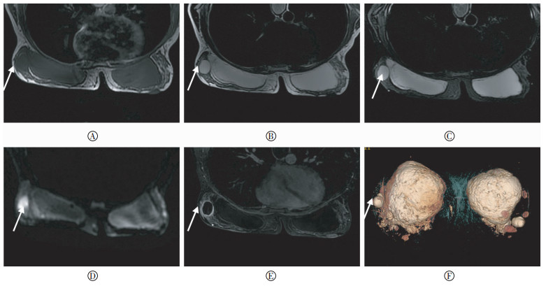
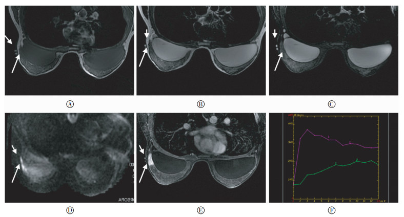
80例160个乳腺中共检出86个病灶,全部经手术病理确诊,结果见表 1。其中,单侧乳腺1个病灶74例;单侧乳腺2个病灶1例;双侧乳腺各1个病灶5例。乳腺病灶性质:乳腺癌30个(图 1)、纤维腺瘤21个、导管内乳头状瘤14个、囊肿10个、脓肿6个(图 2)、乳腺腺病5个。
扫描序列 乳腺癌 纤维腺瘤 导管内乳头状瘤 囊肿 脓肿 乳腺腺病 合计 检出率/% 乳腺癌诊断准确率/% T1加权成像 10 5 3 7 1 2 28 32.56 33.33 T2加权成像 20 10 4 10 6 3 53 61.11 66.67 STIR 20 11 5 10 6 3 55 63.88 66.67 MRH 14 7 2 8 4 1 36 41.66 46.67 DWI 30 16 12 10 6 3 77 89.53 100 LAVA 30 21 14 10 6 5 86 100 100 注:表中,PAHG:聚丙烯酰胺水凝胶;STIR:短时间反转恢复;MRH:MR水成像;DWI:弥散加权成像;LAVA:肝脏容积超快速多期动态增强成像。 表 1 不同MRI序列对PAHG注射隆乳后乳腺病灶(86个)的总检出情况
Table 1. Total detection lesions(86) of different sequences of MR with PAHG injection for augmentation mammoplasty
图 1 患者女性,45岁,聚丙烯酰胺水凝胶注射隆乳术后10年,右侧乳腺外象限扪及肿块,经病理证实为浸润性导管癌Ⅱ级。图中,A:T1加权像呈稍低信号;B:T2加权像呈等信号;C:短时间反转恢复呈等信号;D:弥散加权成像b=800 mm2/s呈稍高信号,病灶边缘模糊;E:肝脏容积超快速多期动态增强3 min图像,显示病灶明显均匀强化;F:肝脏容积超快速多期动态增强时间-信号曲线,呈速升速降型(红色曲线)。右乳腺皮肤表面见鱼肝油胶囊标记(短箭头),对应部位乳腺外上象限见小结节灶(长箭头),大小约2.5 cm×1.1 cm×1.0 cm。
Figure 1. Female patient, 45 years old. 10 years later with polyacrylamide hydrogel injection for augmentation mammoplasty. A mass was touched(with two Cod Liver Oil capsules marker, Short arrow) on the right upper quadrant of the right breast, and was confirmed invasive ductal carcinoma grade Ⅱ by the pathology(long arrow, 2.5 cm×1.1 cm×1.0 cm).

图 2 患者女性,40岁,聚丙烯酰胺水凝胶注射隆乳术后3年,双侧乳腺外象限扪及包块,经手术病理证实为脓肿。图中,A:T1加权像呈稍低信号;B:T2加权像呈高信号;C:短时间反转恢复呈高信号;D:弥散加权成像b=800 mm2/s呈高信号;E:肝脏容积超快速多期动态增强9 min图像,显示病灶环形强化,边缘清晰;F:MR水成像容积再现图,明确显示了结节灶与聚丙烯酰胺水凝胶注射物的立体关系。双侧乳腺见完整PAHG注射填充物。右侧乳腺外上象限见结节灶(箭头),大小约1.9 cm×2.0 cm×2.4 cm。
Figure 2. Female patient, 40 years old. 3 years later with polyacrylamide hydrogel injection for augmentation mammoplasty. Masses were touched in bilateral outer quadrant of the breasts and were diagnosed as abscess by pathology.
-
各序列对乳腺病变的总检出率有显著性差异,R×C交叉表卡方检验显示:χ2=129.428,P<0.05,差异有统计学意义;各序列图像对乳腺癌的诊断准确率也有显著性差异,R×C交叉表分析显示:χ2=51.843,P<0.05,差异有统计学意义(表 1)。
-
乳腺病变总检出率中,除T1加权与MRH、T2加权与STIR的差异无统计学意义外,其他序列之间的差异均有统计学意义。而乳腺癌诊断准确率中,MRH与T1加权、MRH与T2加权、MRH与STIR、T2加权与STIR,以及DWI与LAVA增强序列之间的差异均无统计学意义,其他序列之间的差异有统计学意义(表 2)。
组别 乳腺病变检出率 乳腺癌诊断准确率 χ2值 P值 χ2值 P值 ①与② 14.584 0.000 6.667 0.010 ①与③ 16.974 0.000 6.667 0.010 ①与④ 1.593 0.207 1.111 0.292 ①与⑤ 58.702 0.000 30.000 0.000 ①与⑥ 87.509 0.000 30.000 0.000 ②与③ 0.100 0.752 0.000 1.000 ②与④ 6.729 0.009 2.443 0.118 ②与⑤ 18.145 0.000 12.000 0.001 ②与⑥ 40.835 0.000 12.000 0.001 ③与④ 8.424 0.004 2.443 0.118 ③与⑤ 15.767 0.000 12.000 0.001 ③与⑥ 37.816 0.000 12.000 0.001 ④与⑤ 43.368 0.000 21.818 0.000 ④与⑥ 70.492 0.000 21.818 0.000 ⑤与⑥ 9.497 0.003 0.000 1.000* 注:表中,PAHG:聚丙烯酰胺水凝胶;①:T1加权像;②:T2加权像;③:STIR(短时间反转恢复);④:MRH(MR水成像);⑤:DWI(弥散加权成像);⑥:LAVA(肝脏容积超快速)多期动态增强成像;*:Fisher精确概率法。 表 2 不同MRI序列对PAHG注射隆乳后乳腺病变总检出率和乳腺癌诊断准确率的组内两两比较情况
Table 2. Pairwise comparison within groups for total detection rate of breast lesions and diagnosis accuracy of breast cancer by different MR sequences with PAHG injection for augmentation mammoplasty
-
PAHG是一种用于注射填充、不被人体吸收的水凝胶,被广泛应用于医学美容行业。其主要构成为2.5%~5.0%的聚丙烯酰胺悬浮在95.0%~97.5%的水中[7-8]。过往认为其无毒、无过敏、无致畸特点,但可致纤维包膜形成[9-10]。国内PAHG注射隆乳术绝大多数采用一次性直接注入法。一般在乳腺下皱襞或乳腺上部区域的后间隙注射150~200 mL[11]PAHG。双侧隆乳时,必须确保同一层面、同一腔隙内均匀注入,以获得满意效果。然而许多研究[1-6]认为,PAHG注射隆乳术并发症较多,常见的有乳房硬结、疼痛、胸背疼痛、上肢疼痛、感染、血肿、填充物移位、功能障碍、囊性结节、血肿、炎症及肿瘤等,且有合并肿瘤[4]的潜在可能,在一定程度上为患者带来了较大的心理压力,也是患者要求行MR检查的主要原因。
MRI可多序列、多参数、多平面扫描,且有良好的软组织分辨能力。但不同MRI序列的参数构成特点限定了其成像用途;而病灶本身成分的构成差异等,又使得同一序列图像上与其他组织存在一定差异。因此,优选MR扫描序列以提高病灶检出率和诊断符合率等显得十分必要。目前关于PHGA术后多序列MRI检出病灶效能和诊断准确率的比较研究较为少见。而本研究所涉及的乳腺MR扫描序列较为全面,目的在于比较各序列对病灶的检出效能和诊断准确率,以期为相关病例的MR扫描提供相对准确的序列选择,从而最大限度地避免因序列选择不当而造成的病灶漏检、漏诊和(或)误诊。
本研究中,注射填充物PAHG的MR信号与水类似,表现为长T1、长T2信号,在T1加权图像上呈低信号,T2加权、STIR及MRH序列图像上呈高信号,DWI序列图像上(b=800 mm2/s)呈低信号,增强LAVA扫描序列图像无强化。MR图像可以清晰显示PAHG的范围、所在层面,及其与胸大肌、乳腺组织的关系,并直观表现水凝胶在体内的各种状况,容易发现乳腺皮下、胸肌筋膜内、胸肌间隙或腋下有无游离PAHG,从而判断其有无外渗等并发症[12]。尤其是脂肪抑制MRH技术,通过容积再现后处理得到的三维图像可以清晰显示PAHG的位置和形态[14],但对乳腺病变的检出率低于二维图像。可见,对于拟取出PAHG者,MRI可行完整、直观的术前评估,并提供手术操作路径等关键信息,还可以为术后客观评价手术效果和制定术后复查方案提供依据。
表 1和表 2比较结果表明,T1加权序列对乳腺病变的总检出率和对乳腺癌的诊断准确率均为最低。可能是由于T1加权图像上,病灶与正常腺体组织的信号强度接近,均表现为高信号。因此,难以区分病灶与正常乳腺腺体组织。该序列图像主要用于显示乳腺腺体的解剖结构,并初步判定成像区域内有无血肿存在。T2加权像与STIR序列对乳腺病变的总检出率及乳腺癌诊断准确率的差异无统计学意义。但T2加权图像上大部分病灶与乳腺脂肪组织的信号强度接近,也都表现为高信号。而STIR序列选择性抑制了脂肪组织信号,增加了病灶与乳腺脂肪组织的信号对比度,使得病灶显示更加清晰、突出,从而较为灵敏地检测出较小乳腺病灶。表 1包含的各平扫序列中,DWI序列的乳腺病灶总检出率和乳腺癌诊断准确率均为最高。DWI技术通过检测组织中水分子自由扩散运动的差异,可以敏感、可靠地再现病灶内容物的情况。其表观扩散系数值可定量分析水分子的微观运动情况,可用于乳腺良、恶性病变的鉴别诊断[5, 13]。而动态增强LAVA序列在本研究所有序列中(表 1)对乳腺病灶的总检出率和对乳腺癌的诊断准确率均为最高(100%),与术后病理结果一致。动态增强LAVA序列采取全新的分段选频脂肪反转恢复脂肪抑制技术,不受磁场不均匀的影响,能获得更好的脂肪抑制效果,配合部分K空间填充技术,缩短了采集时间,并采取三维薄层扫描,从而完美捕捉到病灶各时相的强化特点及动态过程,通过分析病灶的强化特点、浸润范围、病灶内及其周围血管灌注情况,有助于确定病灶的数量、大小、位置及形态,能显示平扫序列难以检出的多灶性、多中心病变,结合信号-时间曲线的形态等,能更准确地反映病变的病理状况,也有利于病变的定性诊断。动态增强LAVA可以显示正常组织、病灶在动脉早期、中期、晚期和静脉早期、中期、晚期及延迟期的增强信号特点,但增强扫描时间过短,无法充分显示正常组织与病灶的时间-信号曲线特点。故此本研究中强调乳腺动态增强LAVA扫描时间不能少于9 min,即为满足上述目的,主要用于良、恶性病变的鉴别诊断。
综上所述,本研究乳腺病变的总检出效能显示,LAVA增强序列最高,DWI序列次之,T1加权序列最低。而从乳腺癌的诊断准确率来看,增强LAVA与DWI序列的效能相同,STIR序列次之。笔者认为,对于PAHG注射隆乳患者的术后MR随访中,横轴位STIR、DWI和LAVA增强序列,以及矢状位STIR和LAVA增强序列应列为常规MR扫描序列,以确保病灶的检出率,并尽可能避免漏诊和误诊。此外,本研究通过MR扫描序列优选,一方面可以提高扫描效率,为体质较差和(或)耐受力较差患者达到诊断目的而缩短扫描时间;另一方面,可以尽可能避免对乳腺癌病灶的漏检和(或)误诊,为患者进一步治疗方案的制定提供依据。
当然,影响病灶检出的原因,除患者个体差异、仪器性能、扫描序列及其参数选择外,尚有图像后处理方法和读片经验等。如果结合可疑病灶的表观扩散系数、磁共振波谱成像、定量磁敏感成像等定量分析手段,有可能进一步提高MRI技术对PHGA术后病灶的检出率及其定性诊断的准确率,值得进一步研究。
不同MRI序列检出聚丙烯酰胺水凝胶注射隆乳患者乳腺病变效能的比较研究
The different MRI sequences to detect breast lesions in polyacrylamide hydrogel injection for augmentation patients:a comparative study
-
摘要:
目的 探讨不同MRI序列对聚丙烯酰胺水凝胶(PAHG)注射隆乳后乳腺病变的检出效能。 方法 回顾性分析80例临床怀疑合并乳腺病变的注射隆乳患者的MRI影像资料,扫描序列包括T1加权像、T2加权像、短时间反转恢复(STIR)、弥散加权成像(DWI)、MR水成像(MRH)及肝脏容积超快速(LAVA)三维多期动态增强序列,分析各序列图像中病灶的信号特点、位置、形态、大小和数量,以及时间-信号强度曲线,分别计算不同序列对乳腺病变的总检出率和对乳腺癌的诊断准确率。各序列病灶的总检出率差异采用R×C交叉表卡方检验,组内两两比较采用卡方检验分析。 结果 160个乳腺中共检出86个病灶,其中乳腺癌病灶30个。T1加权像、T2加权像、STIR、DWI、MRH及LAVA增强序列对乳腺病灶的总检出率分别为32.56%(28/86)、61.11%(53/86)、63.88%(55/86)、41.66%(36/86)、89.53%(77/86)、100%(86/86),其中乳腺癌病灶诊断准确率分别为33.33%(10/30)、66.67%(20/30)、66.67%(20/30)、46.67%(14/30)、100%(30/30)、100%(30/30)。对各序列乳腺病变的总检出率行R×C交叉表卡方检验:χ2=129.428,P < 0.05,差异有统计学意义;对各序列乳腺癌病灶诊断准确率行R×C交叉表卡方检验:χ2=51.843,P < 0.05,差异有统计学意义。对各序列乳腺病变总检出率和乳腺癌病灶诊断准确率行组内两两比较分析,除T1加权像与MRH、T2加权像与STIR对乳腺病变总检出率的差异无统计学意义外(χ2=1.593、0.100,P均>0.05),其他均有统计学意义(χ2=6.729~87.509,P均 < 0.05);而MRH与T1加权像、MRH与T2加权像、MRH与STIR、T2加权像与STIR,以及DWI与LAVA增强序列对乳腺癌诊断准确率的差异均无统计学意义(χ2=1.111、2.443、2.443、0.000、0.000,P均>0.05),其他序列的差异有统计学意义(χ2=12.000~30.000,P均 < 0.05)。 结论 DWI和LAVA增强序列对PAHG注射隆乳后乳腺病变的总检出率最高;合理应用MRI序列,有利于提高乳腺癌病灶的诊断准确率。 Abstract:Objective To explore the detective values of various MRI sequences in breast polyacrylamide hydrogel(PAHG) injection for augmentation patients. Methods A total of 80 patients who have been injected for augmentation and who have suspected breast lesions were subjected to MRI scanning. The MRI scan sequence included T1 weighted imaging(T1WI), T2 weighted imaging(T2WI), short time inversion recovery(STIR), diffusion weighted imaging(DWI) and MR hydrography(MRH), as well as liver acquisition with valume acceleration(LAVA) dynamic contrast-enhanced sequence. The main sequence analysis indicators included the detection rate of breast disease and breast cancer, and to analyze statistically. Results A total of 86 lesions were observed in 160 breasts. The breast-lesion detection rates of the T1WI, T2WI, STIR, DWI and MRH, as well as LAVA dynamic contrast-enhanced sequence were 32.56%(28/86), 61.11%(53/86), 63.88%(55/86), 41.66%(36/86), 89.53%(77/86), and 100%(86/86), respectively; the breast-cancer detection rates of these sequences were 33.33%(10/30), 66.67%(20/30), 66.67%(20/30), 46.67%(14/30), 100%(30/30), and 100%(30/30), respectively. Each MRI scanning sequence of breast-lesion detection rate of R×C cross-table analysis, χ2=129.428, P < 0.05; the difference was statistically significant. Each MRI scanning sequence of breast-cancer detection rate of R×C cross-table analysis, χ2=51.843, P < 0.05; the difference was statistically significant. Each MRI scanning sequence detection rate of breast disease and breast cancer in the group were compared pairwise. The detection rates of breast lesions were not statistically significant in T1WI and MRH, T2WI and STIR(χ2=1.593, 0.100, both P>0.05), whereas those in the other scanning sequences were statistically significant(χ2=6.729~87.509, all P < 0.05). The differences in breast-cancer detection rates in MRH and T1WI, MRH and T2WI, MRH and STIR, T2WI and STIR, and DWI and LAVA were not statistically significant(χ2=1.111, 2.443, 2.443, 0.000, 0.000, all P>0.05), whereas those of the other sequences were statistically significant(χ2=12.000~30.000, all P < 0.05). Conclusions The total detection rate of breast lesions with PAHG injection for augmentation mammoplasty by DWI and LAVA-enhanced sequence wasthe highest. Rational use of MRI sequence will help improve the lesion detection rate of patients' breast augmentation PAHG injection and has important clinical value. -
图 1 患者女性,45岁,聚丙烯酰胺水凝胶注射隆乳术后10年,右侧乳腺外象限扪及肿块,经病理证实为浸润性导管癌Ⅱ级。图中,A:T1加权像呈稍低信号;B:T2加权像呈等信号;C:短时间反转恢复呈等信号;D:弥散加权成像b=800 mm2/s呈稍高信号,病灶边缘模糊;E:肝脏容积超快速多期动态增强3 min图像,显示病灶明显均匀强化;F:肝脏容积超快速多期动态增强时间-信号曲线,呈速升速降型(红色曲线)。右乳腺皮肤表面见鱼肝油胶囊标记(短箭头),对应部位乳腺外上象限见小结节灶(长箭头),大小约2.5 cm×1.1 cm×1.0 cm。
Figure 1. Female patient, 45 years old. 10 years later with polyacrylamide hydrogel injection for augmentation mammoplasty. A mass was touched(with two Cod Liver Oil capsules marker, Short arrow) on the right upper quadrant of the right breast, and was confirmed invasive ductal carcinoma grade Ⅱ by the pathology(long arrow, 2.5 cm×1.1 cm×1.0 cm).
图 2 患者女性,40岁,聚丙烯酰胺水凝胶注射隆乳术后3年,双侧乳腺外象限扪及包块,经手术病理证实为脓肿。图中,A:T1加权像呈稍低信号;B:T2加权像呈高信号;C:短时间反转恢复呈高信号;D:弥散加权成像b=800 mm2/s呈高信号;E:肝脏容积超快速多期动态增强9 min图像,显示病灶环形强化,边缘清晰;F:MR水成像容积再现图,明确显示了结节灶与聚丙烯酰胺水凝胶注射物的立体关系。双侧乳腺见完整PAHG注射填充物。右侧乳腺外上象限见结节灶(箭头),大小约1.9 cm×2.0 cm×2.4 cm。
Figure 2. Female patient, 40 years old. 3 years later with polyacrylamide hydrogel injection for augmentation mammoplasty. Masses were touched in bilateral outer quadrant of the breasts and were diagnosed as abscess by pathology.
表 1 不同MRI序列对PAHG注射隆乳后乳腺病灶(86个)的总检出情况
Table 1. Total detection lesions(86) of different sequences of MR with PAHG injection for augmentation mammoplasty
扫描序列 乳腺癌 纤维腺瘤 导管内乳头状瘤 囊肿 脓肿 乳腺腺病 合计 检出率/% 乳腺癌诊断准确率/% T1加权成像 10 5 3 7 1 2 28 32.56 33.33 T2加权成像 20 10 4 10 6 3 53 61.11 66.67 STIR 20 11 5 10 6 3 55 63.88 66.67 MRH 14 7 2 8 4 1 36 41.66 46.67 DWI 30 16 12 10 6 3 77 89.53 100 LAVA 30 21 14 10 6 5 86 100 100 注:表中,PAHG:聚丙烯酰胺水凝胶;STIR:短时间反转恢复;MRH:MR水成像;DWI:弥散加权成像;LAVA:肝脏容积超快速多期动态增强成像。  下载: 导出CSV
表 2 不同MRI序列对PAHG注射隆乳后乳腺病变总检出率和乳腺癌诊断准确率的组内两两比较情况
Table 2. Pairwise comparison within groups for total detection rate of breast lesions and diagnosis accuracy of breast cancer by different MR sequences with PAHG injection for augmentation mammoplasty
组别 乳腺病变检出率 乳腺癌诊断准确率 χ2值 P值 χ2值 P值 ①与② 14.584 0.000 6.667 0.010 ①与③ 16.974 0.000 6.667 0.010 ①与④ 1.593 0.207 1.111 0.292 ①与⑤ 58.702 0.000 30.000 0.000 ①与⑥ 87.509 0.000 30.000 0.000 ②与③ 0.100 0.752 0.000 1.000 ②与④ 6.729 0.009 2.443 0.118 ②与⑤ 18.145 0.000 12.000 0.001 ②与⑥ 40.835 0.000 12.000 0.001 ③与④ 8.424 0.004 2.443 0.118 ③与⑤ 15.767 0.000 12.000 0.001 ③与⑥ 37.816 0.000 12.000 0.001 ④与⑤ 43.368 0.000 21.818 0.000 ④与⑥ 70.492 0.000 21.818 0.000 ⑤与⑥ 9.497 0.003 0.000 1.000* 注:表中,PAHG:聚丙烯酰胺水凝胶;①:T1加权像;②:T2加权像;③:STIR(短时间反转恢复);④:MRH(MR水成像);⑤:DWI(弥散加权成像);⑥:LAVA(肝脏容积超快速)多期动态增强成像;*:Fisher精确概率法。
下载: 导出CSV
-
[1] 王蓼, 胡竺, 水淼, 等.MRI对聚丙烯酰胺水凝胶注射隆乳术后并发症的诊断[J].实用放射学杂志, 2012, 28(8):1211-1213. DOI:10.3969/j.issn.1002-1671.2012.08.015.
Wang L, Hu Z, Shui M, et al.MR imaging in diagnosing the syndrome of polyacrylamide hydrogel injection for augmentation mammoplasty[J].J Pract Radiol, 2012, 28(8):1211-1213. doi: 10.3969/j.issn.1002-1671.2012.08.015[2] 董洁, 田方兴, 唐全.聚丙烯酰胺水凝胶隆乳后无并发症患者生活质量的影响因素[J].中华整形外科杂志, 2013, 29(2):137-139. DOI:10.3760/cma.j.issn.1009-4598.2013.02.015.
Dong J, Tian FX, Tang Q.Factors affecting quality of Life of polyacrylamide hydrogel breast augmentation of patients without complications[J].Chin J Plast Surg, 2013, 29(2):137-139. doi: 10.3760/cma.j.issn.1009-4598.2013.02.015[3] 邢继平, 赵雨千, 乌兰巴特尔, 等.聚丙烯酰胺水凝胶注射隆乳并发纤维囊内远期血肿一例[J].中华整形外科杂志, 2015, 31(1):71-72. DOI:10.3760/cma.j.issn.1009-4598.2015.01.023.
Xing JP, Zhao YQ, Wu LBTE, et al.A case of polyacrylamide hydrogel injection for augmentation with fiber intracapsular hematoma forward[J].Chin J Plast Surg, 2015, 31(1):71-72. doi: 10.3760/cma.j.issn.1009-4598.2015.01.023[4] 黄卫平, 林茜茜, 陈瑾, 等.MRI在聚丙烯酰胺水凝胶注射隆乳术后合并乳腺肿瘤中的诊断价值[J].中华整形外科杂志, 2011, 27(2):136-137. DOI:10.3760/cma.j.issn.1009-4598.2011.02.018.
Huang WP, Lin QQ, Chen J, et al.The MRI diagnostic value in polyacrylamide hydrogel injection for augmentation surgery with breast tumors[J].Chin J Plast Surg, 2011, 27(2):136-137. doi: 10.3760/cma.j.issn.1009-4598.2011.02.018[5] 刘丽华, 龙淼淼, 刘宁, 等.MR扩散加权成像定量诊断聚丙烯酰胺水凝胶隆乳后哺乳期脓肿[J].实用放射学杂志, 2015, 31(4):683-685. DOI:10.3969/j.issn.1002-1671.2015.04.045.
Liu LH, Long MM, Liu N, et al.Quantitative analysis of diffusion weighted imaging for diagnosis of puerperal breast abscess after polyacrylamide hydrogel augmentation mammoplasty[J].J Pract Radiol, 2015, 31(4):683-685. doi: 10.3969/j.issn.1002-1671.2015.04.045[6] 李刚, 苏顺清, 覃达贤, 等.MRI在聚丙烯酰胺水凝胶注射隆乳术后并发症的诊断价值[J].中国临床医学影像杂志, 2013, 24(4):247-250. DOI:10.3969/j.issn.1008-1062.2013.04.007.
Li G, Su SQ, Qin DX, et al.MRI in the diagnosis of complications due to breast augmentation with polyacrylamide hydrogel[J].J Chin Clin Med Imaging, 2013, 24(4):247-250. doi: 10.3969/j.issn.1008-1062.2013.04.007[7] Leung KM, Yeoh GP, Chan KW.Breast pathology in complications associated with polyacrylamide hydrogel(PAAG) mammoplasty[J].Hong Kong Med J, 2007, 13(2):137-140. [8] Christensen LH, Breiting VB, Aasted A, et al.Long-term effects of polyacrylamide hydrogel on human breast tissue[J].Plast Reconstr Surg, 2003, 111(6):1883-1890. DOI:10.1097/01.PRS.0000056873.87165.5A. [9] Bello G, Jackson IT, Keskin M, et al.The use of polyacrylamide gel in soft tissue augmentation:an experimental assessment[J].Plast Reconstr Surg, 2007, 119(4):1326-1336. DOI:10.1097/01.prs.0000254824.13065.3b. [10] Ben Khedher N, David J, Trop I, et al.Imaging findings of breast augmentation with injected hydrophilic polyacrylamide gel:Patient reports and literature review[J].Eur J Radiol, 2011, 78(1):104-111. DOI:10.1016/j.ejrad.2009.09.021. [11] Qiao Q, Wang XC, Sun JM, et al.Management for postoperative complications of breast augmentation by injected polyacrylamide hydrogel[J].Aesthetic Plast Surg, 2005, 29(3):156-161. DOI:10.1007/s00266-004-0099-0. [12] Teo SY, Wang SC.Radiologic features of polyacrylamide gel mammoplasty[J/OL].AJR Am J Roentgenol, 2008, 191(3): W89-95[2016-08-28].https:www.ncbi.nlm.nih.gov/pubmed/?term=18716084. DOI: 10.2214/AJR.07.3616. [13] Unal O, Koparan HI, Avcu S, et al.The diagnostic value of diffusion-weighted magnetic resonance imaging in soft tissue abscesses[J].Eur J Radiol, 2011, 77(3):490-494. DOI:10.1016/j.ejrad.2009.08.025. [14] 卢瑞梁, 蔡华清, 周新韩, 等.聚丙烯酰胺水凝胶注射隆乳术后MR容积再现技术的应用与探讨[J].中国美容整形外科杂志, 2011, 22(12):737-739. DOI:10.3969/j.issn.1673-7040.2011.12.011.
Lu RL, Cai HQ, Zhou XH, et al.Application and investigate of Mr volume rendering technology after augmentation mammoplasty with polyacrylamide hydrogel injection[J].Chin J Aesth Plast Surg, 2011, 22(12):737-739. doi: 10.3969/j.issn.1673-7040.2011.12.011 -

 点击查看大图
计量
- 文章访问数: 2835
- HTML全文浏览量: 1380
- PDF下载量: 2