


-
霍奇金淋巴瘤(Hodgkin lymphoma,HL)是一种罕见的累及淋巴结及淋巴系统的恶性肿瘤。世界卫生组织将HL分为两种主要类型:结节性淋巴细胞为主型霍奇金淋巴瘤(nodular lymphocyte predominant Hodgkin lymphoma,NLPHL)和经典型霍奇金淋巴瘤(classical Hodgkin lymphoma,CHL)[1]。近几十年来,随着个体化治疗方案的实施,HL患者的治疗已有明显进展,现在至少80%的患者可被治愈。治疗后定期复查,及时发现有无肿瘤残留或其他组织浸润,准确评价治疗效果并判断是否复发,对HL患者的预后或下一步治疗方案的选择有至关重要的意义。过去,临床常用B超、CT等影像学方法基于病灶大小、密度等形态学改变来评价HL治疗疗效,但均存在一定的局限性[2]。目前,能同时显示解剖结构和代谢信息的PET/CT已成为HL患者初始分期及治疗结束后疗效评估的重要手段,它对HL的诊断、疗效监测和预后判断具有重要作用,但对HL复发诊断的应用价值需更多临床研究证实[3]。2014年恶性淋巴瘤成像工作小组国际会议发表的共识中,推荐使用Deauville标准进行PET/CT报告分析,并认为这一标准方法简便、重复性好[4]。本研究选择24例临床可疑复发的HL患者进行研究,并基于Deauville标准进行PET/CT报告分析,旨在探讨18F-FDG PET/CT在HL复发诊断中的应用价值。
-
2010年3月至2014年2月在中国人民解放军第二五四医院PET/CT中心检查的HL患者24例,其中,男性14例、女性10例,年龄11~72岁,平均(36±4)岁。所有患者治疗前均经病理或淋巴结穿刺活检证实诊断,其中,CHL 9例、NLPHL 15例。治疗方法:单纯化疗14例、术后化疗4例、同时行放化疗6例。所有化疗患者均化疗4个周期以上,所有患者治疗前均行B超、CT和(或)MRI等传统影像学检查。治疗前后均行PET/CT检查者16例,仅治疗后行PET/CT检查者8例。检查距最近一次治疗结束5~27个月。所有患者或其家属均于检查前签署了知情同意书。
-
显像剂18F-FDG由原子高科股份有限公司医学三部提供,放化纯度 > 95%。显像仪器采用美国GE公司Discovery ST16 PET/CT仪。
-
检查前患者禁食至少6 h以上,监测空腹血糖水平确定 < 7.2 mmol/L后,静脉注射18F-FDG,成人剂量3.70~4.44 MBq/kg(0.10~0.12 mCi/kg),儿童酌减。
注射显像剂后患者在光暗、安静、温暖的房间内休息50~60 min后进行显像,于扫描前排空膀胱、饮用500~800 ml 1%泛影葡胺(湖南汉森制药股份有限公司生产)后开始图像采集。范围从股骨上段至颅底,先行螺旋CT扫描,层厚3.75 mm,再进行PET采集,使用三维扫描,每个床位采集2~3 min,共采集5~7个床位,必要时在注入显像剂后2.5~3 h行局部延迟显像。采集结束后将数据传至Xeleris工作站进行图像融合和图像重建,分别得到全身冠状、矢状、横断面的CT、PET及PET/CT融合图像。
-
PET/CT图像由2名有经验的放射科医师及核医学医师共同阅片,观察CT所示病变部位大小、形态、与周围组织的关系、CT值及相应位置PET所示18F-FDG代谢情况。
-
根据Deauville标准对病灶FDG代谢活跃程度进行计分。该标准采用5分的等级来确定受累部位18F-FDG摄取量(相对于纵隔和肝脏),定义如下,1分:背景以上无摄取;2分:病灶摄取≤纵隔;3分:病灶摄取>纵隔,但≤肝脏;4分:任何病灶摄取程度高于肝脏;5分:任何病灶摄取程度显著高于肝脏和(或)新病灶出现;X分:新的摄取区域不太可能与淋巴瘤相关。其中1~4分为初始受累部位,5分为初始受累部位和(或)淋巴瘤相关的新病灶[5-6]。
-
根据Deauville标准1~2分为阴性,4~5分为阳性[7]。在某些情况下3分可认为是“阴性”,但是,对于根据中期PET检查进行降阶梯治疗时,3分(以纵隔血池摄取量作为参照)被划分在阳性范围内是合适的(即PET检查Deauville标准评分1~2分为阴性,3~5分为阳性)[8]。治疗结束后判断标准为:4~5分代表病灶有残留代谢活性,可以判定为复发;3分则要对比中期PET检查,如果中期评分为1~2分,则判定为复发;1~2分代表原病灶没有明确代谢活性,判定为阴性。
-
应用SPSS13.3软件对数据进行统计学分析,计数资料以率(%)表示,P < 0.05表示差异具有统计学意义。
-
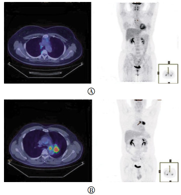
本研究中PET/CT结果显示:1~2分(阴性)者2例;3分者2例,其中1例在降阶梯治疗后2个月,判定为阳性(图 1中A),另1例判定为阴性;4~5分(阳性)者19例;X分者1例,图像显示左上肺结节伴纵隔淋巴结肿大(图 1中B)。

图 1 霍奇金淋巴瘤患者的PET/CT图像
Figure 1. PET/CT images of Hodgkin lymphoma patient
-
24例患者中经病理结果证实17例复发,3例淋巴结反应性增生,1例左肺上叶非小细胞肺癌伴纵隔淋巴结转移,3例未见明确复发。
-
对比病理结果,本研究中共出现3例假阳性,其中,Deauville评分3分者1例,4~5分者2例。假阳性病例18F-FDG摄取增高的淋巴结主要分布在咽旁间隙、下颌下及纵隔内,未见骨髓18F-FDG摄取增高。Deauville评分与病理结果的相关性对照见表 1。
病理结果 Deauville评分 1~2分 3分 4~5分 复发 0 0 17 未复发 2 2 2 注:根据影像学判断,评分为X分的患者病灶不太可能与淋巴瘤相关,故不纳入相关性研究。 表 1 Deauville评分与病理结果对照(n=23)
Table 1. The correlation between the score of Deauville and pathologic results(n=23)
-
根据Deauville标准,18F-FDG PET/CT对HL复发的诊断效能分别为:灵敏度100%(17/17)、特异度50%(3/6)、阳性预测值85%(17/20)、阴性预测值100%(3/3)、准确率87%(20/23)。根据影像学判断,评分为X分的患者病灶不太可能与淋巴瘤相关,故不纳入统计学处理。
-
HL是一种罕见的累及淋巴结及淋巴系统的恶性肿瘤。其发病年龄呈双峰曲线,两个发病高峰年龄段为15~40岁和55岁以上;性别分布男性多于女性,但结节硬化型的性别分布差别很小。世界卫生组织分类将HL分为两种主要类型:NLPHL和CHL。CHL又分为4种亚型:结节硬化型、混合细胞型、少淋巴细胞型和富含淋巴细胞型。在西方国家,CHL占所有HL患者的95%,NLPHL仅占5%。CHL的特点为炎性背景中存在Reed-Sternberg细胞,相比之下NLPHL缺乏Reed-Sternberg细胞,其特点为存在淋巴细胞为主型细胞,有时被称为爆米花样细胞。
近几十年来,随着医学科学技术发展的进步、个体化治疗方案的实施及新型药物的研发,HL患者的缓解率和治愈率得到明显提高,但如何准确评价治疗效果并判断是否复发,各个国家的标准尚不完全统一,并在不断更新中。2007年国际协作项目对国际工作小组指南进行了修改,在缓解定义中加入了免疫组织化学、流式细胞术及PET检查,修改后的指南肯定了在一定程度上PET扫描能进一步区分CT检测出的残留肿物的性质。国际工作组在2009年出台了Deauville标准,根据对受累部位18F-FDG摄取量的肉眼评估,解释了治疗中期和治疗末的PET检查结果。2014年发表的恶性淋巴瘤成像工作小组国际会议的共识,推荐使用Deauville标准进行PET/CT报告分析,并认为这一标准方法简便、重复性好[4]。研究证实,在HL、弥漫性大B细胞淋巴瘤和滤泡型淋巴瘤中应用Deauville标准法,不同观察者之间具有很好的一致性[9]。
恶性淋巴瘤患者治疗后定期复查,及时发现有无肿瘤残余组织或复发、新发病灶,对患者的预后及进一步治疗方案的选择有着至关重要的意义。淋巴瘤患者在治疗结束后,PET/CT对于辨别肿瘤坏死与残留具有较高的准确率[3, 10]。大约有80%的霍奇金病和40%的非霍奇金淋巴瘤患者放化疗结束后体内仍存在残余病灶,但并不是所有的残余病灶都是肿瘤性病变[11]。肿瘤放化疗可导致肿瘤周围组织水肿、纤维化和坏死,临床表现和CT等常规显像很难区分肿瘤治疗后残余与放化疗损伤,PET/CT则能够更加准确地辨别残留肿块是否具有肿瘤活性,从而更加准确地评价疗效。PET/CT在HL和弥漫性大B细胞淋巴瘤治疗后疗效评价中的作用最确切,国内一项研究显示,18F-FDG PET/CT诊断HL化疗后病灶的灵敏度、特异度、阳性预测值、阴性预测值及准确率分别为97.87%、94.12%、96.84%、96.00%和96.55%[12]。
尽管18F-FDG PET显像被认为是目前非侵入性鉴别肿瘤复发和治疗后坏死的最佳方法,有研究还认为18F-FDG PET/CT结果可作为淋巴瘤结束治疗的影像学评估标准,然而亦有报道称其准确性是有限的[13]。PET/CT并不能取代病理活检,当肿瘤太小、恶性程度低时,PET/CT可表现为假阴性;而某些淋巴结炎、淋巴结增生、病毒感染、肉芽肿以及骨髓和胸腺的增生性反应均可导致假阳性[12]。本研究中出现3例假阳性患者,病理活检诊断结果均为淋巴结反应性增生,18F-FDG摄取增高的淋巴结主要分布在咽旁间隙、下颌下及纵隔内,可能是由于患者化疗后免疫力较低、病毒感染或炎性病变等因素导致,这也初步证实了基于Deauville标准进行18F-FDG PET/CT对HL的复发诊断同样存在假阳性。
HL复发诊断的金标准是再活检病理诊断,即淋巴结活检或骨髓活检等,但临床研究表明,在HL和PET显像为高摄取的弥漫性大B细胞淋巴瘤中,PET/CT检查可以代替骨髓活检,即PET显像骨髓局限异常代谢增高,提示骨髓浸润[4]。而PET阴性的弥漫性大B细胞淋巴瘤患者需要进行骨髓活检,活检可以发现PET不能发现的微小病灶。对于其他类型淋巴瘤的弥漫性骨髓浸润,PET的灵敏度有限,需要进行活检分期[4]。目前,PET/CT在淋巴瘤复发监测中的价值尚存争议,有数据表明,PET/CT可疑复发的患者,通过活检病理发现假阳性率高达33%[14],因此PET/CT作为随访手段可能导致过多活检。关于PET/CT随访监测的推荐仅来自于专家的个人经验,缺乏数据支持。但是,笔者认为对于临床可疑复发的HL患者,PET/CT检查具有重要的临床价值,能够准确判断残留或新发肿块性质,精确指导淋巴结或骨髓再活检的部位,还可以发现临床无症状的隐匿病灶,协助淋巴瘤的再分期。
本研究对HL治疗后可疑复发患者进行18F-FDG PET/CT检查,根据Deauville标准对淋巴结和骨髓进行评分,从而判断是否复发,对照活检病理结果,证实了18F-FDG PET/CT对HL治疗后的复发诊断具有重要价值,有助于及时发现复发肿瘤或新发病灶以及肿瘤的临床再分期。但本研究入组病例较少,得到的结果可能存在较大的偶然性,需要在以后的研究中进一步证实。
基于Deauville标准探讨18F-FDG PET/CT在霍奇金淋巴瘤复发诊断中的应用价值
Value of 18F-FDG PET/CT on diagnosis of Hodgkin lymphoma recurrence using Deauville criterion
-
摘要:
目的 基于Deauville标准探讨18F-FDG PET/CT在霍奇金淋巴瘤(HL)复发诊断中的应用价值。 方法 基于Deauville标准, 回顾性分析24例治疗结束后5~27个月, 临床可疑复发的HL患者, 对其18F-FDG PET/CT显像结果进行评分判断, 并与淋巴结活检和(或)骨髓活检病理结果进行对照分析。 结果 病理结果证实17例复发、3例淋巴结反应性增生、1例肺癌并纵隔淋巴结转移、3例未见明确复发。18F-FDG PET/CT诊断HL复发的灵敏度、特异度、阳性预测值、阴性预测值及准确率分别为100%、50%、85%、100%和87%。 结论 基于Deauville标准18F-FDG PET/CT在HL复发诊断中具有重要的临床应用价值。 -
关键词:
- 霍奇金病 /
- 氟脱氧葡萄糖F18 /
- 正电子发射断层显像术 /
- 体层摄影术, X线计算机 /
- 复发诊断 /
- Deauville标准
Abstract:Objective To investigate the diagnostic value of 18F-FDG PET/CT on Hodgkin lymphoma(HL) recurrence according to Deauville criterion. Methods Retrospective analysis was performed on 24 HL cases for a period of 5-27 months after treatment. These patients were clinically suspected of recurrence. Results of 18F-FDG PET/CT imaging according to Deauville criterion, lymph node biopsies, and(or) bone marrow biopsy pathology were analyzed and compared. Results Seventeen cases of recurrence were confirmed by pathologic findings. Three cases of lymph node reactive hyperplasia, 1 case of lung cancer with mediastinal lymph node metastasis, and 3 cases without recurrence were recorded. Sensitivity, specificity, positive predictive value, negative predictive value, and accuracy were 100%, 50%, 85%, 100%, and 87% for 18F-FDG PET/CT on HL recurrence diagnosis. Conclusion 18F-FDG PET/CT is of important clinical value on the diagnosis of HL recurrence according to the standard of Deauville criterion. -
表 1 Deauville评分与病理结果对照(n=23)
Table 1. The correlation between the score of Deauville and pathologic results(n=23)
病理结果 Deauville评分 1~2分 3分 4~5分 复发 0 0 17 未复发 2 2 2 注:根据影像学判断,评分为X分的患者病灶不太可能与淋巴瘤相关,故不纳入相关性研究。  下载: 导出CSV
-
[1] Swerdlow SH, Campo E, Harris NL, et al. WHO Classification of Tumours of Haematopoietic and Lymphoid Tissues[M]. 4th ed. Lyon: IARC, 2008. [2] Cerci JJ, Trindade E, Pracchia LF, et al. Cost effectiveness of positron emission tomography in patients with Hodgkin's lymphoma in unconfirmed complete remission or partial remission after first-line therapy[J]. J Clin Oncol, 2010, 28(8):1415-1421. DOI:10.1200/JCO.2009.25.4367. [3] Baba S, Abe K, Isoda T, et al. Impact of FDG-PET/CT in the management of lymphoma[J]. Ann Nucl Med, 2011, 25(10):701-716. DOI:10.1007/s12149-011-0549-0. [4] Barrington SF, Mikhaeel NG, Kostakoglu L, et al. Role of imaging in the staging and response assessment of lymphoma:consensus of the International Conference on Malignant Lymphomas Imaging Working Group[J]. J Clin Oncol, 2014, 32(27):3048-3058. DOI:10.1200/JCO.2013.53.5229. [5] Meignan M, Gallamini A, Haioun C, et al. Report on the Second International Workshop on interim positron emission tomography in lymphoma held in Menton, France, 8-9 April 2010[J]. Leuk Lymphoma, 2010, 51(12):2171-2180. DOI:10. 3109/10428194.2010.529208. [6] Meignan M, Gallamini A, Itti E, et al. Report on the Third International Workshop on Interim Positron Emission Tomography in Lymphoma held in Menton, France, 26-27 September 2011 and Menton 2011 consensus[J]. Leuk Lymphoma, 2012, 53(10):1876-1881. DOI:10.3109/10428194.2012.677535. [7] Barrington SF, Qian W, Somer EJ, et al. Concordance between four European centres of PET reporting criteria designed for use in multicentre trials in Hodgkin lymphoma[J]. Eur J Nucl Med Mol Imaging, 2010, 37(10):1824-1833. DOI:10.1007/s00259-010-1490-5. [8] Dann EJ. PET/CT adapted therapy in Hodgkin disease:current state of the art and future directions[J]. Curr Oncol Rep, 2012, 14(5):403-410. DOI:10.1007/s11912-012-0250-z. [9] 覃春霞, 兰晓莉. PET/CT在恶性淋巴瘤疗效评估中的应用[J].临床内科杂志, 2015, 32(3):166-169. DOI:10.3969/j.issn.1001-9057.2015.03.006.
Qin CX, Lan XL. Application of PET/CT in evaluation of curative effect of lymphoma[J]. J Clin Int Med, 2015, 32(3):166-169. doi: 10.3969/j.issn.1001-9057.2015.03.006[10] Kasamon YL, Jones RJ, Wahl RL. Integrating PET and PET/CT into the risk-adapted therapy of lymphoma[J]. J Nucl Med, 2007, 48 Suppl 1:S19-27. [11] 陈伟华, 冷吉艳. 18F-FDG PET-CT显像对恶性淋巴瘤诊断和分期中的临床应用价值[J].中国实验诊断学, 2010, 14(5):682-684. DOI:10.3969/j.issn.1007-4287.2010.05.016.
Chen WH, Leng JY. Clinical utility of 18F-FDG PET-CT in diagnosis and staging of lymphoma[J]. Chin J Lab Diagn, 2010, 14(5):682-684. doi: 10.3969/j.issn.1007-4287.2010.05.016[12] 林端瑜, 唐明灯, 李生栩, 等. 18F-FDG PET-CT在霍奇金淋巴瘤疗效评价中的临床价值[J].国际放射医学核医学杂志, 2012, 36(6):352-357. DOI:10.3760/cma.j.issn.1673-4114.2012.06.008.
Lin DY, Tang MD, Li SX, et al. A preliminary study on clinical value of 18F-FDG PET-CT in evaluation of efficacy of Hodgkin lymphoma[J]. Int J Radiat Med Nucl Med, 2012, 36(6):352-357. doi: 10.3760/cma.j.issn.1673-4114.2012.06.008[13] Castellucci P, Zinzani P, Pourdehnad M, et al. 18F-FDG PET in malignant lymphoma:significance of positive findings[J]. Eur J Nucl Med Mol Imaging, 2005, 32(7):749-756. DOI:10.1007/s00259-004-1748-x. [14] 宋玉琴, 朱军. PET/CT在淋巴瘤中的应用价值[J].中华医学杂志, 2012, 92(46):3241-3242. DOI:10.3760/cma.j.issn.0376-2491.2012.46.001.
Song YQ, Zhu J. The application value of PET/CT in lymphoma[J]. Natl Med J China, 2012, 92(46):3241-3242. doi: 10.3760/cma.j.issn.0376-2491.2012.46.001 -

 点击查看大图
图(1)表(1)
计量
- 文章访问数: 2886
- HTML全文浏览量: 1546
- PDF下载量: 2