


 下载:
下载:
-
冠状动脉-肺动脉瘘(coronary artery-pulmonary fistula,CPF)是冠状动脉主干或其分支与肺动脉的异常相通,形成冠状动脉捷路,是一种少见的先天性冠状动脉畸形。CPF临床表现缺乏特异性,易漏诊、误诊,但随着多排螺旋CT血管成像成为冠状动脉疾病的常规筛查手段,检出CPF患者逐渐增多。本研究回顾性分析了20例CPF患者的影像及临床资料,旨在探讨该病的CT特征及其诊断价值。
-
收集本院2010年7月至2015年6月接受多排螺旋CT冠状动脉血管成像(multi-slice spiral CT coronary angiography,MSCTCA)检查的20例CPF患者资料,其中,男性13例、女性7例,年龄1~85岁,平均年龄(50.4±20.9)岁。临床表现胸闷、胸痛者13例,心前区不适2例,心电图疑前壁心肌梗死1例,头痛晕厥1例,体检发现1例,小儿患者2例;所有CPF患者均无其他心脏畸形。所有患者皆于检查前签署了《造影剂使用知情同意书》。
-
采用美国GE公司的Lightspeed 64排螺旋CT扫描仪。成人扫描前控制心率<70次/min,>70次/min者予口服倍他乐克(阿斯利康药业有限公司,中国)25~50 mg。扫描范围自气管分叉至心脏膈面下方20 mm,吸气后1次屏气完成扫描。使用非离子对比剂碘帕醇(370 mg I/ml,上海博莱科信谊药业有限责任公司),用双筒高压注射器经肘静脉注入,首先采用小剂量的20 ml对比剂和20 ml生理盐水在主动脉根部行同层动态扫描,绘制出时间-密度曲线,算出主动脉的强化峰值时间,根据该值设定延迟时间,再行大剂量对比剂进行全冠状动脉扫描,对比剂剂量为60~80 ml,盐水40 ml,速率5 ml/s。扫描参数:机架转速0.35 s/转,视野250 mm×250 mm,矩阵512×512,层厚0.625 mm,层间距0.625 mm,管电压120 kV,管电流500~650 mA,螺距依据检查时心率自动匹配。
小儿患者扫描前1~1.5 h口服倍他乐克1~2 mg/kg控制心率,不能配合者给予口服10%水合氯醛(惠州市中心人民医院,中国)0.2~0.3 ml/kg,镇静后平静呼吸时进行扫描,并用捆缚带适当固定患儿胸部。采用回顾性心电门控技术,扫描范围自气管隆突下至心脏膈面下方20 mm。使用非离子对比剂碘帕醇(370 mg I/ml),用双筒高压注射器经下肢静脉注入,注射剂量为1.0~2.0 ml/kg,速率0.5~1.5 ml/s,再以相同速率注入盐水10 ml,延迟14~17 s扫描。扫描参数:机架转速0.35 s/转,视野200 mm×200 mm,矩阵512×512,层厚0.625 mm,层间距0.625 mm,管电压80~100 kV,管电流100~200 mA(视体重而定)。
原始数据采用标准重建函数重建,重建时相选择40%~50%和70%~80%的心动周期(R-R间期),每间隔5%进行重建,从中筛选出最佳图像用于血管评价。数据传至ADW4.4后处理工作站(美国GE公司),挑选影像质量最佳的时相行容积再现(volume rendering,VR)、多平面重组(multi-planar reconstruction,MPR)、曲面重组(curved planar reconstruction,CPR)和最大密度投影(maximum intensity projection,MIP)。
-
所有MSCTCA图像均由2位超过6年诊断心血管CTA经验的医师进行评价,观察冠状动脉的成像质量,异常血管的起源、数目、走行、形态、瘘口情况、肺动脉粗细,分别给出诊断意见,意见分歧时协商达成共识。
-
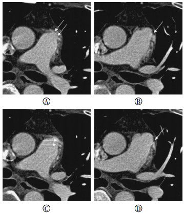
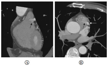
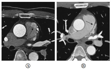
CPF的MSCTCA影像表现:①异常血管起源:左冠状动脉分支或左圆锥支11例,左冠状动脉分支及右圆锥支(或左右圆锥支)7例,右圆锥支1例,双侧冠状动脉多分支1例。②异常分支血管数目:1支冠状动脉分支起源异常12例,2支异常7例(图 1),2支以上异常1例。③异常血管走行与形态:纡曲扩张血管团19例,单纯扩张血管1例,匍匐于肺动脉壁20例(图 1中A);合并囊状动脉瘤5例,梭形动脉瘤1例。④瘘口的位置、数目和大小:主肺动脉左侧壁14例(图 1~4),左前壁1例,前壁2例,左侧壁和前壁1例,左前壁和右侧壁1例,未见明确瘘口1例。有1个瘘口者11例,有2个瘘口者5例,有3个瘘口者2例,有6个瘘口者1例,筛孔状瘘口1例。可测量瘘口33个,大小0.8~5.2 mm,平均(2.36±1.13)mm。⑤增强扫描瘘口的CT表现:冠状动脉异常血管与肺动脉间管壁缺损(开窗征)17例(图 2),瘘口肺动脉侧对比剂呈喷射状流入(射血征)8例(图 3),呈烟雾状弥漫(烟雾征)3例(图 4中A),呈带状紧贴肺动脉内侧壁(飘带征)1例(图 4中B),瘘口肺动脉侧与异常血管管腔密度相当(等密度征)10例(图 2)。⑥肺动脉粗细:与升主动脉管径比较,肺动脉轻度增粗1例。

图 1 容积再现示冠状动脉-肺动脉瘘的迂曲扩张血管
Figure 1. Volume rendering showed tortuous dilatation of blood vessels of coronary artery-pulmonary fistula

图 2 冠状动脉-肺动脉瘘多发瘘口患者的CT图像
Figure 2. CT images of multiple fistula in a patient with coronary artery-pulmonary fistula

图 3 冠状动脉-肺动脉瘘的曲面重组、最大密度投影CT图像
Figure 3. CT images showed curved planar reconstruction and maximum intensity projection of coronary artery-pulmonary fistula

图 4 冠状动脉-肺动脉瘘患者主肺动脉内对比剂造影的CT图像
Figure 4. CT images showed contrast media into the main pulmonary artery of coronary artery-pulmonary fistula
-
CPF形成的原因多为先天性,少数为后天性[1],在临床上绝大部分为先天性,是由于胚胎期心肌中血管窦间隙的发育异常所致。CPF是冠状动脉瘘的最常见类型,文献报道CPF占冠状动脉瘘的50%~90.2%[2-4],占MSCTCA检查的0.15%~0.44%[3, 5-6],其检出率高低不等,推测与入选研究的人群有关,不同的医院重点收治心脏疾病的患者不同,可能会造成筛选的偏移。
CPF临床表现缺乏特异性,其症状、体征等临床表现与瘘口大小、病史长短所致的血流动力学改变有关,瘘口小、分流量少时,可出现不典型轻度胸闷、胸痛或无明显症状,甚至体检时才发现;当瘘口大或合并异常血管扩张、动脉瘤形成,导致窃血量大,尤其在舒张期,更易导致心肌缺血[6],产生症状的年龄一般在18~20岁[7],而中老年人易误诊为冠心病。本研究中,40岁以上患者占85%(17/20),除1例为体检发现外,临床表现以胸闷、胸痛、心前区不适为主,大多数拟诊为冠心病,所以临床上如果能够通过方便、快捷、无创的方法早期发现和诊断,将对治疗CPF具有重要的指导意义。
-
MSCTCA不仅可清楚显示异常血管的起源、走行、形态,明确其与肺动脉的解剖关系,而且结合MPR等技术,绝大多数可以明确瘘口位置、数目和大小。VR可直观、立体、准确显示异常血管网,明确是否合并动脉瘤或瘤样扩张,但不能显示瘘口情况。本研究中的病例异常血管起源主要由左冠状动脉分支或左圆锥支参与,而右侧冠状动脉参与较少,这与文献报道的右冠状动脉参与较多是不同的[8],推测原因可能与筛查人群或统计病例源不同有关。MPR图像效果有赖于操作者的技巧,对于瘘口的显示最好,可以测量其大小、显示管腔情况,但对于走行纡曲的血管显示能力有限,结合MIP可以在一定程度上显示扭曲血管的管腔和解剖位置。CPR虽然能拉直血管,显示管壁、管腔,但对于扭曲血管的解剖定位显示欠佳。因此,显示CPF最好的图像后处理技术是VR、MPR、MIP。MSCT对诊断CPF虽然有较高的准确性,但也存在不足,首先难于显示较小瘘口,特别是筛孔状瘘口;其次显示瘘口与适当的扫描时间、对比剂注射时间长短、肺动脉对比剂排空快慢等因素有关,只有在适当扫描时间上,瘘口肺动脉侧对比剂基本排完,冠状动脉侧压力较高,对比剂较浓,才可见对比剂漏入肺动脉内,否则除非直接显示管壁缺损口外CT只能诊断血管异常;再者MSCT对患者心率、呼吸要求较高,心率较快或心律不齐患者重组图像往往显示不佳,也可能出现漏诊,特别是瘘血管纤细、瘘口较小时漏诊可能性较大。
-
CPF的CT征象与畸形血管团的形态、范围、瘘口的大小、CT扫描的时间等有关,VR可立体、直观显示异常血管网,明确异常血管与周围结构的三维关系,CT表现为血管走行纡曲、扩张,异常血管网沿主肺动脉壁爬行,称之为附壁征。本研究中的病例100%显示扩张纡曲血管团和附壁征,对CPF具有高度的提示作用,可作为拟诊CPF的首要征象。轴位图像与MPR可发现瘘口,瘘口多位于主肺动脉左侧壁,并可测量大小,本研究中仅有1例未能确定瘘口,可能因瘘口小而不能显示。瘘口在MPR上表现为管壁中断,两边血流直接相通,在不同的扫描时间表现为瘘口两边对比剂的密度、压力不同,故CT上可以出现不同的特征性表现:开窗征、射血征、烟雾征、飘带征、等密度征。①开窗征:瘘的最直接征象,MPR重组位置恰当,表现为管壁中断,可以直接测量两端的距离,此征象在本研究病例中占95%(19/20)。②射血征:在适当扫描时间上瘘口肺动脉侧对比剂基本排完,冠状动脉侧压力较高,对比剂较浓,通过瘘口时表现为水柱状或喷泉状射向肺动脉侧,多为瘘口较小。此征象对诊断CPF具有特征性,在本研究病例中占40%(8/20)。③烟雾征:表现为瘘口的肺动脉侧见漏出对比剂呈烟雾状弥散,其机理可能基于扫描时间上肺动脉对比剂量较冠状动脉明显减少或排完,并且瘘口较大或畸形血管团较粗大,冠状动脉侧血流压力不太高,瘘入肺动脉的对比剂流速较多、较慢,被肺动脉血冲散所致。此征象在本研究病例中仅占15%(3/20)。④飘带征:表现为无明显对比剂的肺动脉管壁内侧见带状对比剂影,管壁外侧见畸形血管团,其机理可能为瘘口多而细,呈筛孔状,瘘入对比剂被肺动脉血流局限于管壁旁,轴位显示呈条状高密度,冠状动脉造影可显示漏入肺动脉的对比剂。本研究中仅见1例,占5%(1/20)。⑤等密度征:主要与扫描时间相对过早、对比剂注射时间过长或肺动脉对比剂排空慢等因素有关,表现为瘘口两侧血液密度相当,肉眼无法分辨,只能靠管壁低密度线中断而诊断。本研究病例50%(10/20)显示此征。以上各征象显示率与文献报道[5]相近,本研究病例新发现飘带征,其中开窗征、射血征、烟雾征、飘带征对诊断CPF具有一定的特征性。
-
CPF的诊断方法有常规冠状动脉血管造影、超声心动图、MSCTCA、磁共振血管成像等,常规冠状动脉造影是诊断CPF的“金标准”,能准确显示CPF起源血管、形态、瘘口位置和分流程度,并作出适当治疗,但其属于有创性,也缺乏三维空间结构的显示,对于较小的瘘口较难诊断,所以越来越多的无创检查手段正部分取代造影检查[9]。超声心动图无创、方便,可重复性强,对显示血流动力学改变有较大优势,但其容易受操作者的经验技术、分辨率、胸廓骨和肺气影响,而且对于分流量较小者容易漏诊,文献报道超声对瘘口的漏诊误诊率达52.6%[10],本研究CPF病例中同时行超声心动图检查者15例,结果漏诊11例,漏诊率达73.3%(11/15)。随着MSCT的时间、空间分辨力的提高使得MSCTCA成为一种常规检查手段,而且通过后处理技术能够清晰准确地显示冠状动脉的解剖结构、与周围组织的空间联系和病理改变,有助于CPF瘘口的显示,甚至可通过选取恰当的扫描时间和后处理技术发现较小瘘口或拟诊筛孔样瘘口的存在,对于临床的诊治具有极大的应用价值。
综上所述,MSCTCA作为无创、简便、准确、安全、可重复性强的影像学检查方法,可清晰显示CPF独特的影像特征,具有极大临床应用价值,甚至可作为CPF的首选检查方法。
64排螺旋CT冠状动脉血管成像诊断冠状动脉-肺动脉瘘的应用价值
Clinical value of coronary artery-pulmonary artery fistula revealed by 64-slice spiral CT coronary angiography
-
摘要:
目的 评价64排螺旋CT三维重组技术诊断冠状动脉-肺动脉瘘(CPF)的应用价值。 方法 回顾性分析20例行64排螺旋CT冠状动脉血管成像的CPF患者的资料, 其图像行容积再现、多平面重组、曲面重组、最大密度投影, 分析CPF的检出率、异常血管的CT特征及多排螺旋CT的诊断意义。 结果 CPF血管起源:左冠状动脉分支或左圆锥支11例, 左冠状动脉分支及右圆锥支(或左右圆锥支)7例, 右圆锥支1例, 双侧冠状动脉多分支1例。异常血管形态:迂曲扩张血管网19例, 单纯扩张血管1例, 匍匐于肺动脉壁20例; 合并囊状动脉瘤5例, 梭形动脉瘤1例。肺动脉瘘口的位置:主肺动脉左侧壁14例, 左前壁1例, 前壁2例, 左侧壁和前壁1例, 左前壁和右侧壁1例, 未见明确瘘口1例。瘘口的CT特征:开窗征17例, 射血征8例, 烟雾征3例, 飘带征1例, 等密度征10例。 结论 多排螺旋CT重组技术可清晰显示CPF的异常血管及瘘口特征, 为临床诊治提供重要信息, 可作为诊断的首选方法。 -
关键词:
- 冠状动脉疾病 /
- 肺动脉 /
- 体层摄影术, X线计算机 /
- 动脉-动脉漏
Abstract:Objective To evaluate the value of the three dimensional multi-slice spiral CT in the diagnosis of coronary artery-pulmonary artery fistula(CPF). Methods Retrospectively analyzed the multi-slice spiral CT images of 20 CPF patients diagnosed by coronary CT angiography.All of the cases were used in volume rendering, multiplanar reformation, curved planar reformation, and maximum intensity projection.Moreover, the incidence, the features of the abnormal vessels revealed by multi-slice spiral CT, and the diagnostic significance of CPF were retrospectively summarized. Results The origin of CPF vessels was the branch of left coronary artery or left conus branch in 11 cases, the branch of left coronary artery and right conus branch in 7 cases, the right conus branch in 1 case, and the multi-branch of left and right coronary artery in 1 case.The abnormal vessels demonstrated a tortuous dilatation in 19 cases and a tubular dilatation in 1 case.CPF extended into the pulmonary artery wall in 20 cases; companied with aneurysm in 5 cases and fusiform aneurysm in 1 case.Moreover, the pulmonary artery fistulas were located in the left wall of the main pulmonary artery in 14 cases, in the left anterior wall in 1 case, in the anterior wall in 2 cases, in the left and anterior wall in 1 case, and in the anterior and right wall in 1 case; pulmonary artery fistulas were absent in 1 case.The CT features of the pulmonary artery fistula concluded a window sign in 17 cases, an ejection sign in 8 cases, a smoke sign in 3 cases, a streamer sign in 1 case, and an isodensity sign in 10 cases. Conclusion The 3D-MSCT can clearly visualize abnormal vessels and fistula.Thus, this method can be considered as the primary choice in CPF diagnosis and treatment. -
图 1 容积再现示冠状动脉-肺动脉瘘的迂曲扩张血管
Figure 1. Volume rendering showed tortuous dilatation of blood vessels of coronary artery-pulmonary fistula
图 2 冠状动脉-肺动脉瘘多发瘘口患者的CT图像
Figure 2. CT images of multiple fistula in a patient with coronary artery-pulmonary fistula
图 3 冠状动脉-肺动脉瘘的曲面重组、最大密度投影CT图像
Figure 3. CT images showed curved planar reconstruction and maximum intensity projection of coronary artery-pulmonary fistula
-
[1] 孔淑红, 岳学旺, 孔生.冠状动脉瘘的CT诊断价值[J].中国临床医学影像杂志, 2015, 26(2):134-136.
Kong SH, Yue XW, Kong S.Value of CT in the diagnosis of coronary artery fistula[J].J Chin Clin Med Imaging, 2015, 26(2):134-136.[2] 周长圣, 张龙江, 王怡宁, 等.双源CT冠状动脉成像冠状动脉瘘的检出率及分型诊断[J].中华医学杂志, 2012, 92(47):3336-3340.DOI:10.3760/cma.j.issn.0376-2491.2012.47.008.
Zhou CS, Zhang LJ, Wang YN, et al.Incidence and classification diagnosis of coronary artery fistula at dual-source CT coronary angiography[J].Natl Med J Chin, 2012, 92(47):3336-3340. doi: 10.3760/cma.j.issn.0376-2491.2012.47.008[3] Yun H, Zeng MS, Yang S, et al.Congenital coronary artery fistulas:dual-source CT findings from consecutive 6624 patients with suspected or confirmed coronary artery disease[J].Chin Med J, 2011, 124(24):4172-4177. [4] Abdelmoneim SS, Mookadam F, Moustafa S, et al.Coronary artery fistula:single-center experience spanning 17 years[J].J Interv Cardiol, 2007, 20(4):265-274.DOI:10.1111/j.1540-8183.2007.00267.x. [5] 李景雷, 黄美萍, 崔燕海, 等.冠状动脉-肺动脉瘘多排螺旋CT征象分析[J].暨南大学学报:自然科学与医学版, 2013, 34(2):221-225.DOI:10.3969/j.issn.1000-9965.2013.02.021.
Li JL, Huang MP, Cui YH, et al.The analysis of CT features of coronary artery-pulmonary artery fistula[J].Jinan Univ Natur Sci & Med, 2013, 34(2):221-225. doi: 10.3969/j.issn.1000-9965.2013.02.021[6] 曹志坚, 许茂盛, 陈智能, 等.冠状动脉瘤样扩张征象在CT诊断冠状动脉瘘中的意义[J].医学影像学杂志, 2015, 25(4):625-627.
Cao ZJ, Xu MS, Chen ZN, et al.The significance of aneurismal dilatation in diagnosing coronary artery fistula by CT angiography[J].J Med Imaging, 2015, 25(4):625-627.[7] 杨姗, 曾蒙苏, 恽虹, 等.冠状动脉瘘的CT血管成像表现[J].中华心血管病杂志, 2011, 39(8):739-742.DOI:10.3760/cma.j.issn.0253-3758.2011.08.012.
Yang S, Zeng MS, Yun H, et al.Imaging feature of computed tomography angiography in patients with coronary artery fistulas[J].Chin J Cardiol, 2011, 39(8):739-742. doi: 10.3760/cma.j.issn.0253-3758.2011.08.012[8] Kim SY, Seo JB, Do KH, et al.Coronary artery anomalies:Classification and ECG-gated multi-detector row CT findings with angiographic correlation[J].Radiographics, 2006, 26(2):317-334.DOI:http://dx.doi.org/10.1148/rg.262055068. [9] Woo JJ, Koh YY, Ha SI, et al.Non-invasive recognition of generalized coronary arteriosystemic fistulae by contrast echocardiography and multidetector CT[J].Int J Cardiovasc Imaging, 2011, 27(5):749-753.DOI:10.1007/s10554-011-9866-6. [10] 于喜红, 杨瑞, 刘继伟.双源CT结合超声对小儿先天性冠状动脉瘘的诊断价值[J].中国医学计算机成像杂志, 2015, 21(1):38-42.
Yu XH, Yang R, Liu JW.Value of dual-source computed tomography coronary angiography and echocardiography in diagnosis of congenital coronary artery fistula in children[J].Chin Comput Med Imaging, 2015, 21(1):38-42. -

 点击查看大图

图(4)
计量
- 文章访问数: 2573
- HTML全文浏览量: 1457
- PDF下载量: 3