


-
上肢深静脉血栓(upper extremity deep venous thrombosis,UEDVT)可分为特发性和受挫性血栓形成。解剖及外力因素在受挫性血栓形成中起着重要的作用,而导致特发性血栓的原因目前仍不清楚[1]。近年来,UEDVT的发病率有上升趋势,UEDVT约占所有深静脉血栓(deep venous thrombosis,DVT)事件的1%~4%[2]。国外UEDVT的发病人数占住院人数的0.002%~0.007%[3]。未及时治疗的UEDVT可导致致命性的肺动脉血栓栓塞等并发症,其发生率高达36%[4]。近50%的患者缺乏DVT的典型症状和体征,因此,单凭临床症状诊断DVT常常较困难[5]。本研究旨在探讨多层螺旋CT(multislice spiral CT,MSCT)成像技术在上肢CT静脉血管成像(CT venography,CTV)中的应用,以期为临床确诊UEDVT提供客观依据。
-
收集我院2010年6月至2012年3月行MSCT上肢静脉检查的患者56例,其中,男性32例、女性24例,年龄35~66岁,中位年龄45岁。临床症状包括:41例患者患侧上肢肿胀疼痛,12例患者患侧上肢皮肤有青紫色改变,1例患者伴发轻微的胸闷、胸痛,2例患者可见上肢浅静脉曲张,12例患者上肢骨折。
-
对比剂采用欧乃派克[Omnipaque,通用电气药业(上海)有限公司,350 mg I/ml]35 ml,与0.9%生理盐水65 ml混合成100 ml混合液。自患侧手背静脉注入,注药流率3 ml/s,延迟时间为25 s。CT扫描采用美国GE公司Lightspeed 16螺旋CT扫描仪。LF9000高压注射器为美国LF公司产品。
-
采用MSCT上肢静脉直接成像技术,患者取仰卧位,头先进,将患侧上肢上举,掌心向上放于检查床正中,使上肢冠状面与检查床平行,上肢的正中矢状线与指示灯纵轴线重合,基准线定于肱骨内外髁连线。上肢活动受限者可将患侧上肢放于身体旁,尽量稍外展。
-
管电压120 kV,管电流200~250 mA。采用螺旋扫描,扫描层厚5 mm,螺距1.675:1,进床速度16.75 mm/s,扫描时间(15±3)s。扫描方向为顺静脉血流方向扫描。扫描范围自掌骨近端至主动脉弓水平。
-
图像在美国GE AW4.3工作站进行处理,对上肢静脉血管采用容积再现(volume rendering,VR)、最大密度投影(maximum intensity projection,MIP)、多平面重组(multi planner reformation,MPR)、曲面重建(curve planner reformation,CPR)等多种方式后处理。
-
① 上肢深静脉血管全程显影;②测量位置深静脉血管轴位管腔内密度均匀;③测量位置深静脉血管轴位管腔周边无伪影;④被测量深静脉血管管腔内对比剂浓度可以满足图像后处理的需要。
-
分别在A、B、C 3个层面测量轴位原始图像静脉管腔内的CT值:①A点:肱骨内外髁连线向近心侧5 cm水平测量头静脉和肱静脉管腔内CT值。②B点:肱骨内外髁连线向近心侧8 cm水平测量头静脉和肱静脉管腔内CT值。③C点:锁骨下静脉管腔内CT值。
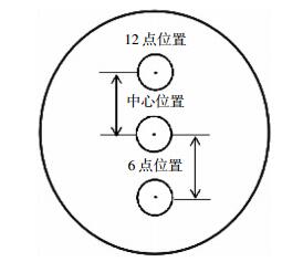
静脉CT值测量:将测量标记ROI分别置于选定测量层面血管腔内的3个位置进行测量,包括中心点位置及垂直方向上腹侧(12点方向)和背侧(6点方向)距中心点等距位置的CT值(图 1)。
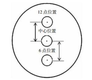
图 1 静脉管腔内3个测量位置的示意图
Figure 1. Schematic diagram of 3 selected location within the venous lumen
-
采用SPSS 17.0统计软件包,对上肢静脉选定层面的静脉管腔内测量的数据采用单因素方差分析,以P < 0.05表示差异有统计学意义。
-
56例患者均成功完成MSCT上肢静脉直接成像检查,其中,右上肢26例,左上肢30例。影像学表现为上肢静脉自掌骨近心端至上腔静脉全程深、浅静脉主干显影良好,充盈饱满,管腔内密度均匀,无充盈缺损及狭窄且管腔周围无伪影产生。
-
56例完成MSCT上肢静脉直接成像技术的患者中,右上肢发现病变14例,左上肢21例,阳性率为62.5%(35/56)。血栓累及部位单发于锁骨下静脉9例、腋静脉6例、肱静脉3例,同时累及多条上肢静脉17例(图 2)。
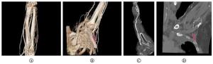
图 2 上肢深静脉血栓的MSCT图像 图中,A:上肢静脉VR图示正常尺、桡静脉及其属支;B:上肢静脉VR图示左锁骨下静脉栓塞(红色箭头);C:上肢静脉MPR图示肱静脉、尺静脉全程;D:MPR图示左锁骨下静脉栓塞(红色箭头)。MSCT:多层螺旋CT;VR:容积再现;MPR:多平面重组。
Figure 2. MSCT images of upper extremity deep vein
-
56例患者同期行彩色多普勒超声检查,右上肢发现病变14例,左上肢19例,阳性率为58.9%(33/56)。血栓累及部位单发于锁骨下静脉8例、腋静脉6例、肱静脉4例,同时累及多条上肢静脉15例(表 1)。
上肢深静脉 CTV CDUS 锁骨下静脉 9(16.07) 8(14.29) 锁骨下、腋、肱静脉 8(14.29) 6(10.71) 锁骨下、肱静脉 0 1(1.79) 腋静脉 6(10.71) 6(10.71) 腋、肱静脉 9(16.07) 8(14.29) 肱静脉 3(5.35) 4(7.14) 表中,CTV:CT静脉血管成像;CDUS:彩色多普勒超声。 表 1 CTV和CDUS检查56例上肢深静脉受累情况[例数(%)]
Table 1. The affected situation of upper extremity deep vein of 56 cases using CT and CDUS examination[n(%)]
-
由表 2中数据得出,肱骨内外髁连线5 cm处各测量位置中静脉管腔内CT值的差异无统计学意义(肱静脉管腔:F=0.004,P>0.05;头静脉管腔:F=0.102,P>0.05)。肱骨内外髁连线8 cm处各测量位置中静脉管腔内CT值的差异无统计学意义(肱静脉管腔:F=0.007,P>0.05;头静脉管腔:F=2.271,P>0.05)。锁骨下静脉层面各测量点之间的CT值差异无统计学意义(F=0.001,P>0.05)。统计结果提示顺静脉血管方向不同位置静脉管腔内对比剂充盈均匀。
测量位置 肱静脉5 cm 头静脉5 cm 肱静脉8 cm 头静脉8 cm 锁骨下静脉 12点位置 797.9±15.49 795.3±31.31 653.2±33.46 673.5±29.90 457.9±30.42 中心位置 797.5±17.81 799.7±35.56 652.4±30.64 656.2±30.93 457.8±30.35 6点位置 797.5±18.00 799.1±35.59 652.0±30.73 655.9±31.64 458.2±30.28 F值 0.004 0.102 0.007 2.271 0.001 P值 > 0.05 > 0.05 > 0.05 > 0.05 > 0.05 表 2 上肢深、浅静脉不同水平CT值测量结果(HU)
Table 2. CT values at different levels of upper extremity deep and shallow vein(HU)
-
由表 2数据分析得出,上肢静脉远端主干管腔内显像剂浓度高于近心端管腔内显像剂浓度,各层面之间CT值的差异有统计学意义(F=1441.52,P < 0.05)。提示顺静脉血流方向显像剂浓度逐渐减低。
-
UEDVT形成曾被认为是罕见并呈良性过程的疾病。1970年后由于使用经静脉起搏和留置中心静脉导管的逐渐增多,UEDVT的发生率也呈逐渐增高的趋势[6]。创伤骨折后患肢因疼痛和制动造成静脉流速缓慢,组织创伤、肿胀和炎症浸润也阻碍了静脉回流,手术后血液高凝状态又增加了静脉血栓的风险[7]。创伤骨折致下肢DVT形成已有很多文献报道[8],肱骨段以上骨折或多发上肢骨折患者,亦有并发DVT风险,对这类患者行骨折切开复位内固定术前评估上肢深静脉通畅性,对于降低麻醉、手术风险,防止致命性肺栓塞有着重要的参考意义[9]。UEDVT可根据解剖位置进行分类,其中最常发生血栓的部位为腋静脉或锁骨下静脉。
-
MSCT上肢静脉检查技术可以快速、准确地完成CTV。扫描范围自手背静脉网至上腔静脉可一次完成,能很好地观察肱静脉及上肢深、浅静脉血管的连续性。结合多种图像后处理技术,包括MPR、CPR和VR技术等,可以多方位、多模式地显示上肢静脉血管。其中,VR技术可以同时显示解剖参照结构的影像,如肱骨、锁骨等,对外科的术前准备有重要意义。VR成像时血管的重建完全依赖管腔内对比剂的充盈度,若注入血管内对比剂浓度过低,则不能反映血管管腔内的真实情况;若注入血管内对比剂浓度过高,不仅会产生伪影影响VR显示,而且会产生“边流现象”造成鉴别诊断的困难。文献[10]研究者和笔者根据经验将对比剂采用欧乃派克35 ml、0.9%生理盐水65 ml混合成100 ml的混合液进行注药,不仅维持了血管内有效的对比剂浓度,而且降低了对比剂过敏的风险。将扫描后的图像应用VR和MIP技术处理,血管显影良好,使用MSCT上肢静脉成像技术能反映血管腔内的真实情况。在三维重组的同时仍需要兼顾横轴位图像的仔细观察。
-
血管彩色多普勒超声检查具有无创、方便快捷、不需要对比剂、价格低廉的特点,可作为上肢静脉疾病检查的首选筛查方法。但是,多普勒超声诊断的准确性也受患者的身体状况干扰,如骨创伤后患肢肿胀、疼痛及强迫体位,超声探头不能压迫受伤的上肢;当被观察的血管周围有重叠的胸廓骨骼阻挡,受血流声束夹角大小和仪器灵敏度等因素影响,也会对静脉血栓诊断造成困难,特别是多普勒超声对锁骨下静脉的观察就存在一定的局限性。而随着MSCT技术的发展,MSCT血管成像检查技术已能满足显示小血管病变的要求,相对于数字减影血管造影更加安全方便、无创伤性,因此越来越多地应用于血管病变的诊断。MSCT上肢静脉直接成像具有扫描时间短、图像密度分辨率高、无操作者依赖性等特点,可有效避免骨骼重叠的影响。对比多普勒超声检查技术,MSCT上肢静脉直接成像对DVT诊断的灵敏度、特异度及准确性均较高,重组图像可直接显示病变部位、范围、程度及侧支循环等情况。本研究结果表明,MSCT上肢静脉直接成像技术对锁骨下静脉、腋静脉血栓的显示率高于彩色多普勒超声检查技术,特别是对骨创伤、上肢制动患者可在短时间内完成大范围扫描,清晰显示DVT部位、范围,避免患者体位、骨骼及周围软组织对观察深静脉的干扰[11],并可同时观察周围骨质情况。因此,可提高骨创伤患者鉴别诊断的准确性,为临床术中或术后治疗方案的制定提供依据。
本研究结果提示上肢不同层面各点间对比剂充盈均匀,自肱静脉及头静脉的静脉远端至近心端的3个测量层面中,管腔内CT值的差异无统计学意义。由此可以看出静脉各层面血管显像剂充盈均匀,说明使用该检查技术能够实现上肢深、浅静脉的全程显示。之所以选择血管层面的中心位置及距中心位置12点方向和6点方向等距位置为中心的测量点,原因是:(1)避免因对比剂与深静脉内血液发生的“边流现象”而影响鉴别诊断。(2)上肢不同层面血管各层面之间CT值的差异有统计学意义,由此可以看出上肢静脉主干血管内对比剂被回心血液稀释,各层面之间有差异,顺静脉血流方向CT值逐渐减低。
综上所述,MSCT上肢静脉直接成像的优势在于可以较大范围显示上肢静脉全程,清晰显示侧支循环,明确血管狭窄、阻塞的原因,了解周围结构情况,辨别血栓形成时间等。该技术相对安全方便、创伤小,减少了碘对比剂用量,减轻了患者的经济负担。应用该技术能提高上肢静脉血栓的诊断率,为临床选择适当的治疗方法提供了可靠的依据。特别是对上肢创伤骨折后不能采用超声探头压迫检查的患者,以及对怀疑锁骨下静脉栓塞的患者,均推荐采用MSCT上肢静脉直接成像检查。
MSCT上肢静脉直接成像技术的应用
The application of MSCT in upper limb vein direct imaging
-
摘要:
目的 探讨多层螺旋CT(MSCT)成像技术在上肢CT静脉血管成像中的诊断价值。 方法 回顾性分析56例行上肢CT静脉血管成像患者的检查资料, 并对上肢静脉血管采用容积再现、最大密度投影、多平面重组、曲面重建等多种方式后处理, 选定A、B、C(肱骨内外髁连线向近心侧5 cm处头静脉和肱静脉的轴位图像;肱骨内外髁连线向近心侧8 cm处头静脉和肱静脉的轴位图像;锁骨下静脉的轴位图像)层面分别测量管腔内CT值。测量数据采用SPSS 17.0统计软件包分析, 对上肢静脉选定层面的静脉管腔内测量的数据采用单因素方差分析, 以P < 0.05表示差异有统计学意义。 结果 56例完成MSCT上肢静脉直接成像的患者中, 右上肢发现病变14例, 左上肢21例, 阳性率为62.5%(35/56), 同期行彩色多普勒超声检查, 阳性率为58.9%(33/56)。肱骨内外髁连线5 cm处各测量位置中静脉管腔内CT值的差异无统计学意义(肱静脉管腔:F=0.004, P>0.05;头静脉管腔:F=0.102, P>0.05)。肱骨内外髁连线8 cm处各测量位置中静脉管腔内CT值的差异无统计学意义(肱静脉管腔:F=0.007, P>0.05;头静脉管腔:F=2.271, P>0.05)。锁骨下静脉层面各测量点之间的CT值差异无统计学意义(F=0.001, P>0.05)。统计结果提示顺静脉血管方向不同位置静脉管腔内对比剂充盈均匀。上肢静脉远端主干管腔内对比剂浓度高于近心端管腔内对比剂浓度, 各层面之间CT值的差异有统计学意义(F=1441.52, P < 0.05)。提示顺静脉血流方向对比剂浓度逐渐减低。 结论 MSCT上肢静脉直接成像的优势在于可以较大范围显示上肢静脉全程, 清晰显示侧支循环, 明确血管狭窄、阻塞的原因, 了解周围结构情况, 辨别血栓形成时间等。该技术相对安全方便、创伤小, 减少了碘对比剂用量, 减轻了患者的经济负担。应用该技术能提高上肢静脉血栓的诊断率, 为临床选择适当的治疗方法提供了可靠的依据。 -
关键词:
- 静脉血栓形成 /
- 体层摄影术, 螺旋计算机 /
- 血管造影术
Abstract:Objective To discuss the diagnostic value of multi-slice computed tomography(MSCT) as an imaging technique used for upper vein angiography. Methods A total of 56 cases were referred for upper limb vein check by MSCT from June 2010 to March 2012. The veins were scanned by using the LightSpeed 16 computed tomography(CT) scanner and reconstructed by using volume rendering, maximum intensity projection, multi-planner formation, and curve planner reformation. Three different layers were selected to measure the CT value of A, B, and C(i.e., the axial image of the cephalic and brachial veins between condyles of the humerus at 5 and 8 cm and the axial image of the subclavian vein). Statistical analysis was conducted by using SPSS 17.0, whereas one-way ANOVA F test was used to analyze the measured CT value(P < 0.05 was considered statistically significant). Results Out of 56 patients, 14 cases of right upper limb and 21 cases of left upper limb were detected to have deep vein thrombosis. That is, 62.5%(35/56) of the cases were detected by MSCT as compared with 58.9%(33/56) of the cases detected by color Doppler. The CT value of the axial image of the brachial and cephalic veins between condyles of the humerus approximately 5 and 8 cm near the heart was P>0.05, whereas that of the axial image of the subclavian vein was P>0.05(F=0.001). In different layers of vein filled with contrast medium, the CT values were uniform and the differences of the values exhibited no statistical significance between different layers. The contrast medium concentration in the distal part of the upper limb vein was higher than that near the heart, and the differences of the CT values exhibited statistical significance between different layers(F=1441.52, P < 0.05). Conclusions In conclusion, the MSCT is a superior technology for upper limb vein imaging because it is able to show the upper limb in an extensive range, display the collateral circulation clearly, identify the causes of hemadostenosis, and predict the occurrence of thrombogenesis. Simultaneously, this technology significantly reduces the dosage of iodine contrast agent, lowers the economic burden of patients, improves the rate of correct diagnosis for lesions, and provides reliable bases for selecting correct methods of treatment in clinics. -
Key words:
- Venous thrombosis /
- Tomography, spiral computed /
- Angiography
-
图 1 静脉管腔内3个测量位置的示意图
Figure 1. Schematic diagram of 3 selected location within the venous lumen
图 2 上肢深静脉血栓的MSCT图像 图中,A:上肢静脉VR图示正常尺、桡静脉及其属支;B:上肢静脉VR图示左锁骨下静脉栓塞(红色箭头);C:上肢静脉MPR图示肱静脉、尺静脉全程;D:MPR图示左锁骨下静脉栓塞(红色箭头)。MSCT:多层螺旋CT;VR:容积再现;MPR:多平面重组。
Figure 2. MSCT images of upper extremity deep vein
表 1 CTV和CDUS检查56例上肢深静脉受累情况[例数(%)]
Table 1. The affected situation of upper extremity deep vein of 56 cases using CT and CDUS examination[n(%)]
上肢深静脉 CTV CDUS 锁骨下静脉 9(16.07) 8(14.29) 锁骨下、腋、肱静脉 8(14.29) 6(10.71) 锁骨下、肱静脉 0 1(1.79) 腋静脉 6(10.71) 6(10.71) 腋、肱静脉 9(16.07) 8(14.29) 肱静脉 3(5.35) 4(7.14) 表中,CTV:CT静脉血管成像;CDUS:彩色多普勒超声。  下载: 导出CSV
表 2 上肢深、浅静脉不同水平CT值测量结果(HU)
Table 2. CT values at different levels of upper extremity deep and shallow vein(HU)
测量位置 肱静脉5 cm 头静脉5 cm 肱静脉8 cm 头静脉8 cm 锁骨下静脉 12点位置 797.9±15.49 795.3±31.31 653.2±33.46 673.5±29.90 457.9±30.42 中心位置 797.5±17.81 799.7±35.56 652.4±30.64 656.2±30.93 457.8±30.35 6点位置 797.5±18.00 799.1±35.59 652.0±30.73 655.9±31.64 458.2±30.28 F值 0.004 0.102 0.007 2.271 0.001 P值 > 0.05 > 0.05 > 0.05 > 0.05 > 0.05
下载: 导出CSV
-
[1] Arnhjort T, Nordberg J, Delle M, et al. The importance of the costoclavicular space in upper limb primary deep vein thrombosis, a study with magnetic resonance imaging(MRI) technique enhanced by a blood pool agent[J]. Eur J Intern Med, 2014, 25(6):545-549. doi: 10.1016/j.ejim.2014.05.005 [2] Kommareddy A, Zaroukian MH, Hassouna HI. Upper extremity deep venous thrombosis[J]. Semin Thromb Hemost, 2002, 28(1):89-99. [3] 陈平, 袁庆文.上肢深静脉血栓形成的诊治体会[J].实用临床医学, 2010, 11(10):30-31. doi: 10.3969/j.issn.1009-8194.2010.10.014
[4] Prandoni P, Polistena P, Bernardi E, et al. Upper-extremity deep vein thrombosis. Risk factors, diagnosis, and complications[J]. Arch Intern Med, 1997, 157(1):57-62. doi: 10.1001/archinte.1997.00440220061008 [5] 蔡柏蔷.提高对深静脉血栓形成的认识[J].中华内科杂志, 2000, 39(8):4-5.
[6] Sabharwal R, Boshell D, Vladica P. Multidetector spiral CT venography in the diagnosis of upper extremity deep venous thrombosis[J]. Australas Radiol, 2007, 51 Suppl:SB253-B256. [7] Marshall PS, Cain H. Upper extremity deep vein thrombosis[J]. Clin Chest Med, 2010, 31(4):783-797. doi: 10.1016/j.ccm.2010.06.005 [8] Mai C, Hunt D. Upper-extremity deep venous thrombosis:a review[J]. Am J Med, 2011, 124(5):402-407. doi: 10.1016/j.amjmed.2010.11.022 [9] 李冬妹, 区锦燕, 刘晓捷.上肢创伤骨折与深静脉血栓的关系[J].当代医学, 2012, 18(18):38-39. doi: 10.3969/j.issn.1009-4393.2012.18.023
[10] 傅菲, 李宝玖, 张越, 等.表浅静脉不同结扎法对MSCT下肢深静脉显示效果的研究[J].临床放射学杂志, 2013, 32(8):1138-1141.
[11] 谢振鹰, 张应和.多层螺旋CT血管造影对后胡桃夹综合征的诊断价值[J].国际放射医学核医学杂志, 2011, 35(4):253-255. doi: 10.3760/cma.j.issn.1673-4114.2011.04.015
-

 点击查看大图
计量
- 文章访问数: 2450
- HTML全文浏览量: 1163
- PDF下载量: 3