


-
大剂量电离辐射照射后,机体会产生一系列复杂的损伤效应,称为急性辐射综合征,症状可持续长达数个月。造血组织在机体内承担着重要的生理功能,一旦其功能低下或缺失,将产生严重后果,甚至导致机体死亡[1]。造血组织对辐射高度敏感,辐射后造血系统损伤是限制临床放疗照射剂量的最常见不良反应[2]。本研究采用6 Gy 137Cs γ射线对小鼠进行一次性全身照射,观察辐射损伤后小鼠的体质量、外周血象、外周血网织红细胞百分率和造血祖细胞增殖能力的变化,为造血系统辐射损伤研究提供基础数据。
-
C57BL/6小鼠70只,雄性,无特定病原体级,8周龄,体质量18~22 g,由北京维通利华实验动物有限公司提供,合格证号:SCXK(京)2009-0017,由中国医学科学院生物医学工程研究所动物实验中心提供无特定病原体级条件饲养。饲料为清洁级全价鼠料,由北京科奥协力饲料有限公司提供。
-
137Cs γ射线照射源(型号USD,Autocell40)购自加拿大原子能有限公司(剂量率为1.01 Gy/min);Accuri C6流式细胞仪为美国BD公司产品;MEK-T222K全自动血液分析仪为日本光电工业株式会社产品。甲基纤维素培养基MethoCultRGF M3534购自加拿大STEMCELL Technologies公司;网织红细胞检测试剂购自美国BD Biosciences公司。
-
随机将C57BL/6小鼠分为对照组和照射组,每组35只。照射组进行137Cs γ射线一次性全身照射,吸收剂量为6.0 Gy,源靶距15 cm,吸收剂量率为1.01 Gy/min;对照组接受假照射。实验过程中,所有动物在同等条件下饲养、自由饮水和摄食。
-
分别于照射后1、3、7、10、14、35及56 d从小鼠眼静脉丛取外周血50 μl,加进提前加入抗凝剂的EP管里,用血细胞计数仪测定WBC、红细胞数、血小板数、血红蛋白含量等指标。
-
分别于照射后1、2、3、7、10、14、21、31、35、42、49及56 d从小鼠眼静脉丛取外周血50 μl,加进提前加入抗凝剂的EP管里,取抗凝血5 μl加入500 μl网织红细胞计数试剂,混匀后于4℃避光反应30 min,用流式细胞仪分析网织红细胞百分率。
-
分别于照射后1、3、7、10、14、35及56 d取血后采用颈椎脱臼法处死小鼠,无菌取股骨并制备骨髓单细胞悬液,分别计数每只小鼠骨髓有核细胞数。
-
分别于照射后14、35及56 d在无菌条件下取小鼠单侧股骨,用含2%胎牛血清的PBS冲洗并进行细胞计数后,用MethoCultRGF M3534培养基将骨髓有核细胞混匀(对照组细胞数为2×10⁵个/ml,照射组细胞数为1×106个/ml),缓慢地接种到24孔培养板中,每组设6个平行孔,于37℃、5%CO2培养箱中培养。7 d后计数小鼠骨髓细胞粒单系集落形成数,≥50个细胞群落为阳性克隆,结果以每105个细胞生成的粒单系集落形成数量表示[3]。
-
所有数据均以x±s表示,采用SPSS 19.0统计学软件进行数据分析,组间比较采用t检验,P<0.05表示差异有统计学意义。
-
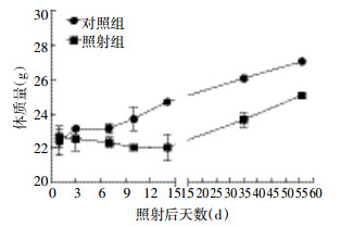
照射后各个时间点照射组小鼠体质量均不同程度地低于对照组,照射组小鼠体质量在照射后呈下降趋势,14 d时体质量降至最低,14 d后体质量缓慢回升(图 1)。
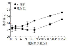
图 1 6 Gy γ射线照射后小鼠体质量的变化
Figure 1. Weight changes of mice after 6 Gy γ-irradiation
-
照射后1 d,照射组小鼠的外周血WBC急剧下降,与对照组比较差异有统计学意义(t=111.9,P<0.05),且持续保持较低水平,照射后7 d,外周血WBC下降至最低值,照射后10 d逐渐开始恢复,照射后56 d,与对照组比较,照射组小鼠外周血WBC尚未得到完全恢复(t=5.27,P<0.05)。
照射后小鼠红细胞数缓慢且持续下降,照射后14 d下降至最低值,与对照组比较差异有统计学意义(t=47.70,P<0.05)。
照射后早期血红蛋白含量缓慢下降,照射后14 d下降至最低值,照射后3、7、10、14 d与对照组比较,差异有统计学意义(t=3.72、8.43、12.34、16.37,P均<0.05)。
照射后早期血小板数缓慢下降,之后迅速下降,照射后10 d下降至最低值,之后缓慢恢复,照射后7、10、14、35、56 d与对照组比较,差异有统计学差异(t=39.10、50.09、17.81、9.97、9.26,P均<0.05)。以上结果详见表 1。
照后时间(d) 组别 WBC
(×109个/L)红细胞数
(×1012个/L)血红蛋白含量(g/L) 血小板数
(×109个/L)1 对照组 7.63±0.03 9.75±1.00 135.33±12.34 839.67±109.44 照射组 1.12±0.66 9.85±0.41 136.67±6.44 791.17±159.52 (t=111.9) 3 对照组 7.25±0.06 9.50±0.46 139.50±5.75 681.50±14.93 照射组 0.38±0.15 8.51±0.48 124.17±7.73 636.00±115.65 (t=96.17) (t=3.83) (t=3.72) 7 对照组 7.37±0.16 9.78±0.03 131.50±1.73 701.50±6.36 照射组 0.20±0.03 7.46±0.66 101.00±7.21 162.67±22.98 (t=106.3) (t=6.38) (t=8.43) (t=39.10) 10 对照组 7.51±0.69 9.35±0.06 134.50±0.58 694.00±28.87 照射组 0.36±0.09 7.04±0.40 96.00±5.79 65.80±17.91 (t=19.66) (t=15.25) (t=12.34) (t=50.09) 14 对照组 7.55±0.06 9.99±0.19 141.67±4.51 708.00±6.93 照射组 0.62±0.20 6.04±0.77 84.83±12.45 161.75±47.33 (t=62.46) (t=47.70) (t=16.37) (t=17.81) 35 对照组 7.65±0.17 9.52±0.26 113.67±3.51 699.7±26.58 照射组 3.18±0.64 7.48±0.34 107.00±4.98 345.0±71.12 (t=15.41) (t=6.54) (t=9.97) 56 对照组 7.40±1.21 8.29±0.42 117.00±4.80 689.70±23.07 照射组 4.22±0.57 7.63±0.41 111.33±4.80 439.4±52.72 (t=5.27) (t=4.33) (t=9.26) 注:表中,括号中t值为照射组与对照组比较的结果,P均<0.05。 表 1 6 Gy γ射线照射后不同时间点小鼠外周血象的变化(n=5,x±s)
Table 1. Changes of peripheral blood cell counts at different time after 6 Gy γ-irradiation(n=5, x±s)
-
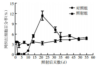
照射后10 d内,小鼠外周血网织红细胞比例出现下降,随后逐渐恢复,而后出现超常时相,最后缓慢恢复正常,与对照组比较,照射后2、3、7、10、14、21和31 d差异有统计学意义(t=18.12、16.89、10.15、8.62、3.16、5.63、3.69,P均<0.05)。照射后35、42、49和56 d,与对照组比较,照射组小鼠外周血网织红细胞比例差异均无统计学意义(t=0.60、2.20、1.01、1.63,P均>0.05),即从照射后35 d开始,小鼠外周血网织红细胞比例基本得到恢复(图 2)。
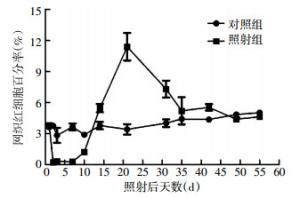
图 2 6 Gy γ射线照射对小鼠外周血网织红细胞百分率的影响
Figure 2. Changes of percentage of reticulocytes at different time after 6 Gy γ-irradiation
-
6 Gy照射后,小鼠骨髓有核细胞数急剧下降,照射后7~10 d下降至最低值,之后逐渐恢复,照射后1、3、7、10、14、35和56 d,照射组小鼠骨髓有核细胞数持续明显低于对照组(t=41.00、19.13、39.01、19.42、24.26、4.16和6.76,P均<0.05)(表 2)。
组别 1 d 3 d 7 d 10 d 14 d 35 d 56 d 对照组 2.64±0.11 2.52±0.23 2.94±0.16 2.86±0.16 2.35±0.16 2.64±0.26 3.03±0.10 照射组 1.04±0.14 0.27±0.06 0.20±0.03 0.72±0.21 0.79±0.22 1.87±0.19 2.46±0.23 t值 41.00 19.13 39.01 19.42 24.26 4.16 6.76 P值 <0.05 <0.05 <0.05 <0.05 <0.05 <0.05 <0.05 表 2 6 Gy γ射线照射后不同时间点小鼠骨髓有核细胞数的变化(×107个/单侧股骨,x±s)
Table 2. Changes of bone marrow mononuclear cells at different time after 6 Gy γ-irradiation(×107/femur, x±s)
-
与对照组比较,照射后14、35和56 d照射组小鼠骨髓有核细胞粒单系集落形成能力分别下降83.45%、71.90%和48.88%(t=16.45、17.78和23.32,P均<0.05)。
-
造血组织是电离辐射最敏感的组织之一,在一定剂量范围内,它的变化速度和进程与机体受照剂量呈正相关。大剂量电离辐射作用后,机体内辐射敏感的造血组织的损伤是严重的。笔者前期的研究结果显示,C57BL/6小鼠在7.2~7.5 Gy 137Cs γ射线照射后30 d生存率低于50%[4-5]。因此,本研究选取照射剂量为6 Gy,用以研究大剂量照射对小鼠造血功能的长期影响。
本研究通过测定小鼠外周血常规的变化,观察了6 Gy 137Cs γ射线一次性全身照射对小鼠造血功能的影响。研究结果显示,在6 Gy照射后,小鼠外周血象出现不同程度的下降,其中WBC下降最早且最显著,其次是血小板数,WBC和血小板数对辐射较敏感且6 Gy照射对小鼠外周血WBC和血小板数造成较严重的影响,恢复缓慢;红细胞数和血红蛋白含量下降缓慢且恢复快,可能是由于哺乳动物红细胞没有细胞核,对辐射的耐受能力强于白细胞。因此,辐射后白细胞的变化可用于诊断和判定预后,并且在治疗上使机体保持较高水平的WBC,对改善机体抗感染、加强防御适应机能具有重要作用。
网织红细胞是一种尚未发育成熟的红细胞,也称年轻红细胞。红细胞在骨髓内生成,成熟后进入血液循环,网织红细胞对电离辐射比较敏感,是反映骨髓造血功能的重要指标之一[6]。研究发现机体受到照射后,网织红细胞出现下降。流式细胞术的广泛应用,为网织红细胞检测提供了快速、简便和准确的方法[7]。本研究结果显示,6 Gy射线照射后,小鼠外周血网织红细胞比例出现下降,而后缓慢恢复并超出正常值,最后逐渐恢复正常。这与使用60Co γ源照射的实验结果一致[7]。
骨髓对辐射高度敏感,辐射引起骨髓损伤必将导致骨髓造血细胞的变化[8-10]。骨髓是主要的造血组织,骨髓有核细胞水平在一定程度上反映了骨髓造血能力。本研究结果表明,照射后小鼠骨髓有核细胞数显著降低,随后缓慢恢复。集落形成实验是功能实验,能检测骨髓造血祖细胞的增殖能力,本研究观察到,6 Gy γ射线照射后,小鼠骨髓有核细胞粒单系集落形成能力显著下降,随后功能明显恢复。
综上所述,本研究动态观察了辐射损伤后小鼠的体质量、外周血象、外周血网织红细胞百分率和造血祖细胞增殖能力的变化,电离辐射对小鼠外周血象、骨髓细胞及有核细胞粒单系集落形成能力均造成了损伤,具有一定的时间效应关系,实验结果丰富了造血系统辐射损伤研究的基础数据,也为后续开展造血系统辐射防护药物的研究提供了一定的参考数据。
6 Gy 137Cs γ射线照射对小鼠造血功能损伤的动态观察研究
Effects of 6 Gy 137Cs γ-irradiattion on the hematopoietic system of mice
-
摘要:
目的 观察6 Gy 137Cs γ射线照射对小鼠造血功能的影响。 方法 取雄性C57BL/6小鼠70只,随机分为对照组和照射组,每组35只。照射组小鼠接受6 Gy 137Cs γ射线一次性全身照射,分别于受照后1、3、7、10、14、35和56 d测定小鼠体质量,并检测外周血象的变化、外周血网织红细胞百分率和骨髓有核细胞数,在受照后14、35和56 d检测小鼠骨髓细胞粒单系集落形成能力。 结果 照射后小鼠体质量下降,14 d后缓慢上升;外周血象中WBC和血小板数迅速下降,分别于照射后7和10 d下降至最低值,而后缓慢回升;骨髓有核细胞数急剧下降,于照射后7和10 d下降至最低值,而后逐渐恢复;外周血网织红细胞百分率经历了下降、恢复、超出正常值范围及恢复至正常值的过程;骨髓有核细胞粒单系集落形成能力显著下降。 结论 6 Gy 137Cs γ射线照射可引起小鼠外周血象的变化并抑制造血祖细胞的增殖能力。 -
关键词:
- 造血系统 /
- 全身照射 /
- 辐射损伤,实验性 /
- 小鼠,近交C57BL
Abstract:Objective To observe the effects of 6 Gy 137Cs γ-irradiation on the hematopoietic system of mice. Methods Seventy male C57BL/6 mice were randomly divided into control and irradiated groups(n=35 each). The irradiation group mice received 6 Gy 137Cs γ-irradiation. Bodyweight, peripheral blood cell counts, bone marrow mononuclear cell counts, and reticulocyte percentage were measured at 1, 3, 7, 10, 14, 35, and 56 d after total body irradiation. CFU-GM was detected at 14, 35, and 56 d after 6 Gy 137Cs γ-irradiation. Results After irradiation, the bodyweight of irradiated mice decreased but slowly increased after 14 d. The peripheral blood of irradiated mice had significant changes in WBC and platelet counts: it respectively decreased to the lowest value at 7 and 10 d post-irradiation and subsequently increased slowly. The reticulocyte percentage decreased, recovered, exceeded the normal range, and returned to normal. Furthermore, the bone marrow mononuclear cell counts decreased sharply, decreased to the minimum value after 7 d post-irradiation, and gradually recovered. The monophyletic cloning ability of irradiated mouse bone marrow nuclear granulocytes also decreased significantly. Conclusion 6 Gy 137Cs γ-irradiation could cause changes in the peripheral blood of mice and inhibit the proliferation of hematopoietic progenitor cells. -
图 2 6 Gy γ射线照射对小鼠外周血网织红细胞百分率的影响
Figure 2. Changes of percentage of reticulocytes at different time after 6 Gy γ-irradiation
表 1 6 Gy γ射线照射后不同时间点小鼠外周血象的变化(n=5,x±s)
Table 1. Changes of peripheral blood cell counts at different time after 6 Gy γ-irradiation(n=5, x±s)
照后时间(d) 组别 WBC
(×109个/L)红细胞数
(×1012个/L)血红蛋白含量(g/L) 血小板数
(×109个/L)1 对照组 7.63±0.03 9.75±1.00 135.33±12.34 839.67±109.44 照射组 1.12±0.66 9.85±0.41 136.67±6.44 791.17±159.52 (t=111.9) 3 对照组 7.25±0.06 9.50±0.46 139.50±5.75 681.50±14.93 照射组 0.38±0.15 8.51±0.48 124.17±7.73 636.00±115.65 (t=96.17) (t=3.83) (t=3.72) 7 对照组 7.37±0.16 9.78±0.03 131.50±1.73 701.50±6.36 照射组 0.20±0.03 7.46±0.66 101.00±7.21 162.67±22.98 (t=106.3) (t=6.38) (t=8.43) (t=39.10) 10 对照组 7.51±0.69 9.35±0.06 134.50±0.58 694.00±28.87 照射组 0.36±0.09 7.04±0.40 96.00±5.79 65.80±17.91 (t=19.66) (t=15.25) (t=12.34) (t=50.09) 14 对照组 7.55±0.06 9.99±0.19 141.67±4.51 708.00±6.93 照射组 0.62±0.20 6.04±0.77 84.83±12.45 161.75±47.33 (t=62.46) (t=47.70) (t=16.37) (t=17.81) 35 对照组 7.65±0.17 9.52±0.26 113.67±3.51 699.7±26.58 照射组 3.18±0.64 7.48±0.34 107.00±4.98 345.0±71.12 (t=15.41) (t=6.54) (t=9.97) 56 对照组 7.40±1.21 8.29±0.42 117.00±4.80 689.70±23.07 照射组 4.22±0.57 7.63±0.41 111.33±4.80 439.4±52.72 (t=5.27) (t=4.33) (t=9.26) 注:表中,括号中t值为照射组与对照组比较的结果,P均<0.05。  下载: 导出CSV
表 2 6 Gy γ射线照射后不同时间点小鼠骨髓有核细胞数的变化(×107个/单侧股骨,x±s)
Table 2. Changes of bone marrow mononuclear cells at different time after 6 Gy γ-irradiation(×107/femur, x±s)
组别 1 d 3 d 7 d 10 d 14 d 35 d 56 d 对照组 2.64±0.11 2.52±0.23 2.94±0.16 2.86±0.16 2.35±0.16 2.64±0.26 3.03±0.10 照射组 1.04±0.14 0.27±0.06 0.20±0.03 0.72±0.21 0.79±0.22 1.87±0.19 2.46±0.23 t值 41.00 19.13 39.01 19.42 24.26 4.16 6.76 P值 <0.05 <0.05 <0.05 <0.05 <0.05 <0.05 <0.05
下载: 导出CSV
-
[1] 路璐, 孟爱民. HSCs辐射损伤机制研究进展[J].生命科学, 2015, 27(2): 161-167.
[2] 李德冠, 樊赛军, 孟爱民.辐射导致长期骨髓抑制的研究进展[J].国际放射医学核医学杂志, 2015, 39(4): 324-327. doi: 10.3760/cma.j.issn.1673-4114.2015.04.011
[3] 路璐, 李德冠, 张俊伶, 等.芝麻酚对4 Gy 137Cs γ射线照射小鼠造血功能的影响[J].中国生化药物杂志, 2014, 34(1): 26-28.
[4] Li D, Lu L, Zhang J, et al. Mitigating the effects of Xuebijing injection on hematopoietic cell injury induced by total body irradiation with γ rays by decreasing reactive Oxygen species levels[J]. Int J Mol Sci, 2014, 15(6): 10541-10553. doi: 10.3390/ijms150610541 [5] Zhang H, Zhai ZB, Wang YE, et al. Resveratrol ameliorates ionizing irradiation-induced long-term hematopoietic stem cell injury in mice[J]. Free Radical Biology and Medicine, 2013, 54: 40-50. [6] 张时民, 李晓京.网织红细胞检测技术的进展和临床应用[J].中国医疗器械信息, 2007, 13(6): 15-23, 77. doi: 10.3969/j.issn.1006-6586.2007.06.005
[7] 王雷, 王治东, 胡海亮, 等.外周血网织红细胞和T淋巴细胞亚群用于辐射损伤快速剂量估算的可行性研究[J].癌变·畸变·突变, 2011, 23(6): 452-455, 459. doi: 10.3969/j.issn.1004-616x.2011.06.012
[8] Shao L, Feng W, Li H, et al. Total body irradiation causes long-term mouse BM injury via induction of HSC premature senescence in an Ink4a- and Arf-independent manner[J]. Blood, 2014, 123(20): 3105-3115. doi: 10.1182/blood-2013-07-515619 [9] Shao L, Luo Y, Zhou D. Hematopoietic stem cell injury induced by ionizing radiation[J]. Antioxid Redox Signal, 2014, 20(9): 1447-1462. doi: 10.1089/ars.2013.5635 [10] Chang J, Feng W, Wang Y, et al. Whole-body proton irradiation causes long-term damage to hematopoietic stem cells in mice[J]. Radiat Res, 2015, 183(2): 240-248. -

 点击查看大图
图(2)表(2)
计量
- 文章访问数: 1894
- HTML全文浏览量: 1025
- PDF下载量: 3