


 下载:
下载:
-
胸腔积液在临床上较为常见,按其发生机制可分为漏出液和渗出液两类,不同性质的胸腔积液可提示有不同的病因和发病机制,对良、恶性胸腔积液的鉴别具有重要的临床意义[1]。磁共振波谱(magnetic resonance spectroscopy,MRS)是利用化学位移现象来测定组成物质的分子成分的检测技术,是目前唯一可测得代谢物的化学成分和含量的检查方法。对体液的MRS研究,国内外一般应用7.0 T及以上场强的仪器,限制了体液波谱研究的临床应用。本研究利用1.5 T临床型磁共振仪对胸腔积液波谱进行研究分析,寻找良恶性胸腔积液的MRS特征及鉴别要点,为临床良恶性胸腔积液的鉴别提供一定的帮助。
-
收集2012年6月至2013年6月在惠州市中心人民医院行胸腔穿刺抽液的胸腔积液标本共46例,所有患者临床进行胸腔积液穿刺引流前均签署了知情同意书。根据胸腔积液的来源将其分为良性胸腔积液和恶性胸腔积液两类,良性胸腔积液中包括结核性和炎性胸腔积液两种类型。46例标本中,原发病确诊为良性者20例,包括肺结核14例、肺炎6例;原发病确诊为恶性者26例,包括肺癌18例、乳腺癌5例、肝癌2例、胃癌1例。
-
每例患者收集胸腔积液标本约50 ml分装于离心管内,常温下置于离心机内,以1500转每分钟(离心半径为6 cm)的转速离心5 min,用注射器抽取上层清液(弃沉渣)并转移至容积为30 ml的球形玻璃容器内,用于分析的标本量为30 ml。MRS检查采用美国GE 1.5 T磁共振扫描仪(GE Signa,General Electric Systems),标准相控阵头部线圈,标本置于线圈中心,周围用泡沫材料固定。波谱扫描采用单体素氢质子波谱-点分辨自旋回波波谱序列(point-resolve echo spin spectroscopy,PRESS),重复时间(TR)=3000 ms,回波时间(TE)=30 ms,激励次数(NEX)=128。首先利用三平面定位图像进行体素定位,体素选择于容器的中心,大小为20 mm×20 mm×20 mm。匀场、抑水和波谱采集扫描均由自动预扫描程序完成,匀场半高宽值≤2,抑水≥99%时满足扫描要求再进行波谱扫描。扫描结束后主机产生P.file文件,从ADW4.3工作站导出,并于实验室导入UNIX系统,通过SAGE软件进行相位和基线校正,得出波谱图像并导出,记录软件计算出的代谢物(包括乳酸、胆碱、肌酸、肌醇和脂质等)的波峰下面积。
-
波谱图像分析由两位具有MRS分析经验的副主任医师共同完成,当意见不一致时通过讨论达成共识。数据利用SPSS 17.0软件进行统计学处理。由于软件处理得出的波谱下相对面积较大,分析前对所得的波峰下面积进行对数处理得出主要代谢峰的相对波峰下面积,结果以x±s表示。对不同类型胸腔积液中同一代谢物的波峰下面积的比较采用单因素方差分析,两两比较采用独立样本t检验,P < 0.05表示差异有统计学意义。
-
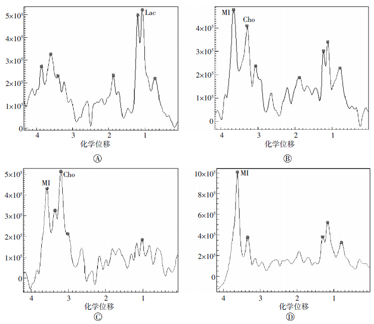
利用PRESS序列采集胸腔积液标本具有较好的匀场效果,其匀场半高宽值≤2,波谱图像具有平稳的基线和清晰的代谢峰。胸腔积液的波谱图中主要的代谢峰有乳酸、胆碱、肌酸、肌醇及脂质等。主要代谢物波峰下面积比较:乳酸:结核性(5.19±1.31)、炎性(6.08±1.56)和恶性胸腔积液(2.40±0.43)的乳酸峰下面积差异有统计学意义(F=8.45,P < 0.01);胆碱:结核性(2.75±0.91)、炎性(3.27±1.21)和恶性胸腔积液(6.76±1.73)的胆碱峰下面积差异有统计学意义(F=2.98,P < 0.01);肌醇:结核性(2.71±1.19)、炎性(2.25±0.81)和恶性胸腔积液(5.83±2.08)的肌醇峰下面积差异有统计学意义(F=38.49,P < 0.01)。结核性和炎性胸腔积液中乳酸(t=-1.04,P > 0.05)、胆碱(t=-0.58,P > 0.05)和肌醇(t=1.19,P > 0.05)波峰下面积比较,差异均无统计学意义。良、恶性胸腔积液波谱图见图 1。

图 1 良、恶性胸腔积液波谱图
Figure 1. Magnetic resonance spectroscopy of benign and malignant pleural effusion
-
胸腔积液是许多疾病的临床表现之一,在我国,结核和恶性肿瘤是引起胸腔积液的两个主要因素,据文献报道,结核性胸腔积液占良性胸腔积液的87%,在恶性胸腔积液中,原发性肺癌的胸腔积液占恶性胸腔积液的68.7%[2]。近年来,国内外学者对胸腔积液进行了大量的研究工作,胸腔积液的多种实验室指标对其性质和病因的分析具有重要意义[3-4]。亦有研究利用超声、能谱CT或PET/CT探讨对良恶性胸腔积液的鉴别诊断价值,但目前尚无明确的诊断标准,尚不能广泛应用于临床[5-8]。MRS技术具有简单、快速、无创等优点,且可从分子水平反映机体组织和器官的代谢信息,对疾病的发病机制及病因分析具有重要价值。基于良恶性胸腔积液不同的病因和代谢基础,利用MRS对良恶性胸腔积液进行鉴别具有一定的临床价值。MRS技术已经在中枢神经系统疾病的诊断和鉴别中发挥了巨大的作用。不同于高场专用实验仪器对极微小量的样本即可进行检测,临床型磁共振对体液等小样本的研究需要达到一定的体积和规则的形状、达到可检测的信号要求和良好的匀场效果后方可进行检测。1.5T临床型磁共振的MRS在技术设计上用于检测组织代谢和生化变化。本研究中,将30 ml胸腔积液标本置于球形玻璃容器中,可达到半高宽≤2的良好匀场效果,并可产生基线平稳、波谱清晰明显的谱线。
本研究发现,良恶性胸腔积液的MRS具有不同的代谢特征,结核性和炎性胸腔积液的乳酸峰均较恶性胸腔积液高且差异有统计学意义,恶性胸腔积液的胆碱和肌醇峰均较结核性和炎性胸腔积液高且差异有统计学意义;而结核性和炎性胸腔积液的乳酸、胆碱和肌醇波峰下面积均未发现明显统计学差异。良恶性胸腔积液不同的MRS特征反映出其不同的病理意义。胸腔积液细胞学检查发现,在良性胸腔积液中,其细胞成分主要为白细胞(包括中性粒细胞、淋巴细胞及嗜酸性粒细胞等)及间皮细胞等,而在恶性胸腔积液中,其细胞成分主要为脱落或转移的肿瘤细胞和一些红细胞及淋巴细胞等[9]。胸腔积液的细胞成分的不同可能是造成其波谱特征不同的原因,在良性胸腔积液中,由于血管通透性的增高及炎性因子释放等因素造成白细胞渗出,白细胞脱离血浆环境后可能因氧含量减少而造成有氧呼吸的减弱及无氧呼吸的增强,进而产生乳酸,这也可能是良性胸腔积液的pH值低于恶性胸腔积液的原因。在恶性胸腔积液中,由于其含肿瘤细胞较多,细胞代谢尤其是细胞膜的代谢速度快,反映细胞膜代谢的标记物胆碱及肌醇浓度会相应升高[10-11]。另外,在本研究中还发现,恶性胸腔积液的乳酸峰虽低于良性胸腔积液,但也会有一定程度的升高,可能与恶性胸腔积液中含有一定量的白细胞及间皮细胞等有关。
综上所述,通过本研究的初步探索,发现良恶性胸腔积液的MRS特征具有一定差异,利用MRS对良恶性胸腔积液的鉴别具有一定的临床应用价值,尤其是在原发灶性质不明确的情况下,利用MRS分析可对胸腔积液良恶性的判断提供参考。本研究仍存在较多不足之处,首先由于本研究所使用的波谱分析软件尚无胸腔积液波谱的基础数据库,导致胸腔积液波谱中仍有较多的代谢峰无法准确定性,也无法实现绝对定量分析;另外,对于极小量标本测试而言,与专用实验仪器比较,临床型仪器在数据采集和分析的精确度等方面存在差距,良恶性胸腔积液中更有意义的生物学标识有待于进一步探究。二是由于病例数限制,本研究对良恶性胸腔积液的波谱特征进行了初步分析,利用MRS对胸腔积液良恶性的判断的可行性和准确性需要大量病例研究进一步验证,不同原发病变引起的胸腔积液之间的波谱学差异需进一步细化。三是良恶性胸腔积液不同的波谱特征差异的原因仍需进一步深入研究。
1.5T磁共振1H-MRS鉴别良恶性胸腔积液的初步研究
A preliminary study of 1H-MRS in diagnosis of benign and malignant pleural effusions
-
摘要: 目的探讨磁共振波谱对良、恶性胸腔积液的鉴别诊断价值。方法收集行胸腔穿刺抽液的胸腔积液标本46例,其中原发病确诊为良性者20例(包括肺结核14例、肺炎6例),原发病确诊为恶性者26例(包括原发性肺癌18例、乳腺癌5例、肝癌2例、胃癌1例)。对胸腔积液标本进行离心处理,然后利用1.5T磁共振对胸腔积液标本进行波谱采集,分析良恶性胸腔积液的波谱特征。结果胸腔积液的波谱图中主要的代谢峰有乳酸、胆碱、肌酸、肌醇及脂质等。主要代谢物波峰下面积比较:乳酸:结核性(5.19±1.31)、炎性(6.08±1.56)和恶性胸腔积液(2.40±0.43)的乳酸峰下面积差异有统计学意义(F=8.45,P < 0.01);胆碱:结核性(2.75±0.91)、炎性(3.27±1.21)和恶性胸腔积液(6.76±1.73)的胆碱峰下面积差异有统计学意义(F=2.98,P < 0.01);肌醇:结核性(2.71±1.19)、炎性(2.25±0.81)和恶性胸腔积液(5.83±2.08)的肌醇峰下面积差异有统计学意义(F=38.49,P < 0.01)。结核性和炎性胸腔积液中乳酸(t=-1.04,P>0.05)、胆碱(t=-0.58,P>0.05)和肌醇(t=1.19,P>0.05)波峰下面积差异无统计学意义。结论良、恶性胸腔积液具有不同的波谱特征,利用磁共振波谱对良、恶性胸腔积液进行鉴别具有一定的临床应用价值。Abstract: Objective To investigate the feasibility of magnetic resonance spectroscopy(MRS) in the diagnosis of benign and malignant pleural effusions. Methods Samples of 46 cases of pleural effusion were collected in clinics after performing pleurocentesis. Among the obtained samples, 20 cases were benign pleural effusion(14 cases of tuberculosis and 6 cases of pneumonia) and 26 cases were malignant pleural effusion(18 cases of primary bronchogenic carcinoma, 5 cases of breast carcinoma, 2 cases of primary hepatic carcinoma, and 1 case of gastric carcinoma). The effusion pleural samples were centrifuged and subjected to 1H MRS examination using a 1.5T MR device. Results The main metabolites in the pleural effusion contain lactate, choline, creatine, myo-inositol(MI), and fatty acids. Area of Lactate: pleural effusion from tuberculosis (5.19±1.31), pneumonia (6.08±1.56) and malignant pleural effusion (2.40±0.43) has statistic significance (F=8.45, P < 0.01). Area of choline: pleural effusion from tuberculosis (2.75±0.91), pneumonia (3.27±1.21) and malignant pleural effusion (6.76±1.73) has statistic significance (F=2.98, P < 0.01). Area of MI: pleural effusion from tuberculosis (2.71±1.19), pneumonia (2.25±0.81) and malignant pleural effusion (5.83±2.08) has statistic significance (F=38.49, P < 0.01). Area of lactate(t=-1.04, P > 0.05), choline(t=-0.58, P > 0.05) and MI(t=1.19, P > 0.05) between pleural effusion from tuberculosis(5.19±1.31) and pneumonia has no statistic significance. Conclusions Benign and malignant pleural effusions show different MRS characteristics. 1H MRS has certain clinical value in the differentiation of benign and malignant pleural effusions.
-
Key words:
- Pleural effusion /
- Magnetic resonance spectroscopy
-
[1] 孙家兴, 于文成, 胡海燕, 等.恶性胸腔积液与结核性胸腔积液的诊断进展[J].现代生物医学进展, 2013, 13(14):2780-2789.
[2] 刘津梅, 尹瑞玲.结核性与癌性胸腔积液的超声鉴别诊断[J].天津医科大学学报, 2010, 16(4):671-673.
[3] 张国庆, 朱庆华, 常利明.检测胸腔积液和血清中肿瘤标记物在良恶性胸腔积液鉴别诊断中的临床价值[J].中国实验诊断学, 2013, 4(20):852-854.
[4] 凡瞿明, 罗关成, 卢小岚, 等.肿瘤标记物联合检测诊断恶性胸腔积液的应用价值[J].国际检验医学杂志, 2013, 4(10):415-417.
[5] 韩志江, 陈文辉, 叶健.结核性胸腔积液和恶性胸腔积液鉴别中CT值的应用[J].中国实用内科杂志, 2006, 12(S2):S128-129.
[6] 马凤, 张志远, 贾永军, 等.宝石能谱CT在结核性胸腔积液和癌性胸腔积液鉴别诊断中的应用[J].中国医学影像学杂志, 2013, 21(5):360-362.
[7] 辛小燕, 朱斌, 陈君坤, 等. CT能谱成像在胸腔渗出液与漏出液定性鉴别中的作用[J].中华放射学杂志, 2011, 45(8):723-726.
[8] Kim BS, Kim IJ, Kim SJ, et al. Predictive value of F-18 FDG PET/CT for malignant pleural effusion in non-small cell lung cancer patients[J]. Onkologie, 2011, 34(6):298-303. [9] 冯恩志, 戚好文, 薛军.胸水淋巴细胞微核率检测在良、恶性胸腔积液鉴别诊断中的意义[J].第四军医大学学报, 2002, 23(6):549-551.
[10] Husarova V, Bittsansky M, Ondrejka I, et al. Correlations of ADHD symptoms with neurometabolites measured by 1H magnetic resonance spectroscopy[J]. Bratisl Lek Listy, 2014, 115(10):635-642. [11] Haris M, Cai K, Singh A, et al. In vivo mapping of brain myo-inositol[J]. Neuroimage, 2011, 54(3):2079-2085. -

 点击查看大图
图(1)
计量
- 文章访问数: 2498
- HTML全文浏览量: 1430
- PDF下载量: 3