


-
MRI是诊断乳腺肿瘤的重要方法,其价值已得到公认[1]。常规MRI诊断乳腺肿瘤有较高的灵敏度,但缺乏特异性,月经周期、激素治疗、增生性病变等因素均可能导致假阳性的发生,导致部分病例在传统MRI成像上难以定性[2-4]。弥散加权成像(diffusion-weighted imaging,DWI)可以反映组织的生物学特征,对鉴别乳腺肿瘤性质具有较高的灵敏度和特异度[5-6]。DWI通常在注射钆对比剂前采集,鲜有给药后进行的报道,本研究旨在探讨1.5 T MRI中乳腺肿瘤患者使用钆对比剂是否对DWI有显著性影响,并分析增强前后表观弥散系数(apparent diffusion coefficient,ADC)及指数表观弥散系数(exponent apparent diffusion coefficient,eADC)值的变化情况及其在乳腺良恶性病变鉴别中的价值。
-
收集2010年6月至2014年6月在我院行乳腺MRI检查的患者共135例,按照以下标准纳入其中40例(共计44个病灶)进行分析:①MRI检查前没有进行过任何化学药物治疗、放射治疗或外科手术;②病灶直径≥1 cm,因为在过小的病灶上测量操作的可重复性较差;③经细针穿刺或外科手术病理证实;④囊性病变不纳入本研究,因其在T1及T2加权图像上有特征性表现,诊断不难,且囊性病变的ADC值很高,这可能导致错误评估良性病变ADC值的均数及范围。本研究中40例患者均为女性,年龄18~80岁,平均(40.4±12.4)岁,44个病灶中,良性肿瘤23个,其中纤维腺瘤17个、导管内乳头状瘤2个、硬化性腺病1个、腺瘤样结节1个、良性叶状肿瘤2个;乳腺癌病灶21个,其中浸润性导管癌16个,浸润性小叶癌5个。肿瘤直径12.5~79.4 mm,平均(27.3±13.5)mm。本研究经我院伦理委员会批准后实施。所有患者在检查前均签署知情同意书。
-
使用荷兰Philips 1.5T Achieva Nova Dual超导型MRI仪,6通道乳腺专用阵相控线圈俯卧扫描,注射器为德国Ulrich medical的Mississippi XD 2000专用高压注射器,对比剂为钆喷酸葡胺(Gd-DTPA,广州康臣药业集团有限公司),剂量0.1 mmol/kg,3 ml/s静脉团注,20 ml生理盐水冲管。常规序列包括T1加权、T2加权、T2压脂、动态增强扫描。扫描参数:轴位T1加权,重复时间498 ms,回波时间10 ms;轴位T2加权,重复时间2922 ms,回波时间120 ms;轴位T2压脂,重复时间4510 ms,回波时间120 ms,精确频率反转恢复(spectral presaturation attenuated inversion recovery,SPAIR)压脂;动态增强扫描采用高分辨率各向同性容积激发成像序列(e-Thrive),重复时间4.8 ms,回波时间2.3 ms,翻转角12°,SPAIR压脂。注射对比剂前进行1次预扫描,启动注射的同时启动连续扫描,共9个动态时相。
DWI采用单次激发平面回波序列,重复时间2000 ms,回波时间63 ms,在X、Y、Z轴3个方向上施加弥散敏感梯度,弥散敏感因子b值=800 s/mm2,SPAIR压脂,常规平扫序列后进行第一次采集,完成动态增强扫描后进行第二次采集,给药前后DWI使用相同参数成像。
-
图像数据使用Extended MR WorkSpace2.6.3.2后处理工作站进行处理。①ROI设置:在增强图像上选取肿瘤强化最明显的部位进行设置,避开出血、囊变及坏死区。在对侧相应位置正常腺体区域设置大小相同的ROI。②分别测量增强前后DWI的信噪比(signal-to-noise ratio,SNR)和对比噪声比(contrast-to-noise ratio,CNR)。计算公式为:SNR=SI/SDnoise,其中SI为组织的信号强度,SDnoise为背景噪声的标准差;CNR=(SItumor-SInormal)/SDnoise,其中SItumor和SInormal分别为病灶和正常腺体信号强度。③测量给药前后乳腺病灶ADC值及eADC值。ADC值计算公式为ADC=-(1/b)In(S/S0),其中,S0和S分别为b值为0和800时的ROI信号强度;eADC值具有不受T2穿透效应影响、SNR较高等优势,其计算公式为eADC=e-bADC。
-
计量资料以均数±标准差(x±s)表示。采用Shapiro-Wilk检验数据正态性,对符合正态分布的数据采用配对t检验,不符合正态分布数据采用Wilcoxon符号秩和检验。使用SPSS19.0软件进行统计学分析,P < 0.05表示差异有统计学意义。
-
表 1为b=0及800时增强前后正常乳腺腺体弥散图像SNR、乳腺病灶弥散图像SNR及CNR。配对t检验结果表明正常乳腺腺体及乳腺病灶弥散图像SNR及CNR在注射钆剂前后差异无统计学意义。
时间点 正常乳腺腺体SNR 乳腺病灶SNR CNR b=0 b=800 b=0 b=800 b=0 b=800 增强前 34.75±3.93 22.71±3.14 53.30±5.26 32.47±4.64 18.55±4.84 9.76±4.04 增强后 35.13±4.00 23.12±3.19 53.97±5.53 32.88±5.41 18.84±5.39 9.76±5.35 t值 0.657 1.360 1.534 0.913 0.480 -0.002 P值 0.515 0.181 0.132 0.366 0.633 0.998 注:表中,SNR:信噪比;CNR:对比噪声比。 表 1 正常乳腺腺体和乳腺病灶MRI弥散加权成像图像增强前后SNR及CNR比较(x±s,n=44)
Table 1. SNR and CNR of the normal breast and the lesions before and after contrast enhancement(x±s, n=44)
-
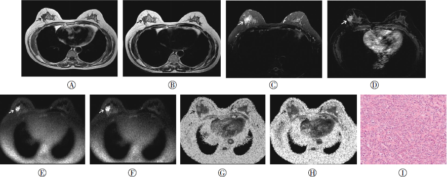
增强前后病灶ADC值及eADC值见表 2。乳腺癌增强后ADC值较增强前降低(图 1),平均降幅10.3%;增强后eADC值明显升高,平均升幅11.3%;配对t检验结果显示两者增强前后差异均有统计学意义。良性肿瘤增强后的ADC值平均降低1.9%;增强后的eADC值平均升高2.0%;配对t检验结果显示两者差异无统计学意义。

图 1 乳腺浸润性导管癌患者磁共振图像和组织病理图。
Figure 1. MRI images and pathological image of patient with invasive ductal carcinoma of breast
时间点 乳腺癌(n=21) 乳腺良性肿瘤(n=23) ADC值 eADC值 ADC值 eADC值 增强前 1.11±0.25 0.40±0.09 1.47±0.27 0.30±0.07 增强后 0.99±0.26 0.44±0.10 1.43±0.24 0.31±0.06 t值 -4.023 4.082 -1.700 1.341 P值 0.001 0.001 0.103 0.194 注:表中,ADC:表观弥散系数;eADC:指数表观弥散系数。 表 2 40例乳腺肿瘤患者MRI弥散加权成像图像增强前后ADC值及eADC值比较(x±s)
Table 2. ADC and eADC value of 40 patients with breast tumors before and after contrast enhancement(x±s)
-
DWI图像信息来自组织中水分子的布朗运动,是目前唯一能观察活体水分子微观运动的无创性检查方法。DWI成像受乳腺组织背景信号的影响很小,因而对乳腺的侵袭性、非侵袭性病变、浸润性导管癌有较高的诊断价值。DWI不同于依赖病变形态和血流灌注的传统成像方式,而是通过提供病理过程中组织水分子流动性的变化信息来提示诊断。
本研究结果显示,乳腺良、恶性病变的平均ADC值在增强后均有减低,乳腺癌增强后ADC值明显下降,最高幅度达31.1%,而乳腺良性肿瘤ADC值的降低与增强前相比差异无统计学意义。Yuen等[7]研究了19例乳腺癌增强后DWI图像,发现增强后其ADC值显著减低,这与本研究结果类似,但Yuen等没有纳入良性肿瘤作为对照。Yamada等[8]在增强后立即行DWI,发现正常脑组织和梗死灶ADC值均显著下降;而Fitzek等[9]在增强后间隔了较长时间的DWI结果则显示两者差异无统计学意义。Li等[10]发现对比剂对ADC值的影响存在时间和病理类型依赖性,富血供的脑膜瘤在增强后ADC值减低较颅内肿瘤明显,持续时间亦较长。对比剂对ADC值的作用机制尚不明确。Yuen等[7]认为是由于对比剂减低了微灌注效应而导致ADC值改变。在生理组织中,微观水分子运动包括了血管内(灌注)和血管外(扩散)水分子的运动,活体内灌注效应对ADC值有一定的影响,因而在微血管密度高的病变如乳腺癌中ADC值可能会被错误地高估。注射钆剂后,血管内钆剂改变了病变局部磁化率,产生的背景磁场梯度叠加或削减了弥散敏感梯度,进而导致ADC值的改变。Janka等[11]的结果支持这一理论,他们使用50 s/mm2为最低b值以减少宏观灌注对ADC值的影响,结果发现微灌注抑制效应仍然存在。鉴于乳腺病变的多样性和异质性,对比剂对DWI作用机制的研究将需要严格设计和精准定量分析。
本组病例中,增强后ADC值下降较大(> 15%)的病例有9例,其中8例为乳腺癌,1例为纤维腺瘤。在这8例乳腺癌中有4例增强前ADC值较高(均 > 1.20×10⁻³ mm2/s),增强后ADC值显著降低,提示增强后DWI能更好地反映乳腺肿瘤的恶性倾向;另外4例增强前的ADC值较低(均 < 1.0×10⁻³ mm2/s),这与Yuen等[7]的结果相左。回顾性观察这4例平扫ADC值较低的乳腺癌的常规图像,发现其中3例乳腺癌病灶在T2加权上信号均偏低,推测与其细胞密度较大有关,但其与增强后ADC值的变化是否存在相关性需要联合病理学研究作进一步探讨。
此外,钆对比剂缩短T2效应可能降低DWI图像的信号强度。但本研究结果注射钆剂前后DWI图像的CNR并无显著性差异,表明缩短T2效应不足以影响ADC值和eADC值,这与Yuen等[7]的结果一致;同时,本研究使用了相对较短的重复时间(2000 ms),也相对削弱了这一效应。
ADC值鉴别乳腺肿瘤性质的能力已得到证实,各家报道其鉴别乳腺良恶性病变的灵敏度(68.8%~97.0%)、特异度(72.7%~96.2%)不等[5-6, 12]。与ADC值相比,eADC值不仅消除了T2穿透效应,同时还保留了扩散图像的信号特点,其数值可通过对ADC值的对数变换而获得,计算公式如下:eADC=e-bADC。由变换公式可以看出,eADC值与ADC值呈负指数相关,eADC值对ADC值的影响因素进行了非线性伸展变换,使得其变异系数具有线性增加的特征。根据这一定义,eADC值并没有显示出明显的优势,但其在图像上的表现较ADC值有明显的优势:eADC图背景抑制较好,SNR较高,肿瘤实质部分显示为高信号,符合临床观察习惯,能够更直观地反映水分子扩散状况,有效确定病变位置和范围。
本研究的不足之处在于未研究特殊病理类型乳腺癌以及增强前后弥散数据的变化情况。文献报道黏液腺癌ADC值高于其他类型乳腺癌及部分良性病变[13],本研究未纳入黏液腺癌病例,未能对其DWI数据变化进行探讨;其次,本研究病例数较少,不适宜做受试者工作特征曲线拟合分析,需要更大样本的研究探讨增强后DWI的诊断效能。
与传统的形态学和动态增强扫描相比,DWI能够为乳腺癌的诊断提供分子水平的信息。静脉注射钆剂前后的DWI图像质量无明显差异,表明可在增强后进行DWI扫描,增强扫描后乳腺癌ADC值明显减低、eADC值明显升高,而良性肿瘤的ADC值及eADC值改变差异无统计学意义,提示增强后DWI能更好地反映肿瘤的恶性倾向。随着MRI技术的进步及DWI成像参数的优化,弥散成像在乳腺癌诊断中的巨大潜力将会得到进一步开发,更好地为临床诊治乳腺癌提供指导。
注射钆剂后弥散加权成像对乳腺肿瘤诊断的影响
Evaluation of the diffusion-weighted imaging after contrast for the characterization of breast tumors
-
摘要: 目的评估1.5 T MRI中乳腺肿瘤患者使用钆对比剂是否对弥散加权成像(DWI)有显著性影响。方法行乳腺MRI检查的女性患者40例(共计44个病灶),分别测量增强前后DWI图像信噪比(SNR)和对比噪声比(CNR)、病灶增强前后的表观扩散系数(ADC)及指数表观扩散系数(eADC)。结果给药前后DWI图像的SNR及CNR差异无统计学意义。乳腺癌给药前后的ADC值(t=-4.023, P=0.001)及eADC值(t=4.082, P=0.001)差异有统计学意义,良性肿瘤给药前后的ADC值(t=-1.700, P=0.103)及eADC值(t=1.341, P=0.194)差异无统计学意义。结论增强后行DWI是可行的,并且有助于提高其鉴别乳腺良恶性肿瘤的能力。Abstract: Objective To evaluate the effects of intravenous gadolinium-DTPA on the diffusion-weighted imaging(DWI) of the breast. Methods A total of 40 women with 44 mass-like breast lesions were examined using 1.5-T MRI. The signal-to-noise ratios(SNRs) of the normal breast and the lesions, the contrast-to-noise ratios(CNRs), apparent diffusion coefficient(ADC), and exponent apparent diffusion coefficient(eADC) values of each lesion were measured from the DWI images before and after contrast enhancement. Results The SNRs and CNRs of the DWI images before and after contrast enhancement were not significantly different. In addition, no significant difference between the ADC and eADC values before and after contrast enhancement was observed in the benign lesions, whereas the opposite was observed in the malignant lesions. Conclusions The application of DWI after contrast enhancement is feasible in characterizing lesions in the breast. After contrast enhancement, the ADC values were significantly lower, and the eADC values were significantly higher in breast carcinomas. DWI after contrast media may improve the diagnosis of malignant breast lesions.
-
Key words:
- Breast neoplasms /
- magnetic resonance imaging /
- Diffusion /
- Gadolinium DTPA
-
图 1 乳腺浸润性导管癌患者磁共振图像和组织病理图。
Figure 1. MRI images and pathological image of patient with invasive ductal carcinoma of breast
表 1 正常乳腺腺体和乳腺病灶MRI弥散加权成像图像增强前后SNR及CNR比较(x±s,n=44)
Table 1. SNR and CNR of the normal breast and the lesions before and after contrast enhancement(x±s, n=44)
时间点 正常乳腺腺体SNR 乳腺病灶SNR CNR b=0 b=800 b=0 b=800 b=0 b=800 增强前 34.75±3.93 22.71±3.14 53.30±5.26 32.47±4.64 18.55±4.84 9.76±4.04 增强后 35.13±4.00 23.12±3.19 53.97±5.53 32.88±5.41 18.84±5.39 9.76±5.35 t值 0.657 1.360 1.534 0.913 0.480 -0.002 P值 0.515 0.181 0.132 0.366 0.633 0.998 注:表中,SNR:信噪比;CNR:对比噪声比。  下载: 导出CSV
表 2 40例乳腺肿瘤患者MRI弥散加权成像图像增强前后ADC值及eADC值比较(x±s)
Table 2. ADC and eADC value of 40 patients with breast tumors before and after contrast enhancement(x±s)
时间点 乳腺癌(n=21) 乳腺良性肿瘤(n=23) ADC值 eADC值 ADC值 eADC值 增强前 1.11±0.25 0.40±0.09 1.47±0.27 0.30±0.07 增强后 0.99±0.26 0.44±0.10 1.43±0.24 0.31±0.06 t值 -4.023 4.082 -1.700 1.341 P值 0.001 0.001 0.103 0.194 注:表中,ADC:表观弥散系数;eADC:指数表观弥散系数。
下载: 导出CSV
-
[1] 巴照贵, 张玉敏, 倪晓丽, 等.乳腺肿块性病变MRI动态增强与扩散加权成像联合诊断方法的探讨[J].实用放射学杂志, 2014(10):1657-1660, 1664.
[2] Schmitz AM, Loo CE, Wesseling JA, et al. Association between rim enhancement of breast cancer on dynamic contrast-enhanced MRI and patient outcome:impact of subtype[J]. Breast Cancer Res Treat, 2014, 148(3):541-551. [3] Medeiros LR, Duarte CS, Rosa DD, et al. Accuracy of magnetic resonance in suspicious breast lesions:a systematic quantitative review and meta-analysis[J]. Breast Cancer Res Treat, 2011, 126(2):273-285. [4] 杨晓棠, 杨继虎, 杜笑松, 等.磁共振扩散加权成像及动态增强MRI在乳腺病变中的应用价值[J].国际放射医学核医学杂志, 2011, 35(3):189-192, 封三.
[5] Imamura T, Isomoto I, Sueyoshi E, et al. Diagnostic performance of ADC for Non-mass-like breast lesions on Mr imaging[J]. Magn Reson Med Sci, 2010, 9(4):217-225. [6] Pereira FP, Martins G, Figueiredo E, et al. Assessment of breast lesions with diffusion-weighted MRI:comparing the use of different b values[J]. AJR Am J Roentgenol, 2009, 193(4):1030-1035. [7] Yuen S, Yamada K, Goto M, et al. Microperfusion-induced elevation of ADC is suppressed after contrast in breast carcinoma[J]. J Magn Reson Imaging, 2009, 29(5):1080-1084. [8] Yamada K, Kubota H, Kizu O, et al. Effect of intravenous gadolinium-DTPA on diffusion-weighted images:evaluation of normal brain and infarcts[J]. Stroke, 2002, 33(7):1799-1802. [9] Fitzek C, Mentzel HJ, Fitzek S, et al. Echoplanar diffusion-weighted MRI with intravenous gadolinium-DTPA[J]. Neuroradiology, 2003, 45(9):592-597. [10] Li X, Qu JR, Luo JP, et al. Effect of intravenous gadolinium-DTPA on diffusion-weighted imaging of brain tumors:a short temporal interval assessment[J]. J Magn Reson Imaging, 2014, 40(3):616-621. [11] Janka R, Hammon M, Geppert C, et al. Diffusion-weighted MR imaging of benign and malignant breast lesions before and after contrast enhancement[J]. Rofo, 2014, 186(2):130-135. [12] Yili Z, Xiaoyan H, Hongwen D, et al. The value of diffusion-weighted imaging in assessing the ADC changes of tissues adjacent to breast carcinoma[J/OL]. BMC Cancer, 2009, 9:18[2014-12-22]. http://www. ncbi. nlm. nih. gov/pmc/articles/PMC2633008. [13] Woodhams R, Kakita S, Hata H, et al. Diffusion-weighted imaging of mucinous carcinoma of the breast:evaluation of apparent diffusion coefficient and signal intensity in correlation with histologic findings[J]. AJR Am J Roentgenol, 2009, 193(1):260-266. -

 点击查看大图
图(1)表(2)
计量
- 文章访问数: 2551
- HTML全文浏览量: 1224
- PDF下载量: 5