


 下载:
下载:
-
骨骼肿瘤在人群中的发病率较低,仅约为0.01%,其中良性骨肿瘤占50%,恶性骨肿瘤占40%,骨肿瘤样病变占10%。肿瘤以及肿瘤样病变均可有FDG摄取[1-2],笔者在日常诊断工作中发现,对于骨骼孤立性FDG摄取增高病灶,如何定性是一个难题,因此本文就本中心收集的经病理证实的22例骨骼孤立性高代谢病灶进行回顾性分析,旨在提高诊断的准确率。
-
收集2003年5月至2013年8月在我中心行PET/CT检查的骨骼病变患者,按以下标准纳入病例:(1)无原发恶性肿瘤病史;(2)全身PET/CT显像只有骨骼病变有异常FDG摄取,且为单发;(3)有病理组织学结果。本研究共纳入22例患者,男性17例、女性5例,年龄5~79岁,中位年龄33.5岁。20例患者的临床表现为病变部位疼痛,其中1例伴有肢体无力;2例表现为包块。所有患者均于活检或手术前行PET/CT检查。
所有患者或其家属均在检查前签署PET/CT检查知情同意书;同时该研究也获得医院及科室的同意。
-
显像仪器为Discovery LS PET/CT扫描仪(GE Healthcare,美国)及mCT128扫描仪(SIEMENS Biography,德国)。显像剂18F-FDG由PET tracer回旋加速器(GE Healthcare,美国)及化学合成模块(北京派特生物技术有限公司,中国)自动合成,放化纯度>95%。患者空腹6 h以上,血糖水平控制在4.7~6.6 mmol/L,平静状态下通过三通管静脉注射18F-FDG 5.5MBq/kg,在暗室内静卧约1 h,排尿后进行PET/CT显像。扫描范围从颅顶至股骨中段,3例患者扫描至双足。PET图像应用CT数据进行衰减校正,重建方法采用有序子集最大期望值迭代法。融合图像通过Xeleris或SyngoMMWP工作站获得。
-
对所有PET图像、CT图像及PET/CT融合图像进行帧对帧对比分析,病灶经2位有PET/CT诊断经验的高年资医师目测确认,采用ROI技术,由计算机计算SUV,取最大值(即SUVmax)。采用SPSS 13.0软件进行统计学分析,计算病灶SUVmax并以均数±标准差(x±s)表示,骨骼良恶性病灶SUVmax的比较采用两独立样本t检验,各病理类型病灶SUVmax的比较采用方差分析;P<0.05表示差异有统计学意义。
-
本研究患者22例,其中淋巴瘤5例(弥漫性大B细胞淋巴瘤2例、间变性大B细胞淋巴瘤2例、B细胞淋巴瘤1例)、嗜酸性肉芽肿5例、浆细胞性骨髓瘤4例、骨肉瘤4例、尤文氏肉瘤2例、Ⅰ级骨巨细胞瘤2例。
-
病灶分布情况为:椎体9例、肋骨3例、胸骨柄2例、四肢骨4例(股骨下段2例、左侧胫骨上端及左侧肱骨头各1例)、盆腔骨3例(骶骨、左侧髂骨及左侧耻骨各1例)、左侧下颌骨1例。
-
病灶对FDG的摄取情况表现为轻度摄取到明显摄取,平均SUVmax为11.08±8.06(2.1~32.6),其中摄取程度与肝脏相近者2例(淋巴瘤及浆细胞瘤各1例);高于肝脏低于脑皮质者9例,其中良性4例(骨巨细胞瘤1例、嗜酸性肉芽肿3例),恶性5例(骨肉瘤及尤文氏肉瘤各2例、浆细胞骨髓瘤1例);与脑皮质相近者11例,其中良性3例(嗜酸性肉芽肿2例、骨巨细胞瘤1例),恶性8例(淋巴瘤4例、浆细胞骨髓瘤及骨肉瘤各2例)。摄取程度均匀者19例、浓淡不均者3例(浆细胞骨髓瘤2例及骨巨细胞瘤1例)。按病理良恶性分类,良恶性病灶FDG摄取的差异无统计学意义(SUVmax:8.86±2.40 vs. 12.12±9.58,t=-1.241,P=0.231)。按病理类型进行分类,各类病灶FDG摄取的差异无统计学意义(F=0.296,df=5,P=0.908)。
-
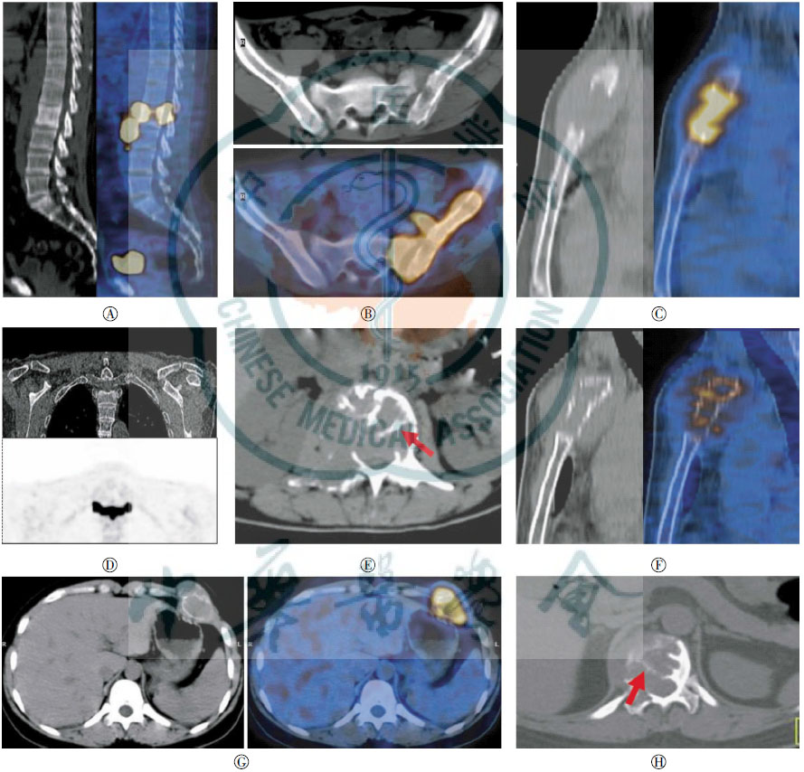
淋巴瘤5例,其中病灶位于胸椎及腰椎各2例、左侧髂骨1例。其同机CT改变有以下3种:(1)成骨硬化3例,表现为骨质密度弥漫性稍增高,椎体形态完整(图 1中A);(2)轻度骨质破坏1例,表现为大病灶,小骨质破坏,呈虫蚀状改变(图 1中B);(3)膨胀性明显溶骨性破坏1例,位于椎体及其附件上,骨质破坏明显,皮质中断,椎体被压缩呈“三角形”。在胸、腰椎病变中同时伴有附件累及3例,向椎管内生长2例。5例均伴有周围明显软组织肿块形成,4例软组织肿块大于骨骼侵犯的范围。

图 1 骨骼肿瘤的PET/CT表现
Figure 1. PET/CT findings of bone tumor
嗜酸性肉芽肿5例,其中病灶位于胸椎、腰椎、胸骨柄、肋骨、左侧耻骨各1例。CT上均表现为明显溶骨性破坏,边缘尚清晰,不膨胀,无骨嵴残留,其中位于胸椎1例累及三个椎体,椎间盘无变窄,椎体呈压缩性骨折。3例突破骨皮质形成周围组织软组织肿块(图 1中C),另2例伴硬化边。
浆细胞性骨髓瘤4例,其中病灶位于胸椎、腰椎、胸骨柄及骶骨各1例。CT上均表现为不同程度溶骨性破坏。胸2椎体累及双侧附件呈对称性破坏,类似“领结征”(图 1中D)。腰3椎体轻度膨胀性骨质破坏累及右侧附件及右侧椎旁组织,破坏区内见残留较粗骨嵴影(图 1中E)。骶骨病灶呈大片状破坏,破坏区内见残留粗大的骨嵴,边缘硬化。胸骨病变呈膨胀性骨质破坏,轮廓基本存在,骨皮质断断续续呈花边状,形成“花边征”(图 1中F),伴椎旁软组织肿块形成。
骨肉瘤4例,其中病灶位于四肢骨干骺端3例、肋骨1例。CT上呈现溶骨性改变为主2例、成骨性改变为主2例(表现为骨质密度不均匀性增高)。4例均伴软组织肿块及骨膜形成。病灶位于四肢骨者向心性沿着长轴方向生长。
尤文氏肉瘤2例,其中病灶位于左侧股骨下段内侧髁及左侧下颌骨各1例。两例均表现为轻度浸润性骨质破坏,周围软组织肿块形成。病灶位于左侧股骨下段内侧髁者偏心性沿着长轴方向生长。
骨巨细胞瘤2例,其中病灶位于左侧第7侧肋骨及胸12椎体各1例。CT上均表现为膨胀性溶骨性破坏,破坏区形成分隔,肋骨病变呈“皂泡征”(图 1中G),胸12椎体破坏区内见纤细骨嵴(图 1中H),边缘略有轻度硬化,病变突破骨皮质形成右侧椎旁软组织肿块并向椎管内生长。
-
本组结果显示,22个骨孤立性良恶性肿瘤对FDG的摄取无明显差异,单凭18F-FDG摄取程度的高低难以对病灶进行定性。这与Costelloe等[3]的报道略有不同,他的研究显示恶性肿瘤对FDG的摄取程度高于所有骨良性肿瘤,但低于侵袭性的骨巨细胞瘤及嗜酸性肉芽肿。Hoshi等[4]也进行了类似的研究,得出的结果与Costelloe等相似,但他以SUVmax=2.6作为良恶性骨肿瘤的分界点时,PET/CT诊断的特异度只有64.7%,假阳性的原因主要也是因为骨巨细胞瘤的高摄取。本组研究结果与上述研究不同的原因估计与病例来源有关,本组只有22例病例,且良性骨肿瘤只有易出现FDG高摄取的骨巨细胞瘤及嗜酸性肉芽肿两种,因而导致摄取无明显差异。文献及本研究结果显示,嗜酸性肉芽肿及骨巨细胞瘤难以在18F-FDG摄取程度上与恶性肿瘤相鉴别。从摄取均匀性方面考虑,浆细胞骨髓瘤及骨巨细胞瘤易出现病灶摄取FDG程度不均一的情况[4-6],估计这与两种肿瘤的异型性明显及生物学特性有关,而其他肿瘤摄取较均匀,这种征象可能有助于鉴别诊断。
全身PET的显像价值在于确认全身骨骼其他部位无其他高代谢病灶存在,也无其他脏器、组织恶性肿瘤存在,另外一个价值在于显示病灶的增殖活跃程度、侵犯范围,以更好地指导活检。本研究中的淋巴瘤病灶的18F-FDG代谢增高程度就很好地反映了肿瘤的恶性程度,1例淋巴瘤病灶摄取程度低,与肝脏相近,提示了该病灶生长缓慢,病理结果是B小细胞淋巴瘤,属于惰性淋巴瘤;而另外4例淋巴瘤摄取明显增高,与脑皮质相近,提示了肿瘤代谢活跃,具有明显的侵袭性,病理结果是弥漫性大B细胞淋巴瘤及间变性大B细胞淋巴瘤,属于侵袭性淋巴瘤。虽然PET能够根据病灶的FDG摄取情况提供部分诊断信息,但鉴别诊断还需结合临床及同机CT的影像学改变来综合判断。
原发骨淋巴瘤好发于40~50岁年龄群,10岁以下较少见,且好发于长骨及脊柱骨。生化检查以微球蛋白、乳酸脱氢酶及铁蛋白水平升高为主。CT改变可分为4种类型:溶骨性破坏、硬化型、囊状膨胀型及混合型。本组骨骼淋巴瘤以硬化型改变为主,见于椎体[7],其他肿瘤病例未见此征象。且椎体淋巴瘤多伴有椎旁明显软组织肿块形成,其长径往往大于骨骼侵犯的范围[8]。同时具备这两种征象应首先考虑到骨淋巴瘤。另外一种典型征象为大病灶伴小骨质破坏[9],其产生原因可能是淋巴瘤细胞通过产生IL-1、IL-6和TNF等细胞因子,引起破骨活动增加,骨质吸收形成“肿瘤通道”,骨髓腔内淋巴瘤通过通道在病变周围形成较大软组织肿块,而骨皮质可以不出现广泛骨质破坏[10]。本组位于髂骨的病灶具有此征象。
骨骼嗜酸性肉芽肿是一种罕见的骨骼肿瘤样病变,活动期具有恶性肿瘤的行为,好发于20岁以下青少年,常见部位是颅骨。生化检查以白细胞、C-反应蛋白及血沉增高为主。CT表现与分期相关,急性期呈明显溶骨性破坏,但不膨胀,少见骨嵴残留[11-12],破坏骨皮质比较彻底,且形成周围软组织肿块。本组有3例有此表现。2例慢性期病变较局限,破坏区边缘因病灶修复而形成硬化边。
浆细胞骨髓瘤好发于50岁以上中老年人,多伴有骨质疏松,实验室检查以本周氏蛋白及免疫球蛋白升高为主。CT均表现为溶骨性改变,部分可呈膨胀性改变,破坏区多不彻底,可见残留的骨嵴影,这种征象需与骨巨细胞瘤进行鉴别。一般来说,浆细胞骨髓瘤残留骨嵴纵向较长,横断面较粗大,与骨应力相关,典型者称之为“微脑征”,而骨巨细胞瘤残留的骨嵴较纤细,与骨应力无关[13-14]。另外,骨巨细胞瘤多表现为偏心性生长;而浆细胞瘤可以为对称性生长,冠状位上形成“领结征”。再结合骨巨细胞瘤的常见发病年龄为20~40岁,可资鉴别。位于胸骨柄的浆细胞骨髓瘤形成一种特殊征象,陈韵等[15]称之为“花边征”,表现为胸骨柄呈膨胀性溶骨性改变,骨皮质断断续续,但轮廓尚可辨。
骨肉瘤及尤文氏肉瘤是骨骼肿瘤中最常见的恶性肿瘤,均好发于青少年,且好发于四肢骨干骺端,但两者起源完全不同。骨肉瘤起源于骨细胞,而尤文氏肉瘤起源于骨髓。因此其影像表现也略有不同,一般骨肉瘤根据其肿瘤细胞成分不同,可为成骨性或溶骨性改变,位于四肢骨者,多有骨皮质破坏、死骨、新生瘤骨、骨膜反应及软组织肿块。而尤文氏肉瘤的肿瘤细胞没有成骨活性,不形成瘤骨或瘤软骨,CT主要表现为髓腔内骨质破坏、软组织肿块及骨膜反应。本组4例骨肉瘤均有典型的表现。但尤文氏肉瘤表现不典型,需与其他疾病进行鉴别。
软组织肿块形成在本组病例中较多见,22例患者中有17例存在这种征象,而且无论是良性或者恶性骨肿瘤,只要破坏骨皮质均有可能形成软组织肿块,因而不能把它当成良恶性鉴别的依据。
本研究结果显示,对于孤立性骨病变且呈现18F-FDG代谢增高者,仅根据骨病灶代谢高低程度难以对病变的良恶性及病种类型进行准确区分,仔细分析病变的CT征象并综合患者的年龄、发病部位及实验室检查资料对疾病的鉴别有较大的帮助。本研究的不足之处是病例数较少,讨论也只是针对现有病例进行,而骨骼肿瘤分类繁多,鉴别诊断常较困难,需拓展病例数并总结出更多信息以进行更好的鉴别。
18F-FDGPET/CT对骨骼孤立性高代谢病灶的初步诊断价值
Preliminary study of 18F-FDG PET/CT in the diagnosis of solitary hypermetabolic lesion of bone
-
摘要:
目的 分析骨骼孤立性高代谢病灶的18F-FDG PET/CT表现,以提高其诊断的水平。 方法 回顾性分析22例经病理学确诊的骨骼孤立性高代谢病灶的PET/CT征象。 结果 22例患者中,淋巴瘤及嗜酸性肉芽肿各5例、浆细胞性骨髓瘤及骨肉瘤各4例、尤文氏肉瘤和Ⅰ级骨巨细胞瘤各2例。病灶平均SUVmax为11.08±8.06(2.1~32.6)。良恶性病灶对FDG摄取的差异无统计学意义(SUVmax:8.86±2.40vs. 12.12±9.58,t=-1.241,P=0.231)。按病理类型进行分类,不同病理类型骨骼病灶对FDG摄取的差异无统计学意义(F=0.296,df=5,P=0.908)。浆细胞骨髓瘤及骨巨细胞瘤易出现FDG摄取不均。同机CT主要表现:溶骨性破坏17例,伴硬化边5例,形成“领结征”、“皂泡征”、“花边征”等典型征象;成骨性改变5例,见于淋巴瘤及骨肉瘤;软组织肿块形成17例;骨膜反应4例。 结论 PET难以对骨骼孤立性病灶进行定性,密切结合同机CT征象有助于诊断。 -
关键词:
- 骨肿瘤 /
- 诊断,鉴别 /
- 正电子发射断层显像术 /
- 体层摄影术,X线计算机 /
- 氟脱氧葡萄糖F18 /
- 孤立性高代谢病灶
Abstract:objective This study aims to investigate the clinical value of 18F-FDG PET/CT in diagnosing solitary hypermetabolic lesion of the bone. Methods Twenty-two patients with solitary FDG uptake in the bone were enrolled. Patient diagnosis was confirmed by biopsy or surgery. PET/CT images were analyzed. Results Of the 22 patients, 5 had primary bone lymphoma and bone eosinophilic granuloma, 4 had plasma cell myeloma and osteosarcoma, and 2 had Ewing's sarcoma and giant cell tumor of the bone. The mean SUVmax of bone lesions is 11.08±06(2.1-32.6). Nineteen lesions had well-distributed FDG uptake. The other three lesions were unevenly distributed. No significant difference in FDG uptake was found between malignant and benign lesions(SUVmax:8.86±40 vs. 12.12±58, respectively; t=-1.241, P=0.231), and among different kinds of bone lesions(F=0.296, df=5, P=0.908). Syn-modality CT images showed that 17 patients have osteolytic changes, 5 of which with sclerosis edge. Some bone lesions presented as "bow tie sign", "oap bubble sign? and lace sign? The other 5 patients, 3 with primary bone lymphoma and 2 with osteosarcoma, presented with ossification changes. Otherwise, soft tissue mass was formulated in 17 patients, and periosteal proliferation developed in 4 patients. Conclusion Syn-modality CT images should be included in the differential diagnosis of solitary hypermetabolic lesion of the bone. -
[1] Buchbender C, Heusner TA, Lauenstein TC, et al. Oncologic PET/MRI, part 2:bone tumors, soft-tissue tumors, melanoma, and lymphoma[J]. J Nucl Med, 2012, 53(8):1244-1252. doi: 10.2967/jnumed.112.109306 [2] Lee HJ, Ahn BC, Lee SW, et al. The usefulness of F-18 fluorodeoxyglucose positron emission tomography/computed tomography in patients with Langerhans cell histiocytosis. Ann Nucl Med, 2012, 26(9): 730-737. [3] Costelloe CM, Chuang HH, Chasen BA, et al. Bone Windows for distinguishing malignant from benign primary bone tumors on FDG PET/CT[J]. J Cancer, 2013, 4(7):524-530. doi: 10.7150/jca.6259 [4] Hoshi M, Takada J, Oebisu N, et al. Overexpression of hexokinase-2 in giant cell tumor of bone is associated with false positive in bone tumor on FDG-PET/CT[J]. Arch Orthop Trauma Surg, 2012, 132(11):1561-1568. doi: 10.1007/s00402-012-1588-2 [5] 柳伟坤, 李向东, 李兴耀, 等.多发性骨髓瘤的18F-FDG PET/CT诊断价值[J].中国医学影像学杂志, 2010, 18(1):89-92. doi: 10.3969/j.issn.1005-5185.2010.01.031
[6] 叶香华, 吴湖炳, 王全师, 等. 18F-FDG PET/CT诊断多发性骨髓瘤[J].中国医学影像技术, 2009, 25(5):897-900. doi: 10.3321/j.issn:1003-3289.2009.05.051
[7] 江少凡, 黄艺婧, 涂蓉.影像学对骨恶性淋巴瘤的诊断价值[J].北方药学, 2013, 10(3):125.
[8] 李景雷, 曾辉, 梁长虹, 等. MRI联合PET诊断原发性骨淋巴瘤[J].中国医学影像技术, 2010, 26(2):319-322.
[9] Krishnan A, Shirkhoda A, Tehranzadeh J, et al. Primary bone lymphoma:Radiographic-MR imaging correlation[J]. Radiographics, 2003, 23(6):1371-1383. doi: 10.1148/rg.236025056 [10] Hicks DG, Gokan T, O'Keefe RJ, et al. Primary lymphoma of bone. Correlation of magnetic imaging features with cytokine production by tumor cells[J]. Cancer, 1995, 75(4):973-980. [11] 张玮曾, 刘福尧.脊柱嗜酸性肉芽肿的螺旋CT表现[J].实用医学影像杂志, 2012, 13(2):108-109. doi: 10.3969/j.issn.1009-6817.2012.02.014
[12] Azouz EM, Saigal G, Rodriguez MM, et al. Langerhans′ cell histiocytosis:pathology, imaging and treatment of skeletal involvement[J]. Pediatr Radiol, 2005, 35(2):103-115. [13] 杜联军, 丁晓毅, 江浩, 等.脊柱骨巨细胞瘤的影像学表现及临床意义[J].实用放射学杂志, 2006, 22(3):300-303. doi: 10.3969/j.issn.1002-1671.2006.03.015
[14] 陶军华, 曹和涛.脊柱孤立性浆细胞瘤的CT和MRI诊断分析[J].中国CT和MRI杂志, 2013, 11(1):95-97, 120. doi: 10.3969/j.issn.1672-5131.2013.01.030
[15] 陈韵, 周永红, 林琼燕.骨孤立性浆细胞瘤影像学表现及相关病理改变[J].实用放射学杂志, 2012, 28(7):1095-1098. doi: 10.3969/j.issn.1002-1671.2012.07.029
-

 点击查看大图
图(1)
计量
- 文章访问数: 2839
- HTML全文浏览量: 1403
- PDF下载量: 4