


-
乳腺癌发病率已占女性恶性肿瘤发病的第一位,而且呈上升趋势[1-2]。骨是乳腺癌最常见的远处转移部位,骨转移约占所有乳腺癌患者的70%[2]。99Tcm-MDP全身骨显像是目前探查乳腺癌骨转移最常用且有效的方法,其灵敏度高,但特异度相对较差,往往需要结合其他影像学检查方能做出判断。近年随着SPECT/CT的逐渐普及,其在临床肿瘤学中的应用日益广泛且重要[3]。本研究旨在评价99Tcm-MDP SPECT/CT同机融合显像对乳腺癌患者骨转移的诊断价值。
-
选择2010年2月至2013年10月在我科进行99Tcm-MDP全身骨显像的乳腺癌患者共1274例,其中难于确诊的乳腺癌良恶性骨病灶患者共856例、1468个病灶;选择其中资料完整的132例乳腺癌患者的210个病灶行SPECT/CT同机融合断层显像进行回顾性研究。入选患者的年龄28~58岁,中位年龄46岁。
-
所有患者均经乳腺癌术后病理或细针穿刺细胞学检查确诊,如患者同时并发其他肿瘤则不入组,且同时符合以下条件之一:①不能明确有无骨转移;②99Tcm-MDP全身骨显像骨骼核素浓集灶小于4个,不能明确诊断为骨转移者。排除标准:①99Tcm-MDP全身骨显像提示广泛骨转移或提示完全正常;②最后没有完全确诊。
-
仪器为美国GE公司Infinia Hawkeye4 SPECT/ CT仪。MDP由江苏省原子医学研究所江原制药厂提供,高锝酸盐由北京原子高科股份有限公司提供。我科自行标记99Tcm-MDP,放化纯度>95%。
-
99Tcm-MDP全身骨显像按照常规方法进行。对不能确诊的病灶即刻行局部SPECT/CT同机图像融合显像,采集方法如下:先行SPECT断层显像,矩阵128×128,连续采集,2个探头各自旋转180°,共采集360°。根据计数率情况每帧定时采集8~10 s。SPECT采集结束后使用同机4排定位CT进行扫描,CT管电压140 kV,管电流2.5 mA,层厚4 mm。
-
SPECT图像不作衰减校正,采用最大期望值迭代法重建,同时获得相应的矢状位、冠状位和横断位图像。CT与SPECT图像重建后,利用GE图像融合软件实现SPECT与CT图像的同机融合。
-
由具有多年读片经验的2名核医学医师单独读片,意见不一致时共同讨论得到一致结果,根据SPECT/CT断层融合显像结果将病灶分为良性和恶性,最后以随访标准诊断有无骨转移。诊断标准[4-6]:①显像剂摄取高于正常髂前上棘,CT见溶骨性或成骨性骨质破坏,并排除其他良性相关性疾病(如结核等),则认为是恶性病灶;显像剂摄取等于或低于正常髂前上棘,或者显像剂摄取增高部位涉及对称的关节,CT见骨质硬化或骨赘位于关节面,则诊断为良性病灶。②骨显像有异常浓集灶,该处为手术部位或有创伤、非病理性骨折史或是骨岛等,诊断为良性病灶;骨显像有异常浓集灶,排除该部位为手术部位或有创伤、非病理性骨折史或是骨岛等良性病变,则诊断为恶性病灶。③CT明显的骨转移影像表现,即使未见显像剂摄取增高,亦诊断为恶性病灶。
-
乳腺癌骨转移诊断金标准为病灶的病理学诊断,但在临床上却并非每个病灶都有病理诊断结果。目前临床诊断多为组织病理学和(或)多种影像综合判断是否为骨移转。本研究随防标准参照参考文献[7]作为诊断骨转移依据。①SPECT/CT显像后1个月追加高性能专用诊断CT、MRI检查进一步明确良恶性。②手术或穿刺病理诊断。③3~6个月以后复查骨扫描或SPECT/ CT显像,无变化或减轻(排除治疗因素)为良性,若病情进一步发展,典型恶性特征出现、病灶增大或加重或增多为恶性。④个别患者需进行PET/CT检查,骨病灶SUV>3.0或出现更多浓集,排除骨损伤或结核等其他良性摄取。
-
采用SPSS 17.0软件进行统计学分析。使用四格表及卡方检验,计算诊断准确率、灵敏度、特异度、阳性预测值及阴性预测值,并对不同部位病灶的诊断准确率进行比较,P<0.05表示差异有统计学意义。
-
132例患者共210个病灶,病灶位于脊椎97个(46.2%)(典型病例见图 1~图 2)、肋骨43个(20.5%)、胸骨32个(15.2%)、骨盆21个(10.0%)、四肢及颅骨17个(8.1%)。

图 1 乳腺癌患者脊柱良性病变的99Tcm-MDP全身骨显像和SPECT/CT显像图。患者女性,70岁,乳腺癌术后5年,99Tcm-MDP全身骨显像示第4颈椎左侧缘浓集灶(1a);SPECT/CT融合显像示第4颈椎左侧横突退变、增生伴放射性浓集(1b),诊断为良性病变;随访4年后见中段颈椎浓集灶减轻(1c)。
Figure 1. Images of spine benign lesions by whole body bone scan and SPECT/CT fusion imaging

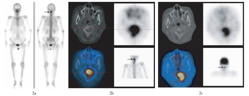
图 2 乳腺癌患者脊柱转移性病变的99Tcm-MDP全身骨显像、SPECT/CT融合显像及PET/CT显像图。患者女性,48岁,乳腺癌术后1年,99Tcm-MDP全身骨显像示第2~3颈椎浓集灶(2a);SPECT/CT融合显像示第2~3颈椎溶骨性骨质破坏伴放射性浓集(2b),诊断为肿瘤骨转移;6个月后18F-FDG PET/CT显像亦示第2~3颈椎溶骨性骨质破坏伴代谢增高(2c)。
Figure 2. Images of the spine metastatic lesions by 99Tcm-whole body bone scan, SPECT/CT fusion imaging and PET/CT imaging
依据随访标准通过高性能专用诊断CT、MRI检查明确病灶87个,以手术或穿刺病理明确病灶23个,通过全身骨显像或SPECT/CT融合显像复查明确病灶88个,通过PET/CT明确病灶12个。所有患者随访确诊时间为SPECT/CT显像后1~12个月,平均5.4个月。最终临床诊断为乳腺癌骨转移病灶87个(41.4%),良性病灶123个(58.6%)。SPECT/ CT正确诊断恶性病灶82个,良性病灶112个,诊断准确率92.4%,对乳腺癌骨转移病灶的诊断灵敏度为94.3%,特异度为91.1%,阳性预测值为88.2%,阴性预测值为95.7%(表 1)。
SPECT/CT诊断 最终临床诊断 合计 恶性 良性 恶性 82 11 93 良性 5 112 117 合计 87 123 210 表 1 SPECT/CT融合显像对乳腺癌骨转移灶的诊断结果
Table 1. Results of SPECT/CT fusion imaging in diagnosing metastatic bone lesions in breast cancer
(个) -
SPECT/CT融合显像对不同部位病灶的诊断准确率不一致,按照诊断准确率由高至低排列,依次为脊椎、骨盆、胸骨、四肢及颅骨、肋骨。对脊椎与肋骨部位病灶的诊断准确率进行比较,其差异有统计学意义(χ2=7.81,P<0.05)(表 2)。
病灶部位 病灶总数 病灶诊断准确数 准确率(%) 恶性 良性 脊椎 97 31 62 95.9 肋骨 43 12 24 83.7 胸骨 32 13 17 93.8 骨盆 21 16 4 95.2 四肢及颅骨 17 10 5 88.2 表 2 SPECT/CT融合显像对不同部位病灶的诊断结果
Table 2. Results of SPECT/CT fusion imaging in diagnosing metastatic bone lesions in different parts of body
-
骨转移是乳腺癌常见的并发症,早期、准确诊断对肿瘤分期、治疗方案选择有重要意义。99Tcm-MDP全身骨显像在鉴别良性骨病与肿瘤骨转移方面比较困难,特别对于乳腺癌患者,由于血液及淋巴回流原因,使骨转移灶特别容易发生在脊椎、肋骨及胸骨等部位。而这些部位亦是良性骨病的好发部位,从而增加了诊断的难度[8]。
本研究中,99Tcm-MDP全身骨显像难以确诊的患者132例共210个病灶,SPECT/ CT准确诊断194个,诊断准确率为92.4%,灵敏度为94.3%,特异度为91.1%,阳性预测值为88.2%,阴性预测值为95.7%。Sharma等[9]报道,SPECT/ CT融合显像诊断乳腺癌骨转移的准确率、灵敏度、特异度、阳性预测值和阴性预测值分别为90%、83%、98%、98%和85%;而顾涛颖等[10]报道的SPECT/CT融合显像诊断乳腺癌骨转移的准确率、灵敏度、特异度、阳性预测值和阴性预测值分别为90.6%、98.9%、95.1%、95.9%和98.7%。与本研究结果基本一致。但是本研究中的灵敏度较Sharma等[9]研究结果偏高,较顾涛颖等[10]研究结果偏低,其原因可能为本研究将诊断为不确定的病灶均考虑为恶性病变所致。另外,本研究所用CT为低剂量定位CT,可能将一些组织结构关系复杂或微小的骨破坏病灶误诊或漏诊,进而导致灵敏度及特异度较顾涛颖等[10]研究结果偏低。
本研究结果发现,SPECT/CT融合显像对不同部位的骨转移灶的诊断准确率是不一致的,对脊椎病灶的诊断准确率高于肋骨,且差异具有统计学意义(χ2=7.81,P<0.05)。分析原因可能为:①CT本身对肋骨的微小的骨质破坏及轻微肋骨骨折灵敏度较低,容易漏诊。②肋骨容易受到隐性外伤的影响,导致其放射性浓集而CT却无明显异常发现,且由于患者自身对这些外伤史的描述不准确,继而导致了这部分病灶的误诊。
SPECT/CT融合显像对于早期转移性病变、CT表现不典型者的诊断也有其局限性,特别是对肋骨处的孤立性病灶。本研究中13例患者的16个误诊病灶中,5个转移性病变误诊为骨骼良性病变,11个良性病变误诊为恶性病变。其中7个为肋骨病灶,另4个为脊椎病灶,其原因可能是肿瘤转移灶处于早期,功能及形态改变不明显,另外椎体退行性变以及内分泌治疗引起的骨质疏松而导致的非特异性浓集亦影响诊断,这与张一秋等[11]的研究结果一致。3个为胸骨与骨盆病灶,胸骨误诊原因为定位CT对轻微胸骨骨质破坏显像欠佳,导致遗漏。而骨盆误诊则与病史询问不详细有关(该患者SPECT/CT融合显像前一周曾进行骨髓穿刺)。2个为四肢及颅骨病灶,也是因为病史询问不详细,误将外伤后浓聚诊断为骨转移。所以详细的问诊及定期的复查对减少误诊是必要的。
综上所述,SPECT/CT特有的功能影像与解剖影像融合的特点克服了全身骨显像特异度差的问题,功能影像与解剖影像融合使诊断达到单独成像所无法企及的深度[12]。其较99Tcm-MDP全身骨显像明显提高了乳腺癌骨转移的诊断准确率,对指导临床治疗方案的制定及预后具有重要意义,有着广阔的临床应用前景。
SPECT/CT融合显像对乳腺癌骨转移的诊断价值
Clinical value of SPECT/CT fusion imaging in diagnosing metastatic bone lesions in breast cancer
-
摘要:
目的 评价SPECT/CT融合显像对99Tcm-MDP全身骨显像难于确诊的乳腺癌骨病灶的鉴别诊断价值。 方法 对99Tcm-MDP全身骨显像难于确诊的132例乳腺癌患者的210个病灶行局部SPECT/CT同机融合断层显像,以临床随访及病理检查获得最终诊断结果,计算SPECT/CT融合显像对骨转移灶的诊断准确率、灵敏度、特异度、阳性预测值及阴性预测值,并对比不同部位病灶的诊断准确率差异。 结果 ① 210个病灶经SPECT/CT融合断层显像正确诊断的恶性病灶82个(39.0%),良性病灶112个(53.3%),诊断准确率为92.4%(194/210),灵敏度为94.3%,特异度为91.1%,阳性预测值为88.2%,阴性预测值为95.7%。②SPECT/CT同机融合断层显像对不同部位的病灶的诊断准确率不一致,脊椎的诊断准确率最高,为95.9%(94/97),肋骨最低,为83.7%(36/43),其差异有统计学意义(χ2=7.81,P<0.05)。 结论 SPECT/CT同机融合显像能够对99Tcm-MDP全身骨显像难于确诊的病灶进行准确诊断,其对不同部位的病灶的诊断准确率有差异,脊椎的诊断准确率最高,肋骨最低。 -
关键词:
- 乳腺肿瘤 /
- 肿瘤转移 /
- 体层摄影术,发射型计算机,单光子 /
- 体层摄影术,X线计算机
Abstract:Objective To evaluate the diagnostic value of SPECT/CT fusion imaging in indeterminate lesions on 99Tcm-MDP whole-body bone scan in patients with breast cancer. Methods 210 bone lesions of 132 patients with breast cancer, for whom the natures of the lesions were unable to be determined by the 99xTcm-MDP whole-body bone scan, were examined by the SPECT/CT fusion imaging simultaneously. The natures of the lesions were eventually confirmed by pathology and clinical follow-up. The accuracy, sensitivity, specificity, positive predictive value and negative predictive value of SPECT/CT fusion imaging for diagnosing bone-metastases were calculated. In addition, the diagnostic accuracy of SPECT/CT fusion imaging for bone lesions in various parts of the body was also analyzed. Results ① Of 210 bone lesions, 82 of bone metastases and 112 benign bone lesions were correctly diagnosed by SPECT/CT fusion imaging. The diagnostic accuracy, sensitivity, specificity, positive prediction value and negative prediction value was 92.4%(194/210), 94.3%(82/87), 91.1%(112/123), 88.2%(82/93), 95.7%(112/117), respectively. ②The diagnostic accuracy of 99Tcm-MDP SPECT/CT for spinal bone lesions were significantly higher than that for rib lesions(95.9% versus 83.7%, χ2=7.81, P < 0.05). Conclusions ① 92.4% bone lesions in patients with breast cancer, which can not determined on 99Tcm-MDP whole-body bone scan, can be diagnosed by SPECT/CT fusion imaging accurately. ②Diagnositic compliance rate of SPECT/CT fusion imaging is different in different parts with obscure lesion, which is the highest in the spine, the minimum in ribs. -
图 1 乳腺癌患者脊柱良性病变的99Tcm-MDP全身骨显像和SPECT/CT显像图。患者女性,70岁,乳腺癌术后5年,99Tcm-MDP全身骨显像示第4颈椎左侧缘浓集灶(1a);SPECT/CT融合显像示第4颈椎左侧横突退变、增生伴放射性浓集(1b),诊断为良性病变;随访4年后见中段颈椎浓集灶减轻(1c)。
Figure 1. Images of spine benign lesions by whole body bone scan and SPECT/CT fusion imaging
图 2 乳腺癌患者脊柱转移性病变的99Tcm-MDP全身骨显像、SPECT/CT融合显像及PET/CT显像图。患者女性,48岁,乳腺癌术后1年,99Tcm-MDP全身骨显像示第2~3颈椎浓集灶(2a);SPECT/CT融合显像示第2~3颈椎溶骨性骨质破坏伴放射性浓集(2b),诊断为肿瘤骨转移;6个月后18F-FDG PET/CT显像亦示第2~3颈椎溶骨性骨质破坏伴代谢增高(2c)。
Figure 2. Images of the spine metastatic lesions by 99Tcm-whole body bone scan, SPECT/CT fusion imaging and PET/CT imaging
表 1 SPECT/CT融合显像对乳腺癌骨转移灶的诊断结果
Table 1. Results of SPECT/CT fusion imaging in diagnosing metastatic bone lesions in breast cancer
(个) SPECT/CT诊断 最终临床诊断 合计 恶性 良性 恶性 82 11 93 良性 5 112 117 合计 87 123 210  下载: 导出CSV
表 2 SPECT/CT融合显像对不同部位病灶的诊断结果
Table 2. Results of SPECT/CT fusion imaging in diagnosing metastatic bone lesions in different parts of body
病灶部位 病灶总数 病灶诊断准确数 准确率(%) 恶性 良性 脊椎 97 31 62 95.9 肋骨 43 12 24 83.7 胸骨 32 13 17 93.8 骨盆 21 16 4 95.2 四肢及颅骨 17 10 5 88.2
下载: 导出CSV
-
[1] 李霓, 郑荣寿, 张思维, 等.中国城乡女性乳腺癌发病趋势分析和预测[J].中华预防医学杂志, 2012, 46(8): 703-707.
[2] Kozlow W, Guise TA. Breast cancer metastasis to bone: mechanisms of osteolysis and implications for therapy[J]. J Mammary Gland Biol Neoplasia, 2005, 10(2): 169-180. [3] 孟德刚, 孙晓光, 黄钢. SPECT/CT在临床肿瘤学中的应用[J].国际放射医学核医学杂志, 2009, 33(4): 214-218.
[4] Utsunomiya D, Shiraishi S, Imuta M, et al. Added value of SPECT/CT fusion in assessing suspected bone metastasis: comparison with scintigraphy alone and nonfused scintigraphy and CT[J]. J Radiology, 2006, 238(1): 264-271. [5] 江勇, 米岚, 余大富, 等. SPECT/CT对脊柱病变鉴别诊断的价值[J].中华核医学杂志, 2011, 31(4): 223-226.
[6] 孟德刚, 孙晓光, 黄刚, 等. SPECT/CT骨显像在多发性骨髓瘤诊断中的临床应用[J].医学影像杂志, 2010, 20(4): 559-562.
[7] 马玉波, 王忠, 顾爱春, 等. SPECT/CT图像融合鉴别诊断疑似骨转移灶良恶性的价值[J].上海交通大学学报, 2010, 30(10): 1246-1250.
[8] Rèmer W, Nèmayr A, Uder M, et al. SPECT-guided CT for evaluating foci of increased bone metabolish classifield as indeterminate on SPECT in cancer patients[J]. J Nucl Med, 2006, 47(7): 1102-1106. [9] Sharma P, Singh H, Kumar R, et al. Bone scintigrsphy in breast cancer: added value of hybrid SPECT-CT and its impact on patient management[J]. Nucl Med Commun, 2012, 33(2): 139-147. [10] 顾涛颖, 石洪成, 陈曙光, 等. SPECT-CT融合图像评价乳腺癌骨转移的增益价值[J].中国临床医学, 2013, 20(5): 665-669.
[11] 张一秋, 石洪成, 陈曙光, 等. SPECT/CT融合图像对脊柱单发病灶鉴别诊断的增益价值[J].中国临床医学, 2010, 17(5): 741-744.
[12] Schillaci O. Hybrid SPECT/CT: a new era for SPECT imaging?[J]. Eur J Nucl Med Mol Imaging, 2005, 32(5): 521-524. -

 点击查看大图

计量
- 文章访问数: 2512
- HTML全文浏览量: 1199
- PDF下载量: 4