


 下载:
下载:
-
脑裂畸形是脑神经元移行异常的一种先天性中枢神经系统疾病,以从侧脑室至半球表面出现裂隙、表面覆有灰质为特点。裂隙可呈开放或闭合状态,可于单侧或双侧大脑中发生。该病的临床症状与裂隙的多少、部位、开放或闭合状态有关。临床上80%的脑裂畸形合并透明隔缺如。本研究对我院近年来诊断或收集的脑裂畸形患者的影像学资料进行回顾性分析,并结合相关文献进行讨论,旨在提高临床对该病影像学表现的认识程度。
-
收集2011年8月至2013年10月在我院被诊断为脑裂畸形的17例患者的临床资料,其中,男性13例、女性4例,年龄18~47岁,平均年龄(29.26±5.21)岁,其中20岁以下3例、20~30岁7例、30岁以上7例。所有患者均有顽固性癫痫,7例合并智力低下、语言障碍和偏瘫。
-
检查仪器为德国西门子公司生产的SOMATOM Emotion16排螺旋CT,行常规颅脑轴位扫描,患者取仰卧位,扫描范围从听眦线至颅顶,层厚2.4 mm,采用脑窗(窗位35 HU,窗宽80 HU)与骨窗(窗位450 HU,窗宽1500 HU)观察。扫描参数为:130 kV,270 mAs,层厚、层距4.8 mm,连续扫描16层,矩阵为512×512。
5例患者均使用深圳市贝斯达医疗器械有限公司BTI-035磁共振成像仪行颅脑平扫,常规横轴位快速自旋回波序列T2加权像:重复时间/回波时间=3800 ms/120 ms,常规横轴位自旋回波T1加权像:重复时间/回波时间=420 ms/20 ms,层厚10 mm,层距1 mm,矩阵:256×192,视野:240×240。
-
由一位从事放射影像学诊断工作20年以上的副主任及以上医师进行读片,并结合相关文献中颅脑的影像学表现进行诊断分析。
-
本研究17例脑裂畸形患者中,裂隙24个,单侧12例、双侧5例,其中同一病例中单侧2处裂隙2例;开放型5例、闭合型12例;横行裂隙位于大脑半球中部中央前后回附近12例、额叶4例、枕叶1例;裂隙内侧端为侧脑室体部外缘14例、侧脑室前角3例,外侧端位于大脑半球外侧凸面中部10例、前部6例、后部1例。
-
脑裂畸形均表现为大脑半球横行裂隙,边缘衬有灰质,裂隙周边衬托的灰质厚薄不均。闭合型裂隙外侧端呈小喇叭或深脑沟样形态,侧脑室外缘或裂隙内侧端呈局限性幕状突起,同侧脑室略扩大12例;开放型裂隙内外侧端呈哑铃状,侧脑室扩大5例,其中2例呈扇形或囊状,侧脑室扩大或积水(图 1~图 4)。

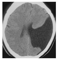
图 1 开放型脑裂畸形患者CT影像图 患者女性,32岁,癫痫,智力低下、右侧偏瘫,CT示开放型裂隙,横跨左侧大脑裂隙呈扇形或囊状,与侧脑室相通,同侧侧脑室扩大,裂隙周边衬托的灰质厚薄不均,部分基底节区和丘脑发育不全,合并透明隔缺如。
Figure 1. CT image of open type schizencephaly

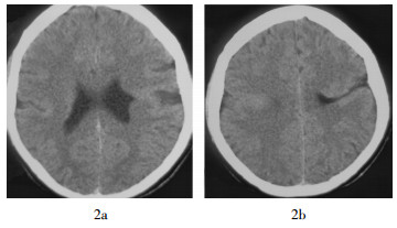
图 2 双侧半球脑裂畸形患者不同扫描层CT影像图 患者男性,18岁,顽固性癫痫。图中,2a:右侧闭合型裂隙,CT示条形灰质层从脑表面延伸到室管膜下附近,与同侧侧脑室不通,伴有透明隔缺如,左侧脑室外壁有幕状突起;2b:左侧开放型裂隙,中央前后回附近一条横行裂隙,边缘衬有厚薄不均匀的灰质层,同侧侧脑室扩大,右侧中央前后回附近见一灰质团块。
Figure 2. CT images of bilateral schizencephaly

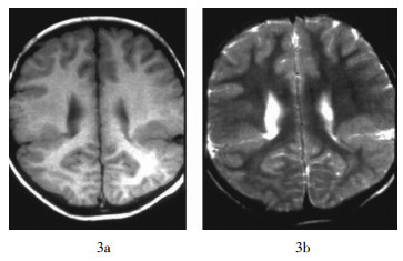
图 3 双侧半球对称性脑裂畸形患者MRI影像图 患者男性,19岁,自幼癫痫、智力低下。图中,T1加权像(3a)和T2加权像(3b)均示右侧开放型裂隙、左侧闭合型裂隙,位于中央后回附近一条横行裂隙,从脑表面延伸到室管膜下,边缘衬有不规则的灰质层团块。
Figure 3. MRI images of bilateral symmetrical schizencephaly

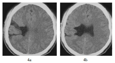
图 4 单侧半球脑裂畸形患者不同扫描层CT影像图 患者女性,25岁,难治性癫痫,自幼智力低下。图中,4a:开放型裂隙,位于右侧中央前后回附近两条横行裂隙,从脑表面延伸到室管膜下,边缘衬有不规则的灰质层,同侧侧脑室扩大,其外壁有幕状突起与裂隙相连,伴有多发性小脑回及透明隔缺如;4b:位于右侧中央前后回附近的两条横行裂隙,其中一条为开放型裂隙,另一条为闭合型裂隙。
Figure 4. CT images of unilateral schizencephaly
-
本研究17例脑裂畸形患者中,合并透明隔缺如10例、灰质异位8例、一侧丘脑和部分基底节缺失2例、多小脑回畸形4例、巨脑回畸形1例、侧脑室三角区白质较少2例、蛛网膜囊肿5例。
-
脑裂畸形系大脑皮层的神经元移行异常最严重的一种类型,大约在胚胎第6周,由脑室壁的单层柱状上皮构成原始的神经管进行分化,形成4个神经胚胎带,由内到外分别为脑室带、脑室下带、中间带及边缘带[1]。脑室带和脑室下带的原始神经细胞分化成各类神经元,又称生发基质层。约第8周开始,生发基质层的成神经细胞逐渐移行穿越中间带及边缘带形成皮质板层,相应的部位分化为各类功能的神经元[2-3]。生发基质层神经元的移行过程主要发生在胚胎第8~16周,整个过程复杂而漫长,期间受到不同程度的干扰,如缺血、感染或照射放射线等,使其出现局灶性紊乱而停留在皮层下不同水平的异常部位,受累的皮质常增厚并伴有神经元排列紊乱,从而导致各种不同类型的脑裂畸形发生[4]。脑裂畸形常伴有透明隔缺如、灰质异位及胼胝体发育不良等,可能与胚胎期干扰因素影响到整体脑组织发育有关,需要进一步探讨。
-
大脑半球内出现横行裂隙,外侧端与软脑膜相连,内侧端与侧脑室相通,同侧脑室呈局限性幕状突起,形成软脑膜-室管膜缝(P-E缝),可单侧或双侧出现,个别病例同侧两处可发生裂隙,邻近的皮层灰质卷入并衬托于裂隙两边,常合并邻近灰质异位。根据裂隙的开放程度和累及区域的大小分为2种类型,其中,闭合型裂隙为一条融合的裂隙,其两边见灰质层相贴,裂隙不含脑脊液,可达脑白质深层,同时常伴有灰质异位,不与侧脑室相连,由于病变侧脑组织的神经元移行障碍,阻碍了邻近脑灰质的发育,使在向脑表面移行的过程中,其灰质向病变部位裂隙陷入并彼此融合,此种类型临床表现为顽固性癫痫发作,智力低下、偏瘫,语言障碍较轻,临床往往易误诊或漏诊;开放型裂隙为横跨大脑半球裂隙分离,皮层内折明显,裂隙与侧脑室相同,其内含脑脊液,内衬有薄膜,其内层为室管膜,外层为软脑膜,受累的大脑半球脑组织常伴有缺失,严重病例呈空洞样与同侧侧脑室和蛛网膜下腔相通,并伴有脑室扩大或脑积水,此类患者临床表现为难治性癫痫,智力低下、偏瘫,语言障碍较为典型。有文献报道不管何种类型的脑裂畸形伴发的癫痫,主要由异位灰质团块所致,功能区脑组织的缺失则为脑瘫和智力低下的主要病因[5]。
-
CT、MRI可清楚显示脑裂畸形的临床解剖特点及伴发脑发育畸形,该病的影像学表现为大脑半球见横行裂隙,从脑表面延伸到室管膜下,裂隙的两侧衬有不规则增厚的灰质层,其表面有软脑膜与室管膜相连。裂隙大多位于中央前后回附近,部分位于额叶和枕叶[6]。闭合型裂隙呈窄缝状或深脑沟样,甚至无明显的裂隙,仅表现为条形灰质层;开放型裂隙则较宽,可呈扇形或囊状,多与侧脑室相通,同侧侧脑室扩大(图 1),其外壁常有幕状突起与裂隙相连。脑表面裂隙外侧端呈喇叭状形态,附近脑回常发育不良,多伴发微小脑回或巨脑回畸形、透明隔缺损、灰质异位、部分基底节缺失及蛛网膜囊肿等。本组病例中2例双侧裂隙位于中央前后回附近,一侧为开放型,而另一侧为闭合型(图 2~图 3);2例单侧2处裂隙,内折的灰质层较单侧一处厚而面大(图 4),此种类型文献报道较少,需要大量的病例进行进一步研究。
-
临床中脑裂畸形需与孤立性灰质异位、脑穿通畸形囊肿、蛛网膜囊肿等疾病进行鉴别:①闭合型脑裂畸形外侧端在脑表面有一凹痕,内侧端深达脑白质深部,有的可至室管膜下,部分患者侧脑室外缘见幕状突起;而孤立性灰质异位无此征象,仅皮质下脑白质区域见异位灰质团块。②开放型脑裂畸形边缘有灰质内折,形态多呈哑铃状;而脑穿通畸形囊肿多呈扇形,最窄处位于一端,边缘无灰质附着,鉴别不难。③蛛网膜囊肿多呈球形,可发生于脑组织的任何部位,边缘无灰质附着,易于鉴别。
脑裂畸形的CT、MRI影像学征象分析
Analysis of the CT and MRI imaging features of schizencephaly
-
摘要:
目的 探讨脑裂畸形的CT、MRI特征性表现及其临床诊断价值。 方法 回顾性分析17例脑裂畸形患者的临床CT、MRI的影像学征象。 结果 17例脑裂畸形患者的CT、MRI图像表现为自皮质至侧脑室的横跨大脑半球的裂隙,裂隙两旁为内折的灰质;裂隙内侧端多为脑室外壁的幕状突起,闭合型裂隙外侧端呈小喇叭或深脑沟样形态,开放型裂隙内外侧端呈哑铃状。17例脑裂畸形患者中,单侧12例、双侧5例,共有24个裂隙;闭合型12例、开放型5例;合并透明隔缺如10例、灰质异位8例、一侧丘脑和部分基底节缺失2例、多小脑回畸形4例、巨脑回畸形1例、侧脑室三角区白质较少2例、蛛网膜囊肿5例。 结论 脑裂畸形具有特征性的影像学表现,CT、MRI检查能够清楚地显示脑裂畸形的病理解剖形态。 -
关键词:
- 体层摄影术,X线计算机 /
- 磁共振成像 /
- 脑裂畸形
Abstract:Objective To investigate the features and clinical diagnosis values of CT and MRI in schizencephaly. Methods The imaging features of CT and MRI were restrospectively analyzed in 17 cases of schizencephaly patients. Results CT and MRI showed the fractures from the cortex to the lateral ventricle which stretch across the brain hemisphere, and there were inflexed grey matter on both sides. In the inner end of fracture, there were some curtain like protuberance of the ventricle walls, the lateral end of closed type fracture presented a trumpet or deep sulci like shape, and the open type fracture presented a dumbbell shape. A total of 24 fractures were found in 17 cases of schizencephaly patients, 12 cases were unilateral, 5 cases were bilateral; 12 cases were closed type, 5 cases were open type; 10 cases complicated with transparent septum defect, 8 cases were heterotopic gray matter, 2 cases were unilateral thalamus and partial basal ganglia defect, 4 cases were polymicrogyria, 1 case was pachygyria, 2 cases were less white matter at the trigone of the lateral ventricles, 5 cases were arachnoid cyst. Conclusion Schizencephaly has distinctive imaging features, CT and MRI examination can clearly display the pathological morphology of schizencephaly. -
Key words:
- Tomography, X-ray computed /
- Magnetic resonance imaging /
- Schizencephaly
-
图 1 开放型脑裂畸形患者CT影像图 患者女性,32岁,癫痫,智力低下、右侧偏瘫,CT示开放型裂隙,横跨左侧大脑裂隙呈扇形或囊状,与侧脑室相通,同侧侧脑室扩大,裂隙周边衬托的灰质厚薄不均,部分基底节区和丘脑发育不全,合并透明隔缺如。
Figure 1. CT image of open type schizencephaly
图 2 双侧半球脑裂畸形患者不同扫描层CT影像图 患者男性,18岁,顽固性癫痫。图中,2a:右侧闭合型裂隙,CT示条形灰质层从脑表面延伸到室管膜下附近,与同侧侧脑室不通,伴有透明隔缺如,左侧脑室外壁有幕状突起;2b:左侧开放型裂隙,中央前后回附近一条横行裂隙,边缘衬有厚薄不均匀的灰质层,同侧侧脑室扩大,右侧中央前后回附近见一灰质团块。
Figure 2. CT images of bilateral schizencephaly
图 3 双侧半球对称性脑裂畸形患者MRI影像图 患者男性,19岁,自幼癫痫、智力低下。图中,T1加权像(3a)和T2加权像(3b)均示右侧开放型裂隙、左侧闭合型裂隙,位于中央后回附近一条横行裂隙,从脑表面延伸到室管膜下,边缘衬有不规则的灰质层团块。
Figure 3. MRI images of bilateral symmetrical schizencephaly
-
[1] 崔光彬, 王玮, 宋立军, 等.脑裂畸形的MRI诊断与鉴别诊断[J].实用放射学杂志, 2007, 23(5): 581-583. doi: 10.3969/j.issn.1002-1671.2007.05.002
[2] 莫瑞嘉, 农明进.脑神经元移行异常的影像学诊断[J].实用放射学杂志, 2004, 20(6): 552-554. doi: 10.3969/j.issn.1002-1671.2004.06.023
[3] 刘可夫, 刘斌, 张家文, 等.脑裂畸形的CT表现[J].放射学实践, 2003, 18(8): 566-567. doi: 10.3969/j.issn.1000-0313.2003.08.009
[4] 曹代荣, 李银官, 倪希和, 等.脑裂畸形的MR影像征象分析(附32例报告)[J].放射学实践, 2002, 17(6): 468-470. doi: 10.3969/j.issn.1000-0313.2002.06.003
[5] 李晓明, 王海, 罗韦华, 等.儿童脑裂畸形的临床与CT分析[J].实用放射学杂志, 2005, 21(6): 661-662. doi: 10.3969/j.issn.1002-1671.2005.06.032
[6] 张文伟, 纪建松, 周利民, 等.闭唇型脑裂畸形的MR诊断[J].医学影像学杂志, 2012, 22(2): 170-172. doi: 10.3969/j.issn.1006-9011.2012.02.009
-

 点击查看大图
计量
- 文章访问数: 3412
- HTML全文浏览量: 2361
- PDF下载量: 5