


-
近年来,通过临床资料、神经解剖及脑功能成像对小脑功能进行研究,发现小脑除了有维持身体平衡、调节肌张力及协调骨骼肌的随意运动外,还参与语言的处理。通过实验任务探讨小脑在语言处理方面的研究国内外都有报道,如国内学者运用听真假词的任务,发现小脑参与汉字词语语义处理[1];国外学者通过词语押韵判断的任务,发现小脑参与语音处理[2],但用静息态来研究小脑在语言方面的功能极为少见,静息态功能磁共振成像(functional magnetic resonance imaging,fMRI)是近几年发展起来的脑功能成像方法,它无需设计繁琐的实验任务,不需考虑受试者执行任务能力的差异,在静息状态下,既可观察远隔脑区之间的时间序列的同步性,又可观察局部脑区自发的神经活动的程度,已经成为目前研究的热点。该研究采用静息态fMRI探讨小脑在语言中的作用机制。
-
选取2012年7月至2013年3月于石河子大学医学院第三附属医院神经内科就诊的脑卒中后失语症患者17例(失语组),其中,男性10例、女性7例,年龄46~74岁,平均年龄(60.5±8.6)岁。入选标准:①首次发病,病程3 d至3个月,CT或MRI显示有明确的左侧大脑半球病灶,无小脑病灶;②发病前言语功能正常,病后失语检查诊断为失语患者;③无小脑疾病,无精神、认知等障碍;④汉族,右利手,受教育年龄>5年。
另选2012年7月至2013年3月在石河子大学医学院第三附属医院体检中心体检的20名健康体检者作为正常对照组,其中,男性12名、女性8名,年龄54~64岁,平均年龄(59.0±7.8)岁。入选标准:①无认知障碍主诉,无神经精神病史及药物滥用史。②无言语障碍、躯体及小脑疾病等;③CT或MRI显示未见明显异常;④汉族,右利手,受教育年龄>5年。
两组受试者都知情同意并签署了知情同意书。
-
患者在做fMRI之前进行西方失语量表(Western Aphasia Battery,WAB)(汉化版)检查[3]。WAB主要包括:①自发语,细分为信息内容和流畅性;②听理解,细分为是或不是问答、单词听理解、执行命令;③复述;④命名,细分为呼名、列举、语句完成、应答。失语检查完成后,可以得到失语商(aphasia quotient,AQ),它为口语障碍程度的可信赖尺度,可反映出失语症的严重度,而且可以作为失语症的好转与恶化的评价指标。其计算方法为:以自发言语分数、口语理解分数除以20,复述分数、命名分数除以10的各项之和乘以2得出。最高分100分,正常值为98.4~99.6,AQ<93.8可评为失语。
-
采用德国Siemens verio 3.0 T磁共振仪,先扫描解剖像,后扫描功能像,三维磁化强度预备快速梯度回波成像获得T1加权结构像:脉冲重复时间/脉冲回波时间2300 ms/2.52 ms,层厚1 mm,层数176;视野256 mm×256 mm,矩阵256×256,体素1.0 mm×1.0 mm×1.0 mm,翻转角9°;静息态fMRI扫描序列采用梯度回波-平面回波序列:脉冲重复时间/脉冲回波时间2330 ms/30 ms,层厚3 mm,层数36,视野240 mm×240 mm,矩阵64×64,体素3.8 mm×3.8 mm×3.0 mm,翻转角90°。静息态功能扫描获得205个时间点,扫描时间为8分15秒。
-
预处理基于Matlab2009b平台,使用SPM8(统计参数图,http://www.fil.ion.ucl.ac.uk/spm)及DPARSF_V2.0(数据处理助理Resting-State fMRI分析工具包,http://resting-fmri.sourceforge.net)和REST_V1.8(Resting-State fMRI数据分析工具包,http://resting-fmri.sourceforge.net)对静息态数据进行系统预处理。考虑到磁场达到稳定需要的时间及被试对环境的适应时间,首先剔除前5个时相的采集图像,对剩余200个时相的数据进行下列预处理:时间校正、头动校正、空间标准化、采用6 mm×6 mm×6 mm半高全宽的高斯核行空间平滑处理、去线性漂移及低频滤波(0.01~0.08 Hz)。
-
滤波后的数据经过傅里叶转换成频率谱,而频率谱与原始时间序列的频率振幅的平方成正比,这样每一体素频率谱的平方根在0.01~0.08 Hz的低频下被计算出来,得到平均平方根作为ALFF,为了达到标准化目的,每个体素的ALFF值都除以全脑均值(mean),即mALFF,得到的全脑mALFF用于双样本的计算。
-
种子点的选取来自于失语患者ALFF值减低的右侧小脑,其X、Y、Z轴的坐标为(42、18、36),ROI的体积是用REST Slice Viewer生成一个覆盖39个体素的Mask(图 1)。将上述Mask应用到正常对照组。

图 1 种子点和ROI的选取图图中,十字交叉点即为种子点,周围体素生成的Mask为ROI,从冠状位、矢状位、轴位3个不同的层面显示种子点及ROI。图像左右按照影像学常规区分。
Figure 1. The selection map of seed point and ROI
-
首先去除协变量,主要为去除头动、全脑信号、白质信号和脑脊液对低频同步振荡信号的影响。然后以右侧小脑中各体素的时间信号的平均值作为种子点的时间信号,通过分析种子点与全脑各体素的时间序列得到Pearson相关图及相关系数。
-
对正常对照者与失语患者的脑区在REST软件里进行双样本t检验,检验水准为P<0.005(Alphasim校正),集簇大小>27体素,结果在REST Slice Viewer显示。对正常对照组以右侧小脑为种子点在REST软件里进行单样本t检验,检验水准为P<0.0001(未校正),集簇大小>40体素。
-
本研究经过预处理,把正常对照组头部运动三维平移超过1 mm、三维旋转超过1°的受试者数据剔除;把失语组头部运动三维平移超过2 mm、三维旋转超过2°的受试者数据剔除。符合上述标准的失语组患者12例,男性8例、女性4例,年龄46~74岁,平均年龄(60.5±8.0)岁;正常对照组20名,男性12名、女性8名,年龄54~64岁,平均年龄(59.0±7.8)岁。
-
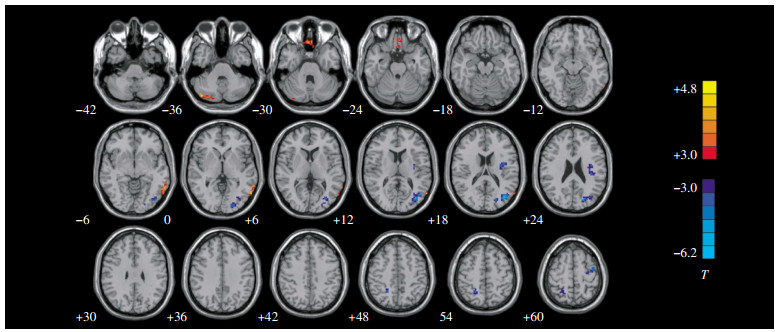
失语患者ALFF值减低的脑区有左侧颞中回、左侧前额叶内侧回、右侧小脑;失语患者ALFF值升高的脑区有左侧枕叶、左侧前额叶、左侧岛叶及右侧楔前叶(P<0.005,Alphasim校正,集簇大小>27体素)(表 1、图 2)。
脑区 Brodmann
分区团块体素大小 MNI坐标 t值 X Y Z 左侧颞中回 21 73 -60 -60 0 4.52 左侧前额叶内侧回 11 79 -3 35 -27 4.59 右侧小脑 - 39 42 -18 -36 4.41 左侧枕叶 19、18、17 220 -45 -69 15 -6.26 左侧岛叶 13 83 -36 -24 27 -5.4 右侧楔前叶 7 43 21 -54 54 -4.39 左侧前额叶 6 35 -39 -3 60 -4.26 注:表中,“-”表示无此分区;P<0.005,Alphasim校正,集簇大小>27体素。 表 1 失语组低频振荡振幅值减低及升高的脑区
Table 1. Brain regions of increased and decreased of amplitude of low frequency fluctuation value in aphasia group

图 2 失语组与正常对照组的低频振荡振幅差异图图中,红色区域表示与正常对照者相比,失语患者ALFF减低的脑区,相关脑区T值越大表示正相关性越强。蓝色区域表示与正常对照者相比,失语患者ALFF升高的脑区,相关脑区T值越大表示负相关性越强。图像下角的数值为MNI坐标Z轴的值。图像左右按照影像学常规区分。
Figure 2. Amplitude of low frequency fluctuation differences between the aphasia and control groups
-
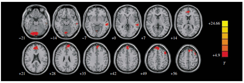
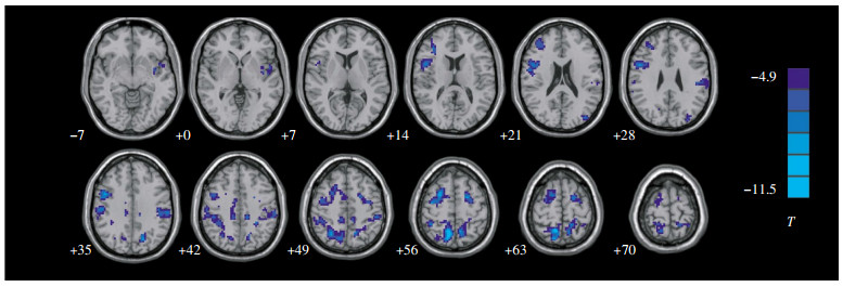
正常对照组静息状态下与右侧小脑呈正相关的脑区有:右侧小脑后部、小脑蚓部、左侧颞中回、左侧额上回内侧面、辅助运动区(图 3)。正常对照组静息状态下与右侧小脑呈负相关的脑区主要有:左侧额中回、左侧中央后回及右侧额中回(P<0.0001,未校正,集簇大小>40体素)(表 2、图 4)。

图 3 正常对照组静息状态下与右侧小脑ROI呈正相关的功能连接图图中,红色区域表示正常对照组静息状态下与右侧小脑ROI呈正相关的脑区。相关脑区T值越大,表示正相关性越强。图像下角的数值为MNI坐标Z轴的值。图像左右按照影像学常规区分。
Figure 3. Positive functional connectivity map with the right cerebellum ROI of the normal control group in resting state
脑区 Brodmann分区 团块体素大小 MNI坐标 t值 X Y Z 右侧小脑后部 - 1540 33 -81 -33 24.7 小脑蚓部 - 85 6 -54 -39 9.9 左侧颞中回 21 83 -63 -30 -6 8.85 左侧额上回内侧面 9 485 -3 45 21 9.47 辅助运动区 6 76 -48 12 51 7.97 右侧额中回 6 1768 27 6 54 -11.49 左侧额中回 6 136 -21 3 51 -10.68 左侧中央后回 2 703 -45 -30 33 -10.78 注:表中,“-”表示无此分区;P<0.005,Alphasim校正,集簇大小>27体素。 表 2 正常对照组静息状态下与右侧小脑ROI呈正负相关的脑区
Table 2. Brain regions of positive and negative functional connec-tivity with the right cerebellum ROI of the normal control group in resting state

图 4 正常对照组静息状态下与右侧小脑ROI呈负相关的功能连接图图中,蓝色区域表示正常对照组静息状态下与右侧小脑ROI负相关的脑区。相关脑区T值越大,表示负相关性越强。图像下角的数值为MNI坐标Z轴的值。图像左右按照影像学常规区分。
Figure 4. Negative functional connectivity map with the right cerebellum ROI of the normal control group in resting state
-
Logothetis等[4]的研究成果显示,BOLD信号与神经突触活动产生的局部场电位的变化相关,认为这种BOLD效应反映了输入和皮层内处理的整合。Nir等[5]发现神经元的自发放电活动有振荡的特性与静息态BOLD-fMRI低频振荡的特征非常吻合,由此认为静息fMRI自发BOLD信号的低频振荡很可能来源于神经元自发放电活动。国内学者开发了一个指数,即低频振幅,来探测局部脑区BOLD信号自发振荡的强度,继而反映局部脑区代谢活动的程度,并应用于儿童注意缺陷多动障碍[6],可知ALFF反映的是局部脑区自发的神经活动的程度。本研究发现失语组的左侧颞中回、左侧前额叶内侧回、右侧小脑的ALFF信号相比正常对照组明显减低,可能与局部的损伤病灶影响这些脑区神经元的活动有关,进而出现语言障碍。Silveri等[7]认为小脑参与认知功能及语言处理的调节,尤其是右侧小脑。Mariën等[8]报道了一例右侧小脑梗死的患者出现认知和语言障碍。Murdoch[9]综述了小脑在语言方面的能力,涉及口语的流利、单词的检索、句法、阅读、书写及综合语言的能力,若小脑受损上述功能会受到影响。这些现象可能源于小脑与大脑皮层解剖连接的破坏,而导致与小脑相连的脑区出现功能降低或丧失。在解剖上,大脑到小脑有两条途径,一条是大脑皮质-脑桥核-对侧小脑,另一条是大脑皮质-红核-下橄榄核-对侧小脑;另外小脑反馈到大脑的路径是小脑齿状核-丘脑(腹外侧核和腹前核)-对侧额叶皮质,可知,当小脑与大脑出现神经机能联系不能时,就会出现小脑反馈到大脑的信息减少或中断,继而导致语言障碍。总之,从右侧小脑损伤的临床表现及与左侧额叶皮层的神经解剖联系和本实验所得出的失语组右侧小脑某一脑区神经活动低于正常对照组,可知,本研究种子点的选取是可行的。
-
本研究显示,正常对照组静息状态下与右侧小脑呈正相关的脑区主要有左侧颞中回、左侧额上回内侧面、辅助运动区。左侧颞中回后部处于听觉与视觉皮层的交界区,接受来自于Wernicke区(颞上回后部)、初级听觉皮层(颞横回)及视觉皮层的信息,参与词汇语句的理解[10]。Turken等[11]发现左侧颞中回与多个有语言功能的脑区存在结构和功能连接,其受损会出现严重而持久的语义理解障碍。Brodman 6区为上语言区,此处在发音和形成语言方面特别重要,Krainik等[12]对20例Brodman 6区有低级胶质细胞瘤的患者进行了切除手术,术后发现患者存在言语障碍。1990年Alexander等[13]提出了“额叶前背外侧环路”,即Brodman 9区和10区发出神经纤维投射到尾状核背外侧,然后投射到苍白球和黑质,再投射到丘脑的腹前核,最后回到前额叶皮质。Gold等[14]认为此环路受损会阻止语义系统的激活,导致词语启动困难,继而导致丧动力性失语。可知,与右侧小脑的种子点存在功能连接的脑区都涉及语言功能。
总之,右侧小脑的解剖连接涉及到对侧的丘脑、额叶皮质;功能连接涉及到左侧颞中回、左侧额上回内侧面、辅助运动区,且本研究显示,左侧大脑半球卒中后的失语患者,其右侧小脑的ALFF信号减低。可知,右侧小脑在语言的形成中起着重要的作用。
静息态功能磁共振成像技术对右侧小脑参与语言功能的应用研究
Analysis of the mechanism of right cerebellum in language using resting state functional magnetic resonance imaging
-
摘要:
目的 通过静息态功能磁共振成像技术来探讨小脑在产生语言中的作用机制。 方法 运用西门子3.0T磁共振仪获得静息态数据,用DPARSF软件对静息态数据进行预处理。首先对12例卒中后失语患者和20例正常对照者进行低频振荡振幅(ALFF)分析,在REST软件中行两样本t检验,得到失语组右侧小脑的某一脑区ALFF信号低于正常对照组,然后把此脑区作为种子点和ROI选取区,在正常对照组中进行单样本t检验,得到功能连接图。 结果 与正常对照组相比,失语组左侧颞中回、左侧前额叶内侧回、右侧小脑的ALFF减低;右侧小脑在正常对照组功能连接的脑区有小脑蚓部、左侧颞中回、左侧额上回内侧面、辅助运动区。 结论 右侧小脑本身及通过影响与其存在功能连接和解剖连接的脑区参与语言的产生。 Abstract:Objective To elucidate the generation mechanism of cerebellum in language by resting state functional magnetic resonance imaging(fMRI). Methods Siemens verio 3.0T MR Scanner was used to obtain all of the subjects of fMRI data. The fMRI data were processed with the software of Data Processing Assistant for Resting-State fMRI Analysis Toolkit. Firstly, 12 patients with poststroke aphasia and 20 normal controls were analyzed by amplitude of low frequency fluctuation(ALFF) and performed with two sample t-test by REST software; Secondly, decreased ALFF of certain brain regions of the cerebellum were selected as the regions of seed point and ROI, and then calculated the single sample t-test in normal controls to obtain function connectivity map. Results As compared with those in normal subjects, the regions showing decreased ALFF in aphasia patients were distributed in left middle temporal gyrus, left medial prefrontal gyrus, right cerebellum. Positive functional connectivity with certain brain regions of the cerebellum ROI was seen in cerebellum tonsil, left middle temporal gyrus, left medial superior frontal gyrus and supplementary motor area. Conclusion Right cerebellum itself and through its impact on the presence of functional connections and structural connections of the brains participate in language production. -
Key words:
- Cerebellum /
- Aphasia /
- Magnetic resonance imaging
-
图 1 种子点和ROI的选取图图中,十字交叉点即为种子点,周围体素生成的Mask为ROI,从冠状位、矢状位、轴位3个不同的层面显示种子点及ROI。图像左右按照影像学常规区分。
Figure 1. The selection map of seed point and ROI
图 2 失语组与正常对照组的低频振荡振幅差异图图中,红色区域表示与正常对照者相比,失语患者ALFF减低的脑区,相关脑区T值越大表示正相关性越强。蓝色区域表示与正常对照者相比,失语患者ALFF升高的脑区,相关脑区T值越大表示负相关性越强。图像下角的数值为MNI坐标Z轴的值。图像左右按照影像学常规区分。
Figure 2. Amplitude of low frequency fluctuation differences between the aphasia and control groups
图 3 正常对照组静息状态下与右侧小脑ROI呈正相关的功能连接图图中,红色区域表示正常对照组静息状态下与右侧小脑ROI呈正相关的脑区。相关脑区T值越大,表示正相关性越强。图像下角的数值为MNI坐标Z轴的值。图像左右按照影像学常规区分。
Figure 3. Positive functional connectivity map with the right cerebellum ROI of the normal control group in resting state
图 4 正常对照组静息状态下与右侧小脑ROI呈负相关的功能连接图图中,蓝色区域表示正常对照组静息状态下与右侧小脑ROI负相关的脑区。相关脑区T值越大,表示负相关性越强。图像下角的数值为MNI坐标Z轴的值。图像左右按照影像学常规区分。
Figure 4. Negative functional connectivity map with the right cerebellum ROI of the normal control group in resting state
表 1 失语组低频振荡振幅值减低及升高的脑区
Table 1. Brain regions of increased and decreased of amplitude of low frequency fluctuation value in aphasia group
脑区 Brodmann
分区团块体素大小 MNI坐标 t值 X Y Z 左侧颞中回 21 73 -60 -60 0 4.52 左侧前额叶内侧回 11 79 -3 35 -27 4.59 右侧小脑 - 39 42 -18 -36 4.41 左侧枕叶 19、18、17 220 -45 -69 15 -6.26 左侧岛叶 13 83 -36 -24 27 -5.4 右侧楔前叶 7 43 21 -54 54 -4.39 左侧前额叶 6 35 -39 -3 60 -4.26 注:表中,“-”表示无此分区;P<0.005,Alphasim校正,集簇大小>27体素。  下载: 导出CSV
表 2 正常对照组静息状态下与右侧小脑ROI呈正负相关的脑区
Table 2. Brain regions of positive and negative functional connec-tivity with the right cerebellum ROI of the normal control group in resting state
脑区 Brodmann分区 团块体素大小 MNI坐标 t值 X Y Z 右侧小脑后部 - 1540 33 -81 -33 24.7 小脑蚓部 - 85 6 -54 -39 9.9 左侧颞中回 21 83 -63 -30 -6 8.85 左侧额上回内侧面 9 485 -3 45 21 9.47 辅助运动区 6 76 -48 12 51 7.97 右侧额中回 6 1768 27 6 54 -11.49 左侧额中回 6 136 -21 3 51 -10.68 左侧中央后回 2 703 -45 -30 33 -10.78 注:表中,“-”表示无此分区;P<0.005,Alphasim校正,集簇大小>27体素。
下载: 导出CSV
-
[1] 掲冰, 杨振燕, 赵小虎, 等.小脑汉字词语处理相关脑区的功能磁共振研究[J].中国医学计算机成像杂志, 2008, 14(3): 200-204. doi: 10.3969/j.issn.1006-5741.2008.03.004
[2] James R, Booth, Lydia Wood, et al. The role of the basal ganglia and cerebellum in language processing[J]. Brain Res, 2007, 1133(1): 136-144. [3] 王荫华.西方失语证成套测验(WAB)介绍(一)[J].中国康复理论与实践杂志, 1997, 3(2): 87-89.
[4] Logothetis NK, Pauls J, Augath M, et al. Neurophysiological investigation of the basis of the fMRI signal[J]. Nature, 2001, 412(6843): 150-157. doi: 10.1038/35084005 [5] Nir Y, Mukamel R, Dinstein I, et al. Interhemispheric correlations of slow spontaneous neuronal fluctuations revealed in human sensory cortex[J]. Nat Neurosci, 2008, 11(9): 1100-1108. doi: 10.1038/nn.2177 [6] Cao X, Cao Q, Long X, et al. Abnormal resting-state functional connectivity patterns of the putamen in medication-naive children with attention deficit hyperactivity disorder[J]. Brain Res, 2009, 1303: 195-206. doi: 10.1016/j.brainres.2009.08.029 [7] Silveri MC, Misciagna S. Language, memory, and the cerebellum[J]. J Neurolinguistics, 2000, 13(2): 129-143. [8] Mariën P, Baillieux H, De Smet HJ, et al. Cognitive, linguistic and affective disturbances following a right superior cerebellar artery infarction: a case study[J]. Cortex, 2009, 45(4): 527-536. doi: 10.1016/j.cortex.2007.12.010 [9] Murdoch BE. The cerebellum and language: historical perspective and review[J]. Cortex, 2010, 46(7): 858-868. doi: 10.1016/j.cortex.2009.07.018 [10] Dronkers NF, Wilkins DP, Van Valin RD Jr, et al. Lesion analysis of the brain areas involved in language comprehension[J]. Cognition, 2004, 92(1-2): 145-177. doi: 10.1016/j.cognition.2003.11.002 [11] Turken AU, Dronkers NF. The neural architecture of the language comprehension network: converging evidence from lesion and connectivity analyses[J]. Front Syst Neurosci, 2011, 5: 1. [12] Krainik A, Lehéricy S, Duffau H, el al. Postoperative speech disorder after medial frontal surgery: role of the supplementary motor area[J]. Neurology, 2003, 60(4): 587-594. doi: 10.1212/01.WNL.0000048206.07837.59 [13] Alexander GE, Crutcher MD, DeLong MR. Basal ganglia-thalamocortical circuits: parallel substrates for motor, oculomotor, "prefrontal" and "limbic" functions[J]. Prog Brain Res, 1990, 85: 119-146. [14] Gold M, Nadeau SE, Jacobs DH, el al. Adynamic aphasia: A transcortical motor aphasia with defective semantic strategy formation[J]. Brain Lang, 1997, 57(3): 374-393. -

 点击查看大图

计量
- 文章访问数: 2939
- HTML全文浏览量: 1626
- PDF下载量: 3