


-
骨肉瘤是最常见的原发恶性骨肿瘤,好发于长骨的干骺端,由肿瘤性成骨细胞、肿瘤性骨样组织和肿瘤骨构成。骨肉瘤易发生肺转移,且一旦出现肺转移往往提示预后不良[1],然而早期发现肺转移并采取有效的手术切除及化疗可明显提高患者的长期生存率[2-4]。由于骨肉瘤肿瘤细胞产生数量不等的新生骨样组织且血供丰富,在99Tcm-MDP骨显像中原发病灶和转移病灶均可表现出对显像剂的高度摄取,因此骨显像不仅可用于了解原发肿瘤的侵犯情况,观察手术前、后化疗疗效,同时还可检出肺或其他软组织的转移病灶[5]。尽管早期已有骨显像用于骨肉瘤肺内转移检出的报道[5-7],但对其诊断效能的评价目前尚缺少大宗病例的分析报道。目前随着SPECT/CT的临床应用,骨肉瘤患者在骨显像检查过程中可同时获得胸部CT诊断信息,这种功能与解剖影像的结合提高了对肺转移诊断的准确率;另一方面,胸部CT也为客观评价骨显像用于骨肉瘤肺转移的诊断提供了一种参考依据。笔者对一组骨肉瘤患者的99Tcm-MDP SPECT/CT的显像结果及临床资料进行了回顾性分析,目的在于观察骨肉瘤肺内转移的发生率,分别评价99Tcm-MDP SPECT/CT显像中的骨显像与胸部CT对肺转移的检出效能及其影响因素。
-
选取2006年4月至2012年12月在我院经手术病理证实、且在术前或术后临床随访中行99Tcm-MDP SPECT/CT显像的178例骨肉瘤患者,其中男性117例、女性61例,患者年龄4~69岁,平均(19.1±9.6)岁。其中术前检查者97例,术后临床随访者81例。在178例患者中可获得显像前后两周内血清碱性磷酸酶(alkaline phosphatase,ALP)测定结果者共有161例。
-
使用Philips Precedence 6.0(荷兰飞利浦公司制造)SPECT/CT仪,所有患者予静脉注射740 MBq 99Tcm-MDP(原子高科股份有限公司提供)3 h后,先行全身前、后位平面骨显像。图像采集条件:低能高分辨准直器,矩阵1024×1024,床速0.14 m/s。骨显像结束后加做胸部CT扫描,扫描过程中要求患者提前5 s屏气,平扫技术参数:球管的电压和电流分别为120 kV和50 mA,矩阵512×512,层厚5 mm,重建图像层厚2 mm。
所有患者或患者家属均于检查前签署了知情同意书。
-
由3名以上的核医学科医师以视觉判断方式集体阅片,首先于骨显像中观察原发骨肿瘤的病变范围及有无其他部位骨或软组织转移,当发现肺内出现异常放射性浓聚灶,在排除肋骨病变的情况下,即认为出现肺内转移;随后观察胸部CT影像,当双肺野内出现异常密度影时,亦考虑为肺内转移。发现肺内病灶时均记录其部位、形态、大小、数目及钙化情况。
-
将SPECT/CT检查结果与组织病理学检查、治疗经过或≥12个月的临床随访进行对照,依据最终临床诊断分析显像结果,显像结果阳性者,若在6月内被证实有肺转移,即为真阳性,反之,则视为假阳性;显像结果阴性者,若在6个月以上的临床随访中未发现肺转移迹象,即为真阴性,反之,则视为假阴性。观察骨显像及CT肺转移在本研究患者中的发生率;分析肺转移病灶的骨显像及CT影像表现,并分别评价两种显像对肺转移的检出效能;分析转移灶钙化对骨显像检出率的影响;分析血清ALP水平在有肺转移与无肺转移患者之间、转移病灶有钙化与无钙化患者之间以及转移灶在骨显像阳性与骨显像阴性患者之间是否存在差异。
-
按照常规统计学方法计算骨显像和胸部CT对骨肉瘤肺转移诊断的灵敏度、特异度和准确率。应用SPSS16.0软件,以χ2检验分析不同显像方法对肺转移检出率的差异,以t检验分析不同组之间血清ALP测量值的差异。P≤0.05表示差异有统计学意义。
-
依据最终临床诊断,178例骨肉瘤患者中共有43例发生了肺转移,占所有骨肉瘤患者的24.2%。其中术前患者中肺转移的发生率为18.6%(18/97例),治疗后临床随访患者中肺转移的发生率为30.9%(25/81例)。
本研究中患者的全身前、后位平面骨显像在显示骨骼原发肿瘤及其他骨骼或软组织转移病灶的同时,发现有19例患者发生了肺内转移,肺内转移灶在骨显像中均表现为点、片状放射性浓聚影,病灶直径分布在0.5~10 cm,以双肺中、下叶及胸膜处多见,其中6例病灶为单发,13例为多发。同机胸部CT检查发现,有肺内病灶者共57例,均呈软组织密度影,部分可见钙化,最大直径分布在0.2~8.8 cm。对照最终临床诊断,骨显像与胸部CT对骨肉瘤肺转移的检出结果评价见表 1。胸部CT对肺转移的检出率显著优于骨显像(χ2=33.2,P < 0.01)。
检查方法 真阳性(例) (例) (例) 假阴性(例) 灵敏度(%) 特异度(%) 准确率(%) 骨显像 19 0 135 24 44.2 100 86.5 胸部CT 43 14 121 0 100 89.6 92.1 表 1 SPECT/CT显像中骨显像与胸部CT对骨肉瘤肺转移的检测结果
Table 1. Bone scan compare chest CT for detection of pulmonary metastasis from osteosarcoma
43例有肺转移患者的骨显像结果显示,阳性的19例中有14例(73.7%)肺内病灶在胸部CT上可见明显钙化(图 1),另5例未见明显钙化;而阴性的24例中有17例(70.8%)病灶表现为无明显钙化的软组织密度影(图 2),另7例病灶内有钙化但未被检出者,其病灶最大直径均≤0.5 cm。χ2检验分析表明,骨显像对有钙化的转移灶的检出率显著高于无钙化者(χ2=8.4,P < 0.01)。

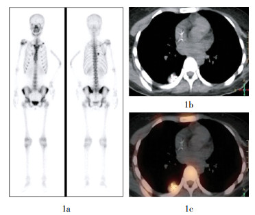
图 1 肺内转移灶摄取显像剂的骨肉瘤患者的骨显像及胸部CT图像患者,女性,19岁,骨肉瘤术后3年。图中,1a:骨显像中右肺中野可见点片状放射性浓聚灶;同机胸部CT见右肺下叶伴有明显钙化的软组织肿物影,1b:示病灶钙化情况,1c:融合图像示钙化病灶放射性摄取情况。
Figure 1. Pulmonary metastasis from osteosarcoma showed high uptake of methylene diphosphonate

图 2 肺内转移灶不摄取显像剂的骨肉瘤患者的骨显像及胸部CT图像患者,男性,15岁,右股骨下段骨肉瘤术后1年余。图中,2a:骨显像中双肺野内未见异常放射性浓聚;但CT见双肺野内多发大小不等结节影,结节呈软组织密度,内无钙化,2b:(肺窗)清晰显示肺内病灶,2c:(纵隔窗)清楚显示病灶内无钙化。
Figure 2. Pulmonary metastasis from osteosarcoma showed no uptake of methylene diphosphonate
骨肉瘤患者的血清ALP测定分析结果显示,发生肺转移患者的平均ALP水平显著高于无肺转移患者(t=2.29,P=0.023);但在有肺转移的患者中,病灶内有钙化与无钙化患者之间(t=0.69,P=0.494)以及病灶骨显像阳性与骨显像阴性患者之间(t=1.19,P=0.243)的平均ALP水平均未见显著差异(表 2)。
患者类型 例数 最大值 最小值 平均值 有肺转移患者 40 2681 50 344±508 无肺转移患者 121 2002 39 206±246 转移灶有钙化患者 20 2681 50 340±691 转移灶无钙化患者 20 1407 58 288±335 转移灶骨显像阳性患者 17 2681 57 454±691 转移灶骨显像阴性患者 23 1407 50 262±308 表 2 骨肉瘤患者的血清碱性磷酸酶测定结果分析(U/L)
Table 2. The serum alkaline phosphatase results of osteosar-coma patients
-
目前骨肉瘤的临床治疗主要采取根治性手术+化疗的综合模式,随着治疗方法的进步,骨肉瘤患者的短期生存率已有了显著的提高,然而,术后局部复发及远处转移,尤其是肺转移仍是导致骨肉瘤患者长期生存率不高的主要原因[1]。本研究中骨肉瘤患者肺转移的发生率为24.2%,提示了影像监测在早期发现肺转移方面的重要性。由于许多肺转移患者无明显症状,临床上通常将胸部CT作为骨肉瘤肺转移早期诊断不可缺少的检查方法,而骨显像也被常规用于骨肉瘤治疗前后的监测,因此使用SPECT/CT技术一站式获得全身骨显像+胸部CT可简化患者的检查程序,并具有较高的经济效价比,此种较理想的检查方法应在有条件的医疗单位加以推广应用。
骨显像一次性大视野成像对骨肉瘤患者全身各部位骨与软组织病灶的检出具有优势,对肺转移的检出也具有一定价值[5-7]。然而,本研究结果显示,骨显像对肺转移诊断的灵敏度仅为44.2%,尽管加做断层显像可能使病灶的检出率有所提高[7],但通过与CT的对比观察可看出,骨显像对肺内转移灶的检出主要与病灶内的矿化程度相关,这并非是通过SPECT断层提高对小病灶的检出率所能够解决的,而加做胸部CT,直接观察肺内是否有占位性病变,可使检出率达到100%,因此,胸部CT仍是诊断骨肉瘤肺转移不可缺少的检查方法。但从另一方面看,尽管CT对肺转移灶的检出具有非常高的灵敏度,但Brader等[8]研究发现,CT所显示的肺内结节中有32%经手术病理证实为良性病变,一些局灶性纤维化、肉芽肿、慢性感染等有时难于与转移灶相鉴别。虽然本研究中多数胸部CT阳性者未能得到肺内病灶的病理结果,但依据随访获得的最终临床诊断发现有14例为假阳性(占CT阳性患者的24.6%),这提示胸部CT用于骨肉瘤肺转移诊断中应注意除外假阳性的情况。由于骨显像中肺内转移灶对显像剂的摄取反映的是肿瘤细胞的成骨特性,因此对转移灶的识别具有很高的特异性,将骨显像与CT联合应用正起到优势互补的作用。本研究中有5例CT未见明显钙化的肺转移灶被骨显像检出的情况,这说明骨显像对于显示肿瘤细胞内的矿化活动较CT更灵敏[9],而一旦发现肺内摄取骨骼显像剂,肺转移诊断即可确立。
Tsuji等[10]的早期临床与动物研究曾证实肺转移灶摄取99Tcm-MDP的程度与转移灶的骨化程度及血清ALP的水平相关,并提出骨显像用于肺转移的诊断适用于选择性患者。本研究结果同样证实了肺转移灶能否被骨显像检出主要与病灶内有无钙化相关,但血清ALP水平的升高却仅与肺转移的发生相关,这提示血清ALP测定可作为肺转移发生的一项预测指标,当临床随访发现血清ALP升高时,应及时行影像学检查;而对于已发生肺转移的骨肉瘤患者来说,血清ALP水平并不能反映转移灶内的钙化情况,因此也与骨显像能否检出无相关性。上述与以往研究结果存在差异的原因可能与研究对象的构成不同有关。
本研究结果分析提示,骨肉瘤患者肺转移的发生概率相对较高,临床上应实行定期监测;使用SPECT/CT技术同时进行全身骨显像和胸部CT检查对骨肉瘤肺转移的检出具有较高的诊断价值,尤其适用于血清ALP升高的患者。
99Tcm-MDP SPECT/CT显像用于骨肉瘤肺转移的诊断
99Tcm-MDP SPECT/CT used in diagnosis of pulmonary metastasis from osteosarcoma
-
摘要:
目的 观察骨肉瘤肺转移的发生率,评价99Tcm-MDP SPECT/CT显像中的骨显像与胸部CT对肺转移的检出效能及其影响因素。 方法 178例骨肉瘤患者使用SPECT/CT行全身骨显像的同时获得胸部CT,以诊断有无肺转移发生。通过回顾性分析,将影像学诊断结果与最终临床诊断进行对照,计算肺转移的发生率;评价骨显像和胸部CT对肺转移的检出效能;分析转移灶钙化对骨显像检出率的影响;分析血清碱性磷酸酶(ALP)水平在有肺转移与无肺转移者之间、转移灶有钙化与无钙化者之间以及转移灶骨显像阳性与阴性者之间的差异。 结果 178例患者肺转移的发生率为24.2%;骨显像与胸部CT对其诊断的灵敏度分别为44.2%和100%;特异度分别为100%和89.6%;骨显像对有钙化转移灶的检出率显著高于无钙化转移灶(χ2=8.4,P < 0.01);患者血清ALP水平与肺转移的发生相关,但与病灶内有无钙化以及病灶是否在骨显像中呈阳性无关。 结论 骨肉瘤患者肺转移的发生率较高,使用SPECT/CT同时行全身骨显像和胸部CT检查对其检出具有较高诊断价值,尤其适用于血清ALP升高的患者。 -
关键词:
- 骨肉瘤 /
- 肿瘤转移 /
- 放射性核素显像 /
- 99m锝亚甲基二膦酸盐
Abstract:Objective To observe the incidence of pulmonary metastasis in osteosarcoma and evaluate the diagnostic efficiency of bone scintigraphy and chest CT in detection of pulmonary metastases and its influential factors. Methods A retrospective study was performed in 178 consecutive patients with osteosarcoma who underwent both bone scintigraphy and chest CT examination simultaneously by using a SPECT/CT instrument. Based on the final clinical diagnosis, incidence of pulmonary metastasis in the group was calculated, and the diagnostic efficacy of bone scintigraphy and chest CT was evaluated, respectively. Detection rate of bone scintigraphy was correlated to the status of calcification in metastatic lesions. Serum alkaline phosphatase (ALP) levels was correlated to the occurrence of pulmonary metastasis, calcification status in metastatic lesions and scintigraphic detections. Results Pulmonary metastasis occurred in 24.2% patients of all the patients. In the detection of pulmonary metastasis, sensitivity of bone scintigraphy and chest CT was 44.2% and 100%, respectively, and specificity was 100% and 89.6%, respectively. Lesion detection on bone scintigraphy was affected by the status of calcification in metastatic tumors(χ2=8.4, P < 0.01). It was found that the serum ALP levels was correlated to the occurrence of pulmonary metastasis, but not to the calcification or scintigraphic detections. Conclusions The risk of pulmonary metastasis in patients with osteosarcoma is higher. Using SPECT/CT to obtain bone scintigraphy and chest CT simultaneously will have a higher diagnostic value for detection of pulmonary metastasis, especially for those patients who with elevated serum ALP. -
图 1 肺内转移灶摄取显像剂的骨肉瘤患者的骨显像及胸部CT图像患者,女性,19岁,骨肉瘤术后3年。图中,1a:骨显像中右肺中野可见点片状放射性浓聚灶;同机胸部CT见右肺下叶伴有明显钙化的软组织肿物影,1b:示病灶钙化情况,1c:融合图像示钙化病灶放射性摄取情况。
Figure 1. Pulmonary metastasis from osteosarcoma showed high uptake of methylene diphosphonate
图 2 肺内转移灶不摄取显像剂的骨肉瘤患者的骨显像及胸部CT图像患者,男性,15岁,右股骨下段骨肉瘤术后1年余。图中,2a:骨显像中双肺野内未见异常放射性浓聚;但CT见双肺野内多发大小不等结节影,结节呈软组织密度,内无钙化,2b:(肺窗)清晰显示肺内病灶,2c:(纵隔窗)清楚显示病灶内无钙化。
Figure 2. Pulmonary metastasis from osteosarcoma showed no uptake of methylene diphosphonate
表 1 SPECT/CT显像中骨显像与胸部CT对骨肉瘤肺转移的检测结果
Table 1. Bone scan compare chest CT for detection of pulmonary metastasis from osteosarcoma
检查方法 真阳性(例) (例) (例) 假阴性(例) 灵敏度(%) 特异度(%) 准确率(%) 骨显像 19 0 135 24 44.2 100 86.5 胸部CT 43 14 121 0 100 89.6 92.1  下载: 导出CSV
表 2 骨肉瘤患者的血清碱性磷酸酶测定结果分析(U/L)
Table 2. The serum alkaline phosphatase results of osteosar-coma patients
患者类型 例数 最大值 最小值 平均值 有肺转移患者 40 2681 50 344±508 无肺转移患者 121 2002 39 206±246 转移灶有钙化患者 20 2681 50 340±691 转移灶无钙化患者 20 1407 58 288±335 转移灶骨显像阳性患者 17 2681 57 454±691 转移灶骨显像阴性患者 23 1407 50 262±308
下载: 导出CSV
-
[1] Wu PK, Chen WM, Chen CF, et al. Primary osteogenic sarcoma with pulmonary metastasis: clinical results and prognostic factors in 91 patients[J]. Jpn J Clin Oncol, 2009, 39(8): 514-522. doi: 10.1093/jjco/hyp057 [2] Letourneau PA, Xiao L, Harting MT, et al. Location of pulmonary metastasis in pediatric osteosarcoma is predictive of outcome[J]. J Pediatr Surg, 2011, 46(7): 1333-1337. doi: 10.1016/j.jpedsurg.2010.12.013 [3] Chen F, Miyahara R, Bando T, et al. Repeat resection of pulmonary metastasis is beneficial for patients with osteosarcoma of the extremities[J]. Interact Cardiovasc Thorac Surg, 2009, 9(4): 649-653. doi: 10.1510/icvts.2009.212498 [4] Bacci G, Rocca M, Salone M, et al. High grade osteosarcoma of the extremities with lung metastases at presentation: treatment with neoadjuvant chemotherapy and simultaneous resection of primary and metastatic lesions[J]. J Surg Oncol, 2008, 98(6): 415-420. doi: 10.1002/jso.21140 [5] 彭京京, 张清, 蔡伯, 等.放射性核素显像在骨肉瘤诊断中的应用[J].中华核医学杂志, 2000, 20(2): 65-67. doi: 10.3760/cma.j.issn.2095-2848.2000.02.007
[6] Körholz D, Wirtz I, Vosberg H, et al. The role of bone scintigraphy in the follow-up of osteogenic sarcoma[J]. Eur J cancer, 1996, 32A(3): 461-464. [7] Pevarski DJ, Drane WE, Scarborough MT. The usefulness of bone scintigraphy with SPECT images for detection of pulmonary metastases from osteosarcoma[J]. AJR Am J Roentgenol, 1998, 170(2): 319-322. doi: 10.2214/ajr.170.2.9456937 [8] Brader P, Abramson SJ, Price AP, et al. Do characteristics of pulmonary nodules on computed tomography in children with known osteosarcoma help distinguish whether the nodules are malignant or benign?[J]. J Pediatr Surg, 2011, 46(4): 729-735. [9] 李原, 王茜, 岳明纲, 等.核素骨显像在骶骨肿瘤术前诊断中的应用[J].中华核医学杂志, 2010, 30: 237-241. doi: 10.3760/cma.j.issn.0253-9780.2010.04.005
[10] Tsuji T, Yamamuro T, Kotoura Y, et al. Uptake of 99Tcm-MDP in lung metastasis from osteosarcoma clinical and animal studies[J]. Nucl Med Commun, 1988, 9(12): 947-954. doi: 10.1097/00006231-198812000-00002 -

 点击查看大图
计量
- 文章访问数: 2428
- HTML全文浏览量: 1179
- PDF下载量: 7