


 下载:
下载:

-
长期以来,金属植入物作为常用的植入体在骨折、骨更换、骨肿瘤切除后人工骨替换等领域发挥了巨大的作用。金属植入物植入人体后临床疗效评估常通过影像学特别是CT来实现,借助CT值来区别正常组织与病变组织,但是金属植入物产生的伪影(失真的CT值)会使图像质量下降从而影响临床疗效的评估,特别是金属植入物周围组织如骨肿瘤等常被忽视。双源CT有望能改善这一现象,双源CT有两套X线成像系统,能同时产生两种不同能量的X线,从而获得同一位置同一时刻的两组图像,对这两组图像进行能谱分析从而融合得到第3组图像[1-2]。过去利用能谱成像分析改善金属植入物影像的定性分析报道较多,但定量分析较少[3-6]。
-
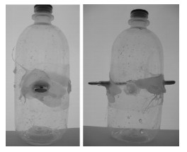
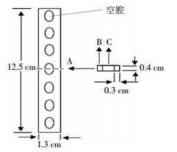
金属植入物为不锈钢植入物,长、宽、高分别为12.5、1.3、0.4 cm,中间有7个凸圆形的空洞(图 1)。把植入体水平插入塑料瓶中,如图 2所示,塑料瓶的中下部分近似为圆柱体,直径为7.5 cm、高度为19.5 cm,植入体两头暴露在空气中,塑料瓶中注满蒸馏水,为防止植入体与塑料瓶的接口处漏水,用热塑料胶进行封口处理。

图 1 金属植入物示意图

图 2 金属植入物CT扫描模型
-
模型扫描在德国西门子公司生产的炫速双源CT机上进行。为防止扫描模型在CT扫描时晃动,在原有的CT弧形扫描床上放置平行扫描床板。2个X线球管的电压分别为80、140 kV,有效管电流量的平均值分别为110、98 mAs,准直器宽度为64×0.6 mm,螺距为0.8,X线管旋转时间为0.5 s/圈,重建层厚为1 mm,层间距为1 mm。沿着金属植入物的纵轴扫描,模型位置不变,重复扫描3次。
-
所得两组图像传输至Syngo Dual Energy软件(购自德国西门子公司)中,利用能谱成像原理,在40~190 keV的电压中选择合适的keV值,进行去金属伪影处理,融合后得到的图像由2名有经验的医师采用双盲法选出他们一致满意的图像即第3组图像,本实验选取的是119 keV,能谱图像加权kV值和加权系数分别是120和0.5。为了便于描述,把当X线球管的电压分别为80、140 kV时得到的图像和通过能谱分析得到的图像分别称为80 kV CT图像、140 kV CT图像和能谱融合图像。
-
把得到的3组图像传输至美国CMS公司生产的XIO 4.4工作站,利用软件中自带的CT值测量工具,沿着图 1中的中间剖面(虚线)从A、B、C 3个方向每隔0.1 cm测量一点的CT值,测量长度为3 cm。测量的剖面中间是空洞,两侧各由0.3 cm宽的钢板组成,A方向是从植入物的右侧开始测量,B方向是从植入物右侧顶部开始测量,C方向是从植入物的空洞正上方开始测量。取3次重复扫描得到CT值的平均值。
-
CT值数据用SAS9.1统计学软件分析。采用随机方差分析比较3种不同方法(80 kV、140 kV和能谱融合)得到的CT值,取值点位置分别距离金属植入物0.1、0.5、1.0、1.5、2.0、2.5、3.0 cm,以P < 0.05为差异具有统计学意义。
-
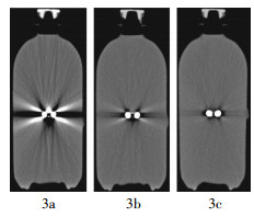
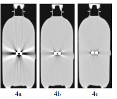
图 3、4为不同窗宽、窗位(600、40 HU和1000、-350 HU)的影像显示结果。图 3a为80 kV CT图像,在金属植入物水平左右两侧有柱状的低密度区域,长度大于3 cm;在植入物的左右斜上方(约45°角)有低密度区域,长度约1.2 cm;与植入物水平呈20°角有4个放射状的高密度区域,离植入物越近密度越高,长度约2.5 cm;在植入物的正上方即空腔的上方看到高密度的水滴状区域;在植入物的正下方即空腔的下方看到低密度三角形的区域;整个图像能清晰看到以植入物为中心的放射状斜纹,能贯穿整个扫描区域。图 3b为140 kV CT图像,在金属植入物水平左右两侧有柱状的低密度区域,随着与植入物距离的增加,密度增加,但在2.5 cm处,该区域密度值与周围水密度值稍有差异;在植入物的左右斜上方(约45°角)有低密度区域,长度约0.5 cm;与植入物水平呈20°角有4个放射状的高密度区域,但高密度区域长度与图 3a相比短很多,约1 cm;在植入物的正上方即空腔的上方密度与较远处水密度相当;在植入物的正下方即空腔的下方,有一个T字形的低密度区域;在扫描区域内,在植入物周围能看到细微放射状斜纹,随着距离的增加,放射状斜纹逐渐消失。图 3c为能谱融合图像,在植入物周围没有高密度区域产生,有宽度约为0.2 cm的环状低密度区域;在金属植入物水平左右两侧有柱状的稍低密度区域产生,随着与金属植入物距离的增加,密度快速增加,在2.5 cm处与周围密度没有差异。调整窗宽、窗位(1000、-350 HU),如图 4,图像观察内容与图 3相似,图 4a中金属植入物周围有严重的金属伪影产生,图 4b中金属植入物周围产生的伪影较轻,图 4c中金属植入物周围产生的伪影最小。

图 3 窗宽、窗位为600、40 HU时不同电压下的CT图像和能谱融合图像 图中,3a:电压为80 kV时的CT图像;3b:电压为140 kV时的CT图像;3c:能谱融合图像。

图 4 窗宽、窗位为1000、-350 HU时不同电压下的CT图像和能谱融合图像 图中,4a:电压为80 kV时的CT图像;4b:电压为140 kV时的CT图像;4c:能谱融合图像。
-
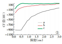
沿着A方向(即金属植入物的右侧方向)(图 5),80 kV CT图像结果:在离植入物0.5 cm内,CT值为(-1010±10)HU,随着距离的增加,CT值也随之增加,在距离金属植入物3.0 cm处CT值为-190 HU;140 kV CT图像结果:在距离植入物0.1 cm处CT值为-359 HU,在0.7 cm内CT值增加速度较快,而后随着距离增加CT值增加较慢,在距离植入物3.0 cm处CT值为-15 HU;能谱融合图像结果:在距离植入物0.1 cm处CT值为-198 HU,而后随着距离的增加CT值快速增加,在0.5 cm处CT值为-3 HU,后面的CT值稳定在(0±5)HU。

图 5 金属植入物的右侧方向CT测量值 图中,曲线a为电压为80 kV时的CT值;曲线b为电压为140 kV时的CT值;曲线c为能谱融合图像下的CT值。
沿着B方向(即植入物右侧顶部方向)(图 6),80 kV CT图像结果:在距离植入物0.1 cm处CT值为-1010 HU,而后CT值快速增加,在0.5 cm处为-8 HU,在0.6、0.7、0.8 cm处CT值分别为22、59、39 HU,而后CT值稍降低,在距离植入物1.4 cm以上区域CT值变化趋于平稳,但曲线仍存在明显的高低不平;140 kV CT图像结果:在距离植入物0.1 cm处CT值为-375 HU,随着距离增加CT值增加,0.6 cm后CT值稳定在(0±5)HU;能谱融合图像结果:在距离植入物0.1 cm处CT值为-226 HU,0.6 cm后CT值稳定在(0±5)HU。

图 6 金属植入物右侧顶部方向CT测量值 图中,曲线a为电压为80 kV时的CT值;曲线b为电压为140 kV时的CT值;曲线c为能谱融合图像下的CT值。
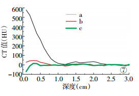
沿着C方向(即植入物的空洞正上方方向)(图 7),80 kV CT图像结果:在距离植入物0.1 cm处CT值为577 HU,随着距离增加CT值快速降低,在1.0 cm处CT值为16 HU,而后CT值变化趋于平稳,但曲线仍存在明显的高低不平;140 kV CT图像结果:在距离植入物0.1 cm处CT值为28 HU,而后CT值呈抛物线发展,先增加后减低,在0.9 cm后CT值为(0±5)HU;能谱融合图像结果:距离植入物0.1 cm处CT值为-69 HU,而后CT值呈抛物线发展,先增加后减低,在0.5 cm后CT值为(0±5)HU。

图 7 金属植入物的空洞正上方方向CT测量值 图中,曲线a为电压为80 kV时的CT值;曲线b为电压为140 kV时的CT值;曲线c为能谱融合图像下的CT值。
-
两两比较80 kV CT图像、140 kV CT图像和能谱融合图像沿着A方向0.1、0.5、1.0、1.5、2.0、2.5、3.0 cm处的CT值,结果发现它们之间的差异有统计学意义(F=46115.2、39825.2、20138.0、15031.4、4105.1、1394.1、2213.4,P均 < 0.001)。沿着B方向,在0.1 cm处,3种图像CT值两两比较发现差异有统计学意义(F=37021.9,P < 0.001);在0.5 cm处,80 kV CT图像与140 kV CT图像和能谱融合图像CT值比较差异有统计学意义,而140 kV CT图像与能谱融合图像CT值比较差异无统计学意义(F=22.09);在1.0、1.5、2.0、2.5、3.0 cm处3种图像CT值的两两比较差异无统计学意义(F=2.3、0.5、3.1、3.8、3.7,P均 > 0.05)。沿着C方向,在0.1、0.5 cm处,3种图像CT值的两两比较差异有统计学意义(F=15294.3、4783.7,P均 < 0.001);在1.0、1.5、2.0 cm处,80 kV CT图像与140 kV CT图像和能谱融合图像CT值比较差异有统计学意义,但140 kV CT图像与能谱融合图像CT值比较差异无统计学意义(F=18.5、171.2、129.1);在2.5、3.0 cm处3种图像CT值的两两比较差异无统计学意义(F=2.6、5.7,P均 > 0.05)。
-
由于CT扫描金属植入物后会产生金属伪影,主要原因是X线球管产生的X射线穿过金属植入物后急剧衰减,使得探测器探测到的投影数据失真,丢失了植入物周围的图像数据[7-8],这是常规CT的一大缺点。由于在A方向植入物的厚度大于B方向植入物的厚度,X线衰减的多,因此在A方向CT值失真严重。图像显示和CT值的测量结果互相印证,由于电压为80 kV时CT图像产生大量明显的放射状斜纹,使得在测量CT值时,产生的CT值不稳定(如图 6、图 7中的a曲线的中后段部分)。随着球管电压的增加(140 kV),图像质量得到了改善,这与电压的增加产生的射线穿透力增强有关,探测器能探测到较多的投影数据信息。因此在扫描有金属植入物的患者时,提高扫描电压能在一定程度上改善影像质量,但提高扫描电压后会产生软组织影像数据的缺失。
双源单能谱CT有望解决常规CT产生金属伪影的缺点[7]。从研究结果看,与植入物距离>0.5 cm时水的CT值与较远处CT值相当,其原理是双源单能谱成像可以任意选两种物质进行物质分离,当用不同能量的X线进行扫描时,两种物质的衰减也会产生变化,不同能量下获得的数据经过计算可获得不同keV值下的图像,调整keV值,可以获得较满意的影像。但是与植入物距离<0.5 cm特别是在0.2 cm内,水的CT值与较远处CT值差距大,改变不同的keV值(40~190 keV)都不能解决CT值偏小的问题,其原因有可能是不同能量的CT扫描植入物周围时获得的投影信息太少,不能给能谱成像计算提供足够的信息数据。因此,双源单能谱CT能改善金属植入物周围的CT值,但与金属植入物的距离在0.2 cm内时与实际值仍有一定差异,在诊断时应注意。
过去对利用能谱成像减少金属伪影的研究取得了不少成果,但都是以实际患者为例,没有以标准的水模体进行定量研究[4-7]。水模体的优势是能保证植入物与水无缝连接,且水的CT值是相对固定的。与扫描轴垂直的金属植入物的厚度、宽度都会影响常规CT图像,对能谱成像影响的大小也是今后继续研究的内容。
总之,CT能谱成像是种新兴技术,在改善金属伪影方面的优势是其他常规CT扫描所不具备的。
双源CT单能谱成像技术在正确评估金属植入物周围组织CT值中的应用
The application of dual-energy CT in the CT quantitative study values of water surrounding metal implants
-
摘要:
目的 探讨双源CT单能谱成像技术在正确评估金属(不锈钢)植入物周围组织CT值中的应用。 方法 对含有金属植入物的水模体使用双源CT双能量扫描,采用双能量扫描程序,一次性扫描采集2个不同能量(80 kV及140 kV)的数据,采用单能谱成像技术对所得原始数据进行处理,得到融合后图像。对金属植入物周围的水进行CT值测量和统计分析。 结果 3种图像(80 kV CT图像、140 kV CT图像、能谱融合图像)中,能谱融合图像的质量最高,140 kV CT图像次之,80 kV CT图像最差。用SAS9.1软件分别比较80 kV CT图像和140 kV CT图像与能谱融合图像沿着金属植入物右侧方向0.1、0.5、1.0、1.5、2.0、2.5、3.0 cm的CT值,结果发现它们之间的差异基本上都有统计学意义。 结论 双源CT单能谱成像技术改善了金属植入物植入后CT检查的图像质量。 -
关键词:
- 体层摄影术,X线计算机 /
- CT值 /
- 金属植入物
Abstract:Objective To evaluate monoenergetic imaging of dual energy CT in the visualization of metal implants(stainless steel implants). Methods Two different data(80 and 140 kV)of water phantom which contained metal implants were collected in one-time scanning using dual-energy scanning sequence. Monoenergetic technique which two different imaging at 80 kV and 140 kV were used for subtraction to removing metal artifacts was applied to generate the new imaging. The CT value of water surrounding metal implants were measured and compared. Results In the three images(80 kV, 140 kV, spectrum image fusion), the latter quality is the highest and the 80 kV scanning images′ quality was the worst. The difference between CT values of 80 kV images(140 kV images) and energy spectrum images along the right direction 0.1, 0.5, 1, 1.5, 2.0, 2.5, 3.0 cm of the metal basically was significant assessed by the SAS9.1 software. Conclusions Dual energy CT can effectively reduce artifacts of metal implants and will provide high image quality in the area with metal implants. -
Key words:
- Tomography, X-ray computed /
- CT values /
- metal implants
-
图 3 窗宽、窗位为600、40 HU时不同电压下的CT图像和能谱融合图像 图中,3a:电压为80 kV时的CT图像;3b:电压为140 kV时的CT图像;3c:能谱融合图像。
图 4 窗宽、窗位为1000、-350 HU时不同电压下的CT图像和能谱融合图像 图中,4a:电压为80 kV时的CT图像;4b:电压为140 kV时的CT图像;4c:能谱融合图像。
-
[1] Johnson TR, Krauss B, Sedlmair M, et al. Material differentiation by dual energy CT: initial experience. Eur Radiol, 2007, 17(6): 1510-1517. doi: 10.1007/s00330-006-0517-6 [2] 林晓珠, 沈云, 陈克敏. CT能谱成像的基本原理与临床应用研究进展.中华放射学杂志, 2011, 45(8): 798-800. doi: 10.3760/cma.j.issn.1005-1201.2011.08.028
[3] Scheffel H, Alkadhi H, Plass A, et al. Accuracy of dual-source CT coronary angiography: First experience in a high pre-test probability population without heart rate control. Eur Radiol, 2006, 16(12): 2739-2747. doi: 10.1007/s00330-006-0474-0 [4] 李晓莉, 冯卫华, 董诚, 等. CT能谱成像技术减除金属植入物伪影的定量实验研究.中华放射学杂志, 2011, 45(8): 736-739. doi: 10.3760/cma.j.issn.1005-1201.2011.08.007
[5] 周长圣, 郑玲, 张龙江, 等.双源CT单能谱成像技术在骨折金属固定术后的应用.中华放射学杂志, 2011, 45(2): 124-127.
[6] 惠萍, 王新江, 崔志鹏, 等. CT能谱成像在消除金属移植物伪影中的应用价值.中华放射学杂志, 2011, 45(8): 740-742. doi: 10.3760/cma.j.issn.1005-1201.2011.08.008
[7] 任庆国, 滑炎卿, 李剑颖. CT能谱成像的基本原理及临床应用.国际医学放射学杂志, 2011, 34(6): 559-563. doi: 10.3784/j.issn.1674-1897.2011.06.Z0612
[8] Fletcher JG, Takahashi N, Hartman R, et al. Dual-energy and dual-source CT: is there a role in the abdomen and pelvis?. Radiol Clin North Am, 2009, 47(1): 41-57. doi: 10.1016/j.rcl.2008.10.003 -

 点击查看大图
图(7)
计量
- 文章访问数: 2396
- HTML全文浏览量: 1184
- PDF下载量: 3