


 下载:
下载:
-
乳腺癌是危害女性健康的主要恶性肿瘤,近年来乳腺癌的发病率明显上升,且发病年龄呈年轻化趋势,早期诊断是提高乳腺癌患者生存率、降低病死率的关键因素之一。随着功能影像的技术的发展,18F-FDG PET/CT与3.0T MRI在乳腺癌诊断中的价值越来越受到重视,且两种显像方法在乳腺癌原发病灶诊断中各具优势,本研究对38例临床高度怀疑为乳腺癌的患者于一周内分别行18F-FDG PET/CT、3.0T MRI和病理学检查,以探讨18F-FDG PET/CT与3.0T MRI联合显像在乳腺癌原发病灶诊断中的价值。
-
选取2007年2月至2012年9月因临床高度怀疑为乳腺癌而在我院行18F-FDG PET/CT与3.0T MRI检查的38例女性患者。患者年龄25~71岁,检查前未接受任何治疗并均签署了知情同意书。18F-FDG PET/CT、3.0T MRI和病理学检查均在一周内完成。
-
采用美国GE公司的Signa EXCITE HD 3.0T超导磁共振显像仪,用腹部8通道相控阵线圈。线圈由前片和后片两部分组成,由于前片信号吸收率高,故在前片上放置一特制海绵垫(体积为200 cm×44 cm×15 cm,中上1/3处挖一大小约20 cm×18 cm×15 cm的椭圆形洞穴),嘱患者取俯卧位,双侧乳腺自然下垂放置在洞穴内,尽量不要压迫乳腺,后片紧贴检查者背后,利用约束带把前后片连接呈桶状围绕整个胸部,包括腋窝和双侧锁骨上区,头先进并双臂上举。扫描序列和参数如下:①短时反转恢复序列T2加权像:重复时间为4100 ms,回波时间为42~68 ms,反转时间为200 ms,矩阵288×224,激励次数为2;②快速自旋回波T1加权像:重复时间600 ms,回波时间7.5 ms,矩阵416×256,激励次数2,视野36 cm×36 cm,层厚7 mm,层间距1 mm;③弥散加权成像采用平面回波成像序列,b值为1000 s/mm2;④动态增强扫描使用肝脏快速容积成像序列:重复时间为3 ms,回波时间为1.8 ms,翻转角15°,层厚3 mm,层间距0,矩阵288×256,每个序列扫描45 s,间隔3 s,显像剂采用钆-二乙烯三胺五乙酸,剂量为0.1 mmol/kg体重,注射流率为2 ml/s,并用20 ml生理盐水冲洗导管。第一个序列为平扫,第二个序列在注入显像剂15 s后启动扫描,扫描25~30次,总扫描时间≥9 min。动态增强扫描后,数据传送到GE HP工作站(ADW 4.2),运用Functool 2功能软件进行图像后处理,在最大增强斜率伪彩图上病变显示最大、色彩红的层面选择感兴趣区(5~10 mm2),获得病灶的动态增强时间-信号强度曲线。
-
采用荷兰Philips公司GEMINI型PET/CT显像仪,CT为MX8000双排螺旋CT。18F-FDG由广州同位素中心提供,放化纯度大于95%。受检者均禁食4 h以上,给药前常规检测血糖并控制在7.2 mmol/L以下,按体重注入5.18 MBq/kg的18F-FDG,静卧60 min后采用PET进行三维采集,共10个床位,3 min/床位;CT扫描参数为120 kV、180 mAs,扫描层厚10.4 mm,重建层厚5.0 mm,进床速度为5 mm/圈,旋转时间0.75 s,螺距1.0。采用CT进行非均匀透射衰减校正图像重建,数据经Ramla三维方法重建获得衰减校正影像。
-
3.0T MRI结果判断:由两位有经验的MRI室医师结合病灶的形态学和动态增强时间-信号强度曲线进行判断。
18F-FDG PET/CT结果判断:由两位有经验的核医学科医师共同阅片,在浓聚灶部位勾画感兴趣区,由计算机程序自动生成该部位的最大标准化摄取值(maximum of standardized uptake value,SUVmax)。乳腺病灶以SUVmax≥2.0作为良、恶性判断的阈值[1]。
-
采用SPSS13.0统计学分析软件,组间灵敏度、特异度和准确率的比较采用χ2检验,P<0.05表示差异有统计学意义。
-
经病理确诊,38例患者中,24例为乳腺癌患者,14例为乳腺良性肿瘤患者。24例乳腺癌患者均为浸润性导管癌,其中1例为多中心病灶,癌灶最长直径介于0.7~2.8 cm。24例乳腺癌患者中有5例发生腋窝淋巴结转移,其中2例伴内乳淋巴结转移,远处转移3例。
-
38例患者中,MRI诊断真阳性22例,假阴性2例,真阴性11例,假阳性3例,其灵敏度、特异度和准确率分别为91.7%、78.6%和86.8%。
-
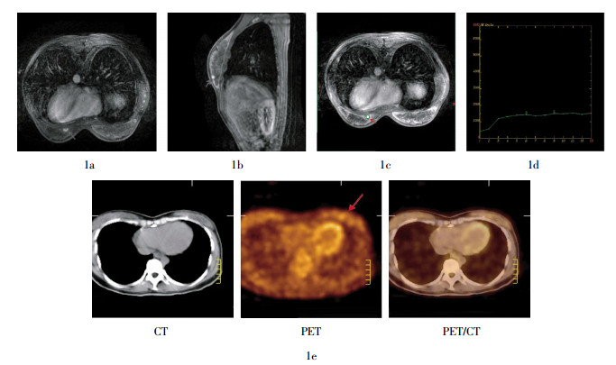
38例患者中,PET/CT检查诊断真阳性21例,假阴性3例,真阴性13例,假阳性1例,其灵敏度、特异度和准确率分别为87.5%、92.9%和89.5%。本研究中的3例PET/CT假阴性患者,其中2例为致密型乳腺癌患者,病灶的最大直径分别为1.2 cm、1.1 cm,最小直径分别为0.8 cm、0.9 cm,病灶SUVmax分别为1.22、1.31,稍高于周围正常乳腺组织(图 1);另1例假阴性患者,病灶最大直径为0.7 cm,未见明显18F-FDG高摄取。

图 1 患者女性,37岁,经病理学确诊为左侧乳腺浸润性导管癌Ⅱ级的致密型乳腺癌患者图中,1a、1b:3.0T MRI增强扫描横断面和矢状面图像,左乳内下象限见一环形明显强化灶,见分叶征,病灶大小1.2 cm×0.8 cm;1c、1d:左乳内下象限病灶3.0T MRI动态增强时间-信号强化曲线,呈“缓慢上升型”,其中,箭头所指区域为感兴趣区;1e:PET/CT融合图像,示双侧乳腺放射性分布弥漫性稍增高,平均标准化摄取值为1.1,左乳内下象限示一局灶性边界欠清的18F-FDG代谢轻度增高灶,最大标准化摄取值为1.22。
-
38例患者中,PET/CT和3.0T MRI联合显像诊断真阳性24例,假阴性0例,真阴性13例,假阳性1例,其灵敏度、特异度和准确率分别为100.0%、92.9%和97.4%。
-
3.0T MRI、PET/CT以及PET/CT和3.0T MRI联合显像在原发性乳腺癌诊断中其灵敏度、特异度和准确率三者之间比较差异无统计学意义(χ2=2.987、1.612和2.955,P均>0.05)。
-
18F-FDG PET是通过细胞对放射性核素标记的FDG的不同代谢水平来判断肿块性质,即所谓的生物学影像技术(或称为分子影像学技术)。PET/CT显像是解剖影像与功能影像的完美结合,一次成像即可以完成全身检查,目前广泛用于肿瘤的诊断、鉴别诊断以及临床分期,其最大优势在于其可以从分子水平检测和识别活体内不同状态下先于组织器官结构变化而发生的代谢改变,达到早期诊断和准确临床分期的目的。有研究表明18F-FDG PET/CT诊断乳腺癌的灵敏度介于80%~93%,特异度介于75%~100%,准确率介于79%~100%[2-4],明显高于乳腺钼靶X线摄影和乳腺超声检查。本研究中18F-FDG PET/CT诊断乳腺癌原发病灶的灵敏度、特异度和准确率分别为87.5%、92.9%和89.5%,与国内、外研究结果基本一致[5-7]。然而,18F-FDG PET/CT在乳腺癌诊断中也存在一些不足之处:①在致密乳腺腺体组织呈弥漫性放射性摄取增高的高本底中探测轻度或中度放射性增高的肿瘤时比较困难;②PET/CT对一些较小的原发病灶(<1.0 cm)或多灶性小病灶的诊断有一定困难。Wahl等[8]报道PET是一种良好的、能定性诊断>1.0 cm的乳腺肿块的方法,而对于<1.0 cm的乳腺肿块,其定性诊断的灵敏度、特异度比较低。本研究中的3例假阴性患者,其中2例为致密型乳腺癌患者,病灶SUVmax分别为1.22、1.31;另1例PET/CT假阴性患者为小原发病灶患者,病灶最大直径为0.7 cm,未见明显18F-FDG高摄取。
越来越多的研究表明,MRI是乳腺影像学综合诊断的必要手段之一,MRI在乳腺疾病诊断中的优点如下:①对发现乳腺病变具有较高的灵敏度;②三维成像使病灶定位更准确;③对乳腺高位、深位病灶显示较好;④对多中心、多灶性病变检出较为灵敏;⑤对于钼靶X线摄影无法显示的部位,如乳房根部、腋窝或病变接近胸壁时,MRI均可显示[9]。但MRI无法揭示乳腺癌患者的远处转移并提供准确的临床分期。
MRI能显著提高早期乳腺癌和多源性乳腺癌的检出率,其以极高的软组织分辨力在发现病灶、确定病灶范围、数量及周边关系等方面明显优于其他影像学诊断手段。国外文献报道,MRI发现乳腺恶性疾病的灵敏度高达94%~100%[10-11],而本研究中3.0T MRI诊断的灵敏度为91.7%,特异度为78.6%,准确率为86.8%,与国外研究基本一致。在本研究中,3.0T MRI诊断乳腺癌的特异度低于18F-FDG PET/CT,这可能与18F-FDG PET/CT是从分子水平来评价病灶的良、恶性有关。
PET/CT和3.0T MRI在乳腺癌原发病灶诊断中各具优势和不足,而18F-FDG PET/CT与3.0T MRI联合显像一方面可以发挥各自的优势,另一方面又可以弥补各自的不足,其在乳腺癌原发病灶的诊断中是否更具有优势?在本研究中,PET/CT与3.0T MRI联合显像诊断原发性乳腺癌的灵敏度、特异度和准确率分别为100.0%、92.9%和97.4%,其诊断灵敏度和准确率均高于单一的PET/CT或3.0T MRI检查,但三者之间比较差异无统计学意义(χ2=2.987、1.612和2.955,P均>0.05),鉴于样本数量有限,本研究结果还有待于进一步的大样本研究来证实。同时,值得注意的是,虽然18F-FDG PET/CT与3.0T MRI在乳腺癌原发病灶诊断效能中差异无统计学意义,但PET/CT在揭示乳腺癌患者远处转移、准确临床分期以及指导临床治疗方案等方面的优越性已得到充分肯定,这为准确指导临床治疗提供了重要依据。
综上所述,18F-FDG PET/CT和3.0T MRI是互为补充的医学影像学方法,两种方法联合显像在乳腺癌原发病灶诊断中具有重要价值;与单独18F-FDG PET/CT或3.0T MRI显像比较,3种显像方法在乳腺癌原发病灶的诊断效能上差异无统计学意义。
18F-FDG PET/CT与3.0T MRI联合显像在乳腺癌原发病灶诊断中的价值
Diagnosis value of 18F-FDG PET/CT and 3.0T MRI combined imaging in detecting the primary focus of breast cancer
-
摘要:
目的 探讨18F-FDG PET/CT与3.0T MRI联合显像在乳腺癌原发病灶诊断中的价值。 方法 对38例临床怀疑为乳腺癌的女性患者于一周内分别行18F-FDG PET/CT、3.0T MRI和病理学检查。 结果 组织病理学检查结果证实,全部患者中,24例为乳腺癌患者,14例为乳腺良性肿瘤患者。3.0T MRI诊断乳腺癌的灵敏度、特异度、准确率分别为91.7%、78.6%、86.8%;PET/CT诊断乳腺癌的灵敏度、特异度、准确率分别为87.5%、92.9%、89.5%;PET/CT和3.0T MR联合显像诊断乳腺癌的灵敏度、特异度、准确率分别为100.0%、92.9%、97.4%;3种显像方法间灵敏度、特异度、准确率差异无统计学意义(χ2=2.987、1.612和2.955,P均>0.05)。 结论 18F-FDG PET/CT和3.0T MRI联合显像在乳腺癌原发病灶诊断中具有重要价值;但与单独18F-FDG PET/CT和3.0T MRI显像比较,3种显像方法在乳腺癌原发病灶的诊断效能上差异无统计学意义。 -
关键词:
- 乳腺肿瘤 /
- 正电子发射断层显像术 /
- 体层摄影术,X线计算机 /
- 氟脱氧葡萄糖F18 /
- 磁共振成像
Abstract:Objective To explore the diagnosis value of 18F-FDG PET/CT and 3.0T MR combined imaging in detecting the primary focus of breast cancer. Methods Thirty eight female patients with highly suspected breast cancer were selected, 18F-FDG PET/CT and 3.0T MRI imaging were performed and their pathological results were gotten within 7 days. Results Twenty four malignant cases and 14 benign cases of the 38 patients were proved by pathology. The sensitivity, specificity and accuracy rate obtained by 3.0T MRI were 91.7%, 78.6% and 86.8%, respectively; they were 87.5%, 92.9% and 89.5% for 18F-FDG PET/CT; and which were 100.0%, 92.9% and 97.4% with 18F-FDG PET/CT and 3.0T MRI combined imaging. There were no significant difference among the three methods in the diagnostic ability(χ2=2.987, 1.612 and 2.955, all P > 0.05). Conclusions The combination of 18F-FDG PET/CT imaging and 3.0T MRI imaging in detecting the primary focus of breast cancer has important value, and comparison with separate 18F-FDG PET/CT imaging or 3.0T MRI imaging, the diagnosis efficiency of three imaging methods in breast cancer without statistics difference. -
[1] 余丰文, 冯彦林, 贺小红, 等. PET-CT与钼靶X线、B超定性诊断乳腺癌的临床对比研究.国际放射医学核医学杂志, 2006, 30(3): 148-151. doi: 10.3760/cma.j.issn.1673-4114.2006.03.007
[2] Wang Y, Yu J, Liu J, et al. PET-CT in the diagnosis of both primary breast cancer and axillary lymph node metastasis: initial experience. Int J Radiat Oncol Biol phys, 2003, 57(2 Suppl): S362-363. [3] Scheidhauer K, Walter C, Seemann MD. FDG PET and other imaging modalities in the primary diagnosis of suspicious breast lesions. Eur J Nucl Med Mol Imaging, 2004, 31(1 Suppl): S70-79. [4] 赵桐, 王永胜, 杨国仁, 等. PET/CT在乳腺癌保乳微创治疗中的应用研究.肿瘤研究与临床, 2004, 16(2): 103-106. doi: 10.3760/cma.j.issn.1006-9801.2004.02.012
[5] 赵继红, 丁军, 徐松柏, 等. 18F-FDG PET/CT与MRI对乳腺癌诊断价值的比较.吉林大学学报:医学版, 2011, 37(4): 746-749.
[6] 唐立岷, 马超. 18F-FDG PET/CT在诊断浸润性乳腺癌和淋巴结转移中的价值.上海交通大学学报:医学版, 2012, 32(6): 761-765.
[7] Walter C, Scheidhauer K, Scharl A, et al. Clinical and diagnostic value of preoperative MR mammography and FDG-PET in suspicious breast lesions. Eur Radiol, 2003, 13(7): 1651-1656. doi: 10.1007/s00330-002-1816-1 [8] Wahl RL, Siegel BA, Coleman RE, et al. Prospective multicenter study of axillary nodal staging by positron emission tomography in breast cancer: a report of the staging breast cancer with PET Study Group. J Clin Oncol, 2004, 22(2): 277-285. [9] 易梅, 程绍玲, 李冬梅.钼靶、MRI在乳腺癌诊断中的应用.中国医药导报, 2008, 5(8): 87. doi: 10.3969/j.issn.1673-7210.2008.08.060
[10] Liberman L, Morris EA, Lee MJ, et al. Breast lesions detected on MR imaging: features and positive predictive value. AJR Am J Roentgenol, 2002, 179(1): 171-178. doi: 10.2214/ajr.179.1.1790171 [11] Al-Khawari H, Kovacs A, Athyal R, et al. Breast magnetic resonance imaging: initial experience in Kuwait. Med Princ Pract, 2009, 18(2): 143-148. doi: 10.1159/000189813 -

 点击查看大图
图(1)
计量
- 文章访问数: 3148
- HTML全文浏览量: 1217
- PDF下载量: 9