


 下载:
下载:
-
近年来,儿童泌尿系统损伤的发生率较过去明显增多,其中尤以肾脏和男性尿道更为多发。随着多层螺旋CT(mutislice spiral computed tomography,MSCT)的临床应用,其快速的多层扫描及强大的图像后处理技术在评估损伤的部位、范围、程度等方面优势明显,同时可以观察其他腹部脏器的情况。本文对43例泌尿系统损伤患儿的影像学资料进行分析,旨在探讨MSCT及图像后处理技术在儿童泌尿系统损伤中的应用价值。
-
收集2007年7月至2010年12月在我院行MSCT检查的泌尿系统损伤患儿43例,其中男性患儿25例、女性患儿18例,年龄5月~13岁,平均年龄7.4岁。损伤原因:车祸、坠落及骑跨伤等。主要临床表现为腹胀、腹部包块、腹部压痛及叩击痛、腹膜刺激症、肉眼血尿、镜下血尿、无尿、出血性休克等。所有患儿家长均签署知情同意书。
-
采用德国SIEMENS公司SOMATOM sensation16层螺旋CT机进行螺旋扫描,扫描参数:管电压为120 kV,管电流量为150 mAs,准直器宽度0.75 mm,螺距1.25,扫描层厚10 mm,重建层厚2 mm,重建间隔1 mm。患儿仰卧定位,轴面扫描,采集原始二维图像20~40层,范围自膈顶至耻骨联合以下。所有患儿均行平扫、双期增强扫描及延迟扫描,动脉期20~30 s、实质期60~90 s,延迟扫描时间为5~30 min。造影剂:碘海醇(300 mgI/ml)由美国GE公司生产,剂量为1.5 ml/kg,注射速率为1.5~2 ml/s,经肘静脉团注射。
-
在Cyngo CT Workplace工作站上对原始图像数据结果行图像后处理,主要包括多平面重组(multi-planar reformation)、最大密度投影(maximum intensity projection)、容积再现(volume rendering)。由两名有经验的副主任医师负责图像分析及诊断。
-
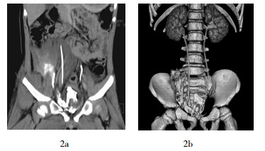
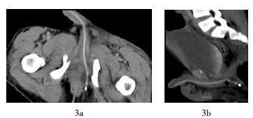
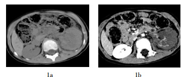
本研究中43例患儿全部经MSCT增强扫描,运用后处理技术,所获得的泌尿系三维重组图像清晰,可清楚显示病变位置、大小、范围以及与周围组织的关系。其中,单纯肾挫伤5例,MSCT表现为肾脏密度不均,肾实质内或肾盂内高密度出血灶,增强扫描肾实质密度增高,血肿呈低密度;肾挫裂伤伴包膜下血肿5例,由于局限于肾包膜内,MSCT表现为半月形或新月形紧贴肾实质表面,相应肾实质的边缘变平,新鲜血肿呈高密度,血肿随时间延长可呈等密度或低密度;肾挫裂伤伴肾周血肿4例,大量血液贮在肾包膜外吉氏筋膜内,或肾前、后间隙内;肾挫裂伤伴肾上腺血肿2例,MSCT表现为肾上腺区高(或低)密度不规则团块,肾上腺显示不清;肾撕裂伴尿外渗13例,MSCT表现为肾实质内线条状、三角形低密度灶,增强延迟扫描可发现造影剂外溢;肾撕裂伴肾蒂损伤3例,MSCT增强延迟扫描表现为节段性梗死或整个肾实质不强化,肾盂内无造影剂分泌(图 1);肾撕裂伴输尿管损伤6例,MSCT表现为肾脏周围积液,周围脂肪间隙毛糙,增强延迟扫描可发现造影剂外漏入腹腔,输尿管内造影剂与外漏造影剂分界不清,远端输尿管未见显影或部分显影,利用三维重组可以清晰显示输尿管破口处的位置;膀胱壁损伤伴膀胱内血凝块3例,MSCT显示均有不同程度的膀胱壁增厚、毛糙,内容物密度不均匀,膀胱脂肪间隙内充满液体密度影,增强延迟扫描可见膀胱充盈形态不规整,造影剂外溢至膀胱外(图 2);骨盆骨折伴后尿道断裂2例,MSCT平扫、增强延迟扫描均显示不清,仅发现骨盆骨折及盆腔软组织肿胀,后经MSCT尿路造影方能确诊,表现为造影剂未沿正常尿道走行,未与膀胱相连,并溢出至周围软组织间隙(图 3)。

-
儿童创伤性疾病中泌尿系统损伤较为常见,受伤原因以车祸最多,击打伤次之,其次为坠落伤等。由于儿童肾脏体积相对成人较大,腹壁及腰部肌肉不发达,肾周筋膜发育差,肾周脂肪薄弱,第11、12肋骨未完全骨化,肾脏活动度相对成人较大,致使儿童泌尿系统对外力的防卫能力较弱,较成人易受到损伤[1]。一般根据病史、临床症状、体征、尿常规检查等,大多可获急诊诊断。但单纯依靠临床体征不能准确判断损伤程度和范围,延误选择正确治疗方案。
儿童泌尿系统损伤常用的检查方法包括超声、静脉尿路造影(intravenous urography)、CT和肾动脉造影等。超声的组织分辨率较高,对伤肾的形态,特别是肾内、肾周血肿及肾血管损伤的观察较为理想,适合外伤者的筛选检查诊断,同时不受患者状态限制,对于危重患者还可做急诊床旁超声检查。但超声难以测定肾裂伤的程度和肾功能,不能对肾外伤进行准确分级,特别是肾脏上极损伤不易显示,也不能了解尿外漏、输尿管有无连续性中断的情况,因此超声并不能成为泌尿系统损伤的理想检查项目。
静脉尿路造影是泌尿系损伤的主要检查方法,可显示集合系统及泌尿道,但肾实质显示不清。在肾损伤患者中,静脉尿路造影常常表现为无明显异常或显影不良,对肾周情况也不能直接显示;当输尿管断裂时对显示损伤部位造影剂外渗、损伤部位以上输尿管扩张效果良好,是诊断输尿管断裂的首选方法。
肾动脉造影可明确肾损伤的部位及血管,可在必要时行出血区域栓塞治疗,适用于疑有肾蒂血管损伤的患者。但肾动脉造影费时长,损伤相对较大,对急、重患者不具有优越性。
泌尿系统损伤复杂多样,需要显示的重点和内容不一,应用MSCT对泌尿系统平扫、肾脏双期及排泄延迟期动态扫描能实现一次检查获得完整泌尿系统影像,充分显示肾盂、输尿管、膀胱的关系,其多期扫描及强大的图像后处理技术在尿路造影方面优势明显,可清晰显示尿路及其周围结构的情况[2]。
MSCT扫描方式及图像采集方式与传统螺旋CT不同,其采用薄层容积扫描及多排探测器接收,十几秒内就可以完成全泌尿系统的检查。利用对比剂经肾脏分泌排泄的原理,通过软件对图像进行多平面重组、最大密度投影及容积再现处理,最后使泌尿系统立体显影。通过三维重组,从不同角度观察肾脏、输尿管、膀胱的情况,不仅能显示病变与周围组织的关系,还能够准确地定位及定性[3]。
MSCT可以准确判断肾损伤的程度和类型,并且能良好显示肾损伤的范围和基本病变特征(挫伤、撕裂、梗死、血肿和尿外渗)以及肾功能的情况。MSCT平扫能反映肾损伤程度范围及出血情况,通过增强检查可清楚显示肾门处动脉及静脉内有无供血障碍,对肾脏及肾血管的损伤提供准确的信息[4],甚至可以通过对比剂外渗来提示有无活动性出血。MSCT不仅对肾损伤的诊断有很高的准确率,并为其分型、分类,以及临床手术方法的选择,提供了可靠的依据[5]。
输尿管损伤最好发的部位是肾盂、输尿管连接部,平扫时可表现为正常或沿受损输尿管走行区的水样低密度影,增强后可见造影剂于输尿管受损处外溢。因输尿管行程长且迂曲,在轴位图像上不能完整观察整条输尿管影[6],此时利用MSCT强大的图像后处理技术,通过后期重组的最大密度投影及容积再现图像可观察绵长迂曲的输尿管空间位置及走行,达到明确诊断的目的。Mulligan等[7]报道,MSCT增强扫描时,在肾实质显影阶段,即使没有发现大量对比剂的外溢,也不能排除肾盂输尿管连接部破裂的可能。而通过增强延迟扫描恰恰弥补了这一缺陷,能够发现对比剂的外溢,从而确诊肾脏集合系统的损伤或肾盂输尿管连接部破裂,既可做出定性诊断,又可做出定位诊断。本研究病例中,6例输尿管损伤患儿经MSCT增强延迟扫描能够比较直观、清楚地显示输尿管创伤后对比剂外溢的部位。
当合并骨盆骨折、失血性休克、会阴部有明显血肿或有排尿困难甚至无尿,应高度怀疑尿道、膀胱损伤或破裂,临床主要通过试插导尿管帮助诊断,但一次插管失败后一般不重复试插,避免加重损伤。此时可以在MSCT增强并延迟扫描的基础上,行CT尿路造影帮助诊断。具体方法为将导尿管或注射器头置于尿道外口附近,在无菌条件下注入造影剂,然后行MSCT扫描[8],通过观察造影剂中断或外溢至尿道周围来判断有无尿道损伤及断裂。本研究中的2例患儿经MSCT尿路造影而明确诊断。
总之,利用MSCT扫描速度快、图像分辨率高、伪影少及图像后处理方法多的优点,一次检查即能够完整、准确地显示泌尿系统损伤的部位、范围、程度,确定其分型,同时可以观察其他腹部脏器情况,也可观察下位肋骨及腰椎是否骨折及移位情况,为临床诊断和治疗提供重要的依据,弥补了其他检查技术的缺陷,因而成为泌尿系统损伤的主要影像学检查方法。
多层螺旋CT在儿童泌尿系统损伤中的应用
Application of mutislice spiral CT in children urinary tract injuries
-
摘要:
目的 探讨多层螺旋CT(MSCT)三维重建技术在泌尿系统损伤中的应用价值。 方法 对43例泌尿系统损伤的患儿行MSCT后,应用三维重建技术对横断面图像进行容积重建(VR)、最大密度投影(MIP)及多平面重组(MPR),获得完整的尿路影像。由两名副主任医师负责图像分析及诊断。 结果 43例患儿中,单纯肾挫伤5例、肾挫裂伤伴包膜下血肿5例、肾挫裂伤伴肾周血肿4例、肾挫裂伤伴肾上腺血肿2例、肾撕裂伴尿外渗13例、肾撕裂伴肾蒂损伤3例、肾撕裂伴输尿管损伤6例、骨盆骨折伴后尿道断裂2例、膀胱壁损伤伴膀胱内血凝块3例。 结论 MSCT具有扫描速度快、覆盖范围广、图像质量高及薄层扫描的特点,对儿童泌尿系统损伤的诊断具有优势。 -
关键词:
- 泌尿系疾病 /
- 体层摄影术,X线计算机 /
- 儿童
Abstract:Objective To investigate the application value of three-dimensional reconstruction technique of mutislice spiral CT(MSCT)in children urinary tract injuries. Methods Forty-three patients with urinary tract injury performed MSCT scan, and three-dimensional reconstruction technique was used for volume rendering, maximum intensity projection and multi-planar reformation, urinary tract imaging was acquired completely. The images were reviewed by two experienced radiologists. Results Of the 43 cases, there were 5 simple contusion of kidney, 5 contusion and laceration of kidney accompanied with subcapsular haematoma, 4 contusion and laceration of kidney accompanied with perinephric haematoma, 2 contusion and laceration of kidney accompanied with adrenal haematoma, 13 shattered kidney accompanied with extravasation of urine, 3 shattered kidney accompanied with renal pedicle injury, 6 shattered kidney accompanied with ureter injury, 2 pelvic fracture accompanied with posterior urethra split, 3 contusion of urinary bladder wall accompanied with blood clot in the urinary bladder. Conclusion MSCT has the characteristic of high scanning speed, wide overlay scope, high image quality and thin slice scan. It is an effective modality in the evaluation of children urinary tract injuries. -
Key words:
- Urologic diseases /
- Tomography, X-ray computed /
- Children
-
-
[1] 黄澄如.小儿泌尿外科学.济南:山东科学技术出版社, 1996:302.
[2] Kemper J, Adam J, Nolte Emsting C. Modern diagnostic assessment of the upper urinary tract using multislice CT urography. Rofo, 2006, 178(11): 1086-1094. doi: 10.1055/s-2006-926957 [3] 汪立娟, 张淑芳, 林华, 等.多层螺旋CT及其图像后处理技术在泌尿系疾病诊断中的临床应用.实用医学影像杂志, 2010, 11(3): 180-182. doi: 10.3969/j.issn.1009-6817.2010.03.017
[4] Goldfarb CR, Srivastava NC, Grotas AB, et al. Radionuclide imaging in urology. Urol Clin North Am, 2006, 33(3): 319-328. doi: 10.1016/j.ucl.2006.03.006 [5] Bhatti AA, Chugtai A, Haslam P, et al. Prospective study comparing three-dimensional computed tomography and magnetic resonance imaging for evaluating renal vascular anatomy in potential living renal donors. BJU Int, 2005, 96(7): 1105-1108. doi: 10.1111/j.1464-410X.2005.05809.x [6] 何亚奇, 张雪林.多层螺旋CT尿路造影在重复肾盂输尿管畸形诊断中的应用.实用放射学杂志, 2008, 24(6): 852-853. doi: 10.3969/j.issn.1002-1671.2008.06.042
[7] Mulligan JM, Cagiannos I, Collins JP, et al. Ureteropelvic junction disruption secondary to blunt trauma: Excretory phase imaging(delayed films) should help prevent a missed diagnosis. UroL, 1998, 159(1): 67-70. [8] Nino-Muricia M, Jeffrey Jr RB, Beaulieu CF, et al. Multidetector CT of the pancreas and bile duct system: value of curved planar re-formation. AJR, 2001, 176 (3): 689-693. doi: 10.2214/ajr.176.3.1760689 -

 点击查看大图
图(3)
计量
- 文章访问数: 2109
- HTML全文浏览量: 855
- PDF下载量: 2