


-
结核病是由结核分枝杆菌感染引起的一种慢性传染病,可累及全身多个脏器。其中,肺部是感染结核菌的最主要脏器。个体一旦感染结核菌后,将终身携带病菌,约有10%~15%的感染者会在一定条件下发展为活动性结核病,成为新患者并继续传染给其他人。目前,临床上最常用的判断结核病灶是否活动的影像学方法是X射线检查和CT检查。近年来,18F-FDG PET-CT广泛应用于肿瘤病灶的评估和肿瘤筛查,但是,18F-FDG并不是肿瘤特异性显像剂,炎性病灶亦可呈阳性显像,有文献报道,18F-FDG摄取可作为结核活动性的一个标志[1-3]。笔者对31例肺结核病例进行回顾性分析,分别运用18F-FDG PET和CT两种影像学方法对结核病变是否活动进行判断对比,旨在对诊断和治疗肺结核病变提供帮助,现报道如下。
-
选取2010年2月1日至2011年3月30日于本院行18F-FDG PET-CT肿瘤相关检查而意外发现肺结核病变的31例患者,其中,男性22例、女性9例,年龄45~82岁,平均年龄65岁。31例患者中,28例为PET-CT检查前有肺结核病史(26例肺结核病史为2年以上,2例肺结核病史为2年以内),另3例为PET-CT检查时发现肺结核病灶,其中1例经术后病理确诊,另外2例经随访结核菌素试验阳性并有典型影像学征象、抗结核治疗后有效确诊。
-
PET-CT为西门子Biography Truepoint HD型仪器(同机CT为64层螺旋CT)。18F-FDG由日本住友公司的HM-10型回旋加速器生产,放化纯度 > 95%。受检者于检查前禁食6 h以上,血糖值< 6.7 mmol/L,按患者体重静脉注射18F-FDG 5.55 MBq/kg,注射后1~1.5 h行常规全身显像。PET图像重建方法为迭代法,CT扫描层距为5 mm,层厚为5 mm,扫描结束后以高分辨骨算法重建肺部CT图像,并与PET图像匹配融合。
-
分别采用高分辨骨算法重建肺部CT图像和PET图像对结核病灶是否活动进行判断。CT图像由两名有5年以上CT诊断经验的影像医师判断,判断标准:肺部病灶完全钙化或以索条影为主、部分伴有钙化者判断为非活动性肺结核;病灶呈斑片影、结节影、磨玻璃影、空洞等征象者判断为活动性肺结核。PET图像由两名核医学科医师对CT所示结核病灶处的PET图像进行判断,判断标准参照文献[4]并修改为:0级:与周围肺野本底无视觉差异;1级:稍高于周围肺野本底但低于纵隔血池水平;2级:高于周围肺野本底并与纵隔血池相似;3级:高于纵隔血池;4级:明显异常浓聚。其中0、1级判断为非活动性病灶,2、3、4级判断为活动性病灶。
-
CT诊断结果为:22例患者为非活动性肺结核者,其中病灶完全钙化者6例(6例皆为愈合病灶),病灶大部分钙化、伴有少许索条影者16例(16例皆为陈旧性病灶);9例患者为活动性肺结核者。
PET诊断结果为:17例患者为非活动性肺结核者;14例患者为活动性肺结核者。
-
CT与PET判断结果一致者26例,判断结果不一致者5例,两者的判断结果比较见表 1。
PET诊断为活动性肺结核者 PET诊断为非活动性肺结核者 合计 CT诊断为活动性肺结核者 9 0 9 CT诊断为非活动性肺结核者 5 17 22 合计 14 17 31 表 1 CT和PET对31例肺结核患者的肺结核病灶活动性判断结果的比较
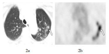
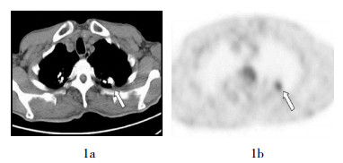
(例) 6例CT诊断为病灶完全钙化的非活动性肺结核者的PET图像均无明显放射性摄取,判断为非活动性病灶。16例CT诊断为病灶大部分钙化、伴有少许索条影的非活动性肺结核者中,5例PET判断为活动性病灶,其中3例病灶为结节的未钙化部分放射性轻度浓聚(图 1),2例为索条影伴有放射性轻度浓聚;其余11例PET判断为非活动性病灶。9例CT诊断为活动性肺结核者的PET图像呈片状或团块状放射性浓聚灶,均判断为活动性病灶,其中5例为近期感染结核,4例为多年前感染结核最近复发,结核病史最长者达55年(图 2)。

-
虽然肺结核的活动性取决于在痰或支气管肺泡灌洗液中是否可以检测到抗酸杆菌,但其阳性率较低,因此,诊断其有无活动性比较困难。胸片的表现也常是不肯定的,近年来用胸部CT来诊断肺结核和判断病灶是否活动越来越广泛。活动性肺结核的基本病理变化主要以渗出、增生和坏死为主,3种变化往往同时存在而以某一种改变为主,而且可以相互转化。这3种病理变化会在CT图像上形成各种各样的征象,如小叶结节影、斑片影、磨玻璃影等,所以CT诊断肺结核的准确率较高[5]。近年来,随着18F-FDG PET-CT应用的越来越多,一些学者发现,18F-FDG PET显像阳性可反映结核病灶的活动性,其原理主要为活动性肺结核病灶中含有大量的类上皮细胞、郎罕巨细胞和淋巴细胞等,这些细胞的葡萄糖代谢旺盛,18F-FDG摄取较高,是结核病变18F-FDG显像阳性的主要原因[3]。
本研究中6例完全钙化的肺结核病灶均无18F-FDG浓聚,9例有典型活动性肺结核病灶者均表现为18F-FDG浓聚,与以往研究结果一致[3-4, 6]。16例CT表现为结节大部分钙化、伴有少许索条影并判断为非活动性病灶者中,有5例PET表现为18F-FDG浓聚,两者判断结果不一致,其原因可能为:结核病灶经过治疗后转向愈合,渗出性的病灶经淋巴道吸收而使病灶缩小或消散,而增生性的病变和小的干酪样坏死灶逐渐纤维化,最后形成瘢痕而愈合,较大的干酪样坏死灶难以全部纤维化,则由周边纤维组织增生将其包裹,继而纤维化和钙化,在X射线和CT上表现为索条影和钙化,一般认为这是非活动性肺结核的典型征象,为陈旧性结核病灶,但是这些纤维化和部分钙化的结核灶内常有少量的结核杆菌残留,而且当机体抵抗力降低时仍可复发进展[7]。作为代谢显像的18F-FDG PET在此类病灶的判断上有非常高的灵敏度,这些残存的结核病灶中的炎性细胞的18F-FDG代谢旺盛,故通过PET可以判断是否有活动性结核病灶存在。在本研究病例中,有4例结核病灶复发者的感染结核病史为30年以上,最长者为55年前感染结核,所以要重视对陈旧性结核病灶的随访,并对伴有活动性结核病灶残余的患者进行相应的抗结核治疗。
本研究结果显示,18F-FDG PET在判断愈合后的结核病灶和完全处于活动期的结核病灶时和CT判断结果一致,但在对陈旧性结核病灶是否有残余活动性病灶的判断上明显优于CT。PET-CT一次显像可以同时形成两种影像,两者结合起来对病灶的判断更加全面,虽然目前PET-CT检查主要用于肿瘤相关检查,但在检查过程中意外发现活动性结核病灶时需建议患者进行抗结核治疗,以防其进一步恶化。已有学者尝试用18F-FDG PET来监测结核治疗效果,特别是全身广泛性结核病灶者,效果良好[4, 8]。
18F-FDG PET和CT判断肺结核病灶活动性的比较
Comparison between 18F-FDG PET and CT in evaluating the activity of pulmonary tuberculosis
-
摘要:
目的 评价18F-FDG PET和CT两种影像学方法对肺结核病灶活动性判断的差异。 方法 对18F-FDG PET-CT显像中发现的31例肺结核病例,分别用CT图像和PET图像对肺结核病灶是否活动进行判断,然后对两种影像学方法的结果进行比较。 结果 两种方法判断结果一致者26例,不一致者5例。6例CT诊断为非活动性肺结核者(愈合病灶),PET均判断为非活动性病灶;16例CT示病灶大部分钙化、伴有少许索条影(陈旧性病灶)并诊断为非活动性肺结核者中,5例PET判断为活动性病灶,其中3例病灶为结节的未钙化部分放射性轻度浓聚,2例为索条影伴有放射性轻度浓聚;9例CT诊断为活动性肺结核者,PET均判断为活动性病灶。 结论 18F-FDG PET在判断愈合后的结核病灶和完全处于活动期的结核病灶时,与CT判断结果一致,但在对陈旧性结核病灶是否存在残余活动性病灶的判断上优于CT。 -
关键词:
- 结核,肺 /
- 氟脱氧葡萄糖F18 /
- 正电子发射断层显像术 /
- 体层摄影术,X线计算机
Abstract:Objective To compare the difference between 18F-FDG PET and CT for evaluating the activity of pulmonary tuberculosis. Methods 18F-FDG PET-CT was performed in 31 pulmonary tuberculosis patients, the activity of the tuberculosis lesions was evaluated by PET and CT images, the results of the two imaging methods were compared separately. Results The results obtained with the two imaging methods were consistent in 26 cases and inconsistent in 5 cases. 6 cases which CT diagnosed as inactive tuberculosis (healed lesions) were also judged as inactive lesions by PET imaging. In 16 cases, CT displayed that most of the lesions were calcified and associated with little streaks and diagnosed as inactive tuberculosis (obsolete lesions), among them PET judged 5 cases as active lesions, of which 3 cases with partly calcified lesion associated with mild radioactive uptake, 2 cases with streaks associated with mild radioactive uptake. 9 cases which CT diagnosed as active pulmonary tuberculosis, PET judged as active lesions too. Conclusions 18F-FDG PET and CT have the same judgment in diagnosing healed and active tuberculosis lesions, while 18F-FDG PET is superior to CT in evaluating the active lesions residue in obsolete lesions. -

表 1 CT和PET对31例肺结核患者的肺结核病灶活动性判断结果的比较
(例) PET诊断为活动性肺结核者 PET诊断为非活动性肺结核者 合计 CT诊断为活动性肺结核者 9 0 9 CT诊断为非活动性肺结核者 5 17 22 合计 14 17 31  下载: 导出CSV
-
[1] Kim IJ, Lee JS, Kim SJ, et al. Double-phase 18F-FDG PET-CT for determination of pulmonary tuberculoma activity. Eur J Nucl Med Mol Imaging, 2008, 35(4): 808-814. doi: 10.1007/s00259-007-0585-0 [2] Goo JM, Im JG, Do KH, et al. Pulmonary tuberculoma evaluated by means of FDG PET: findings in 10 cases. Radiology, 2000, 216(1): 117-121. [3] 赵军, 林祥通, 管一晖, 等.结核病18F-FDG PET图像表现的多样性.中华核医学杂志, 2003, 23增刊: 37-39.
[4] Demura Y, Tsuchida T, Uesaka D, et al. Usefulness of 18F-fluorodeoxyglucose positron emission tomography for diagnosing disease activity and monitoring therapeutic response in patients with pulmonary mycobacteriosis. Eur J Nucl Med Mol Imaging, 2009, 36(4): 632-639. doi: 10.1007/s00259-008-1009-5 [5] Goo JM, Im JG. CT of tuberculosis and nontuberculous mycobacterial infections. Radiol Clin North Am, 2002, 40(1): 73-87, viii. doi: 10.1016/S0033-8389(03)00110-6 [6] 刘进军, 苗积生.肺结核病18F-FDG符合线路显像研究.上海医学影像, 2001, 10(3): 177-178. doi: 10.3969/j.issn.1008-617X.2001.03.007
[7] 李玉林, 唐建武.病理学. 6版.北京:人民卫生出版社, 2005: 368-375.
[8] Tian G, Xiao Y, Chen B, et al. FDG PET/CT for therapeutic response monitoring in multi-site non-respiratory tuberculosis. Acta Radiol, 2010, 51(9): 1002-1006. doi: 10.3109/02841851.2010.504744 -

 点击查看大图
图(2)表(1)
计量
- 文章访问数: 2334
- HTML全文浏览量: 1198
- PDF下载量: 2