


-
肾上腺血运丰富,是恶性肿瘤的常见转移部位之一,CT、MRI、B超等检查对肾上腺病灶的定位诊断具有很高的灵敏度,但均不能准确定性及判断肿瘤全身转移情况。PET-CT是融合了功能定性与解剖定位的影像技术,能对患者进行全身代谢显像,对恶性肿瘤的肾上腺转移灶具有较高的诊断价值,并能同时发现原发病灶及其他转移灶。本研究通过对在我院行18F-FDG PET-CT的77例肾上腺病变患者的PET-CT影像特征及临床资料进行综合分析,探讨18F-FDG PET-CT在肾上腺转移癌中的诊断价值及其最佳诊断标准。
-
选取2009年9月至2011年8月在我院行18F-FDG PET-CT检查并诊断为肾上腺转移癌及良性病变的77例患者(共99个肾上腺病灶),其中,男性47例、女性30例,年龄29~86岁,平均(61.6±12.8)岁。
将77例患者分为2组:肾上腺转移癌组和肾上腺良性病变组,肾上腺转移癌组共计59例患者(共78个肾上腺病灶),其中原发肿瘤为肺癌者41例,乳腺癌者5例,胃肠道肿瘤者3例,胰腺癌、肝胆肿瘤及原发灶不明者各2例,淋巴瘤、恶性黑色素瘤、宫颈癌及恶性胸腺瘤患者各1例;肾上腺良性病变组共计18例患者(共21个肾上腺病灶),其中肾上腺皮脂腺瘤患者13例,皮质脂肪瘤患者4例,良性嗜铬细胞瘤患者1例。所有病例均由术后病理或影像学及临床随访确诊。
-
Biograph 64 HD PET-CT仪由德国Siemens公司生产,其中CT为64层螺旋CT。18F-FDG由美国GE公司的Minitrace回旋加速器生产并通过自动合成模块自动合成,放化纯>95%。
-
检查前患者空腹超过4 h,按5.55 MBq/kg静脉注射18F-FDG。随后患者在暗室静息状态下平卧50~60 min,排尿后进行18F-FDG PET-CT显像。PET发射扫描每床位扫描1.0~1.5 min,CT参数:管电压120 kV,管电流98~115 mA,层厚5 mm。并根据扫描部位厚度及密度进行管电流实时动态自动调节。所有图像重建采用有序子集最大期望值迭代法。将PET和CT图像传送到工作站进行图像对位融合显示。
-
由2名副主任医师职称以上的有多年工作经验的医师对所有检查结果采用互盲法进行判断,PET-CT结果由定量(SUVmax测定)和目测相结合的方法分析判断得出。
肾上腺转移瘤的PET-CT诊断参考标准:
(1)肾上腺恶性肿瘤PET-CT诊断标准:有原发恶性肿瘤和(或)伴有其他部位转移灶且:①肾上腺病灶SUVmax>3.1和(或)大于正常肝脏的SUVmax;②平均CT值≥10 HU[1](除外液化坏死);③复查患者结节明显增大或化疗后结节缩小超过20%[2];④与既往影像学检查结果对比,PET-CT检查中发现新病灶(属既往检查中漏诊的病灶除外)。
(2)肾上腺良性肿瘤PET-CT诊断标准:①平均SUV小于正常肝脏和(或)SUVmax<2.5;②平均CT值<10 HU(除外液化坏死);③复查肿块6个月内无明显变化。
符合以上一项或多项标准则诊断成立的可能性大。
-
采用SPSS13.0软件进行统计学处理,分别采用成组两样本的t检验、受试者工作特征曲线(receiver operator characteristic curve, ROC)分析和相关性分析。P<0.05为差异有统计学意义。
-
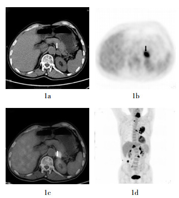
59例肾上腺转移癌患者共78个肾上腺病灶中,有65个病灶的放射性摄取显著增高(SUVmax>3.1),明显高于正常肝组织(图 1),有13个病灶的大小(短径)<1.0 cm,且SUVmax<3.1,但只有3个病灶的SUVmax低于正常肝组织。59例肾上腺转移癌患者中,57例患者同时伴有其他转移灶(图 1);47例原发肿瘤病灶表现为高代谢,10例原发病灶已切除,2例原发病灶不明但有全身多器官转移。

良性病变18例共21个肾上腺病灶中,有19个病灶未见放射性摄取增高(SUVmax<3.1)或低于正常肝组织(图 2),有2个病灶SUVmax>3.1且高于正常肝组织,但术后病理结果显示这2个病灶均为肾上腺皮脂腺瘤;9例患者有恶性肿瘤病史或存在其他部位的恶性肿瘤病灶,但其肾上腺占位均为良性(图 2)。

2组患者肾上腺病灶的大小(短径)、SUVmax及病灶SUVmax与正常肝脏的SUVmax比值结果见表 1。肾上腺转移癌的病灶SUVmax、病灶SUVmax与正常肝脏SUVmax的比值均高于肾上腺良性病变,差异均有统计学意义;而转移癌与良性病变的病灶大小(短径)之间的差异并无统计学意义。
组别 病灶数 病灶大小
[短径(cm)]病灶SUVmax 病灶SUVmax与正常肝脏SUVmax比值 肾上腺转移癌组 78 1.52±1.05 7.05±5.26 2.89±2.49 肾上腺良性病变组 21 1.66±0.91 2.27±1.35 0.79±0.47 t值 0.529 7.199 6.983 P值 >0.05 <0.01 <0.01 表 1 肾上腺转移癌及良性病变的病灶大小(短径)、SUVmax及病灶SUVmax与正常肝脏SUVmax比值对比
-
2组患者的肾上腺病灶分布见表 2。肾上腺转移癌半数以上发生在左侧肾上腺,其次为双侧肾上腺;而肾上腺良性病变好发于单侧,且左右两侧发病率相似,双侧发病较少见。
组别 病例数 左侧病灶 右侧病灶 双侧病灶 肾上腺转移癌组 59 30(50.8) 10(17.0) 19(32.2) 肾上腺良性病变组 18 8(44.4) 7(38.9) 3(16.7) 表 2 肾上腺转移癌及良性病变的病灶分布比例
[例数(%)] -
采用ROC曲线分析所有肾上腺病灶的SUVmax及其与正常肝脏SUVmax的比值,根据各点所对应的灵敏度和误诊率得出区分良恶性病变的最佳临界值;以肾上腺病灶SUVmax>3.1作为良恶性诊断临界值,其诊断灵敏度、特异度和准确率分别为83.3%、90.5%和84.9%;以肾上腺病灶SUVmax与正常肝组织SUVmax的比值>1作为良恶性诊断标准,则诊断灵敏度、特异度、准确率分别为96.2%、90.5%和95.0%。肾上腺转移癌病灶的大小(短径)与其SUVmax的相关性分析,相关系数为0.501。
-
肾上腺血运丰富,是瘤栓的常驻地[3],全身各部位原发癌转移到肾上腺的转移率高达26%~50%,仅次于肺、肝、骨,居第四位[4],因此,准确诊断肾上腺转移瘤具有重要的临床意义。肾上腺转移瘤原发肿瘤位于肾上腺外,除少数因双侧转移导致肾上腺功能低下外,大部分无特异性临床症状,内分泌检查亦无异常,因此常出现漏诊或误诊。
CT和MRI对肾上腺疾病的定位诊断率可高达100%,但其定性诊断较差,依靠病灶大小、HU值、信号强度及强化表现等来判断,有很多病变不能定性[2]。例如,在CT诊断中,往往以肾上腺病灶的大小(直径>5 cm)作为良恶性诊断标准之一,而本研究中的数据表明,肾上腺转移灶与良性病灶的大小之间的差异并无统计学意义。PET-CT是全身的代谢显像与CT形态学检查相结合的一种先进的显像方法,不仅能够测定肾上腺病灶的大小、密度及CT值等形态学指标,还能显示其葡萄糖等物质的代谢状况。Boland等[5]最早报道了18F-FDG PET诊断肾上腺疾病的研究,得出其灵敏度和特异度均为100%。近来的研究表明,18F-FDG PET-CT对肾上腺疾病诊断的灵敏度达93%~100%,特异度达80%~100%,定性准确率为94 %以上[6-9],在本研究中,18F-FDG PET-CT诊断肾上腺转移癌总的灵敏度、特异度、准确率分别为96.2%、90.5%和95.0%,与文献报道相似。同时,PET-CT的全身显像技术对原发肿瘤病灶及肾上腺外的其他转移病灶均能同时进行显示与诊断。
肾上腺转移癌的原发恶性肿瘤以乳腺癌、肺癌、肾癌和恶性黑色素瘤为多,本研究中也以肺癌最多,这与本次统计的病例中肺癌患者所占比例较大有关。肾上腺转移瘤的转移途径主要为血行播散和淋巴系统播散,因此,当恶性肿瘤出现肾上腺转移时,多伴有其他部位的转移,本研究数据中,59例转移患者中有57例同时伴有其他转移灶,有47例原发肿瘤病灶表现为高代谢,10例原发病灶切除术后,2例原发病灶不明但有全身多器官转移。当然恶性肿瘤患者的肾上腺病灶也可能是良性病灶,本研究数据显示,9例患者有恶性肿瘤病灶,但其肾上腺占位均为良性病灶,因此在诊断肾上腺转移时应谨慎。
目前,国内外对PET-CT诊断肾上腺良恶性病变的标准主要有两种:肾上腺病灶SUVmax>3.1和肾上腺病灶的SUVmax与正常肝脏组织的SUVmax比值>1[1, 3, 10-11]。但SUVmax的影响因素较多,不仅受患者体重、血糖浓度等生理因素影响,同时亦受到采集模式、重建算法及衰减校正方式、部分容积效应、分析者感兴趣区设置等的影响[3]。本研究的数据显示,以肾上腺SUVmax>3.1作为肾上腺病灶良恶性的诊断临界值,其对应的诊断灵敏度、特异度和准确率分别为83.3%、90.5%和84.9%,其中有13个恶性病灶(16.7%)被漏诊,原因可能是因为这13个病灶的大小(短径)均<1.0 cm,因此,肾上腺SUVmax的测量受部分容积效应的影响较大。有2个良性病灶(9.5%)被误诊,其中1例患者双侧肾上腺占位但无内分泌症状,SUVmax为7.7,术后病理为肾上腺皮脂腺瘤,术后3月再发,再次手术,病理经国内多家医院会诊均为皮脂腺瘤(但结合生物学行为恶性不除外),经半年随访无再发。另1例SUVmax为3.2,患者无肿瘤病史但有内分泌症状,考虑为有功能皮脂腺瘤,经一年随访确诊。
由于肝细胞内葡萄糖-6-磷酸酶的活性较高,所以18F-FDG不会堆积在肝细胞内,正常人群肝摄取强度比较恒定,个体间、不同时间的肝18F-FDG浓聚程度变化很小[12],因此,以肾上腺病灶SUVmax与正常肝组织的SUVmax比值>1作为良恶性判断标准,则可避免SUVmax测定中的大部分影响因素(如患者体重、血糖浓度、采集模式、重建算法及衰减校正方式等)。本研究中的数据表明,此标准与单纯以肾上腺SUVmax>3.1作为诊断标准相比,其诊断的灵敏度(96.2%)和准确率(95.0%)均明显提高,特异度(90.5%)则相同,其中只有3个(3.8%)恶性病灶被漏诊,有2个良性病灶被误诊,原因是这2个良性病灶的SUVmax均大于3.1,因此,与正常肝组织的SUVmax的比值均大于1。在应用肾上腺病灶SUVmax与正常肝组织的SUVmax比值作为判断标准时,应注意当患者存在肝脏良性病变时(如:肝包虫病、肝脓肿、肝细胞腺瘤、肝脏炎性假瘤、肝结核瘤、肝内结节病、肝硬化再生结节、肝局灶性结节样增生、弥漫性脂肪肝伴正常肝岛等),可能出现18F-FDG代谢增高[13-14],从而使比值下降造成漏诊,因此,检查前需详细了解患者病史及其临床资料。肾上腺转移瘤以单侧病变较多,其中以左侧最多(50.8%),分布比例依次为左侧>双侧>右侧,这可能与左侧肾上腺体积及血供大于右侧有关[3]。
18F-FDG PET-CT显示了全身各部位的葡萄糖代谢状态,与CT的形态学图像相融合,能灵敏诊断有无原发恶性肿瘤病灶及其他转移病灶,并且肾上腺转移癌与良性病灶在葡萄糖代谢及形态学方面具有差异性,因此,通过分析全身显像结果,再结合肾上腺病灶的大小、代谢特点、SUVmax及其与正常肝组织的SUVmax的比值等进行综合分析,可使得18F-FDG PET-CT在诊断肾上腺转移癌方面具有很高的灵敏度及准确率。
18F-FDG PET-CT在肾上腺转移癌诊断中的价值及其诊断标准探讨
Diagnostic value and diagnostic criteria of 18F-FDG PET-CT in adrenal metastasis
-
摘要:
目的 研究18F-FDG PET-CT对肾上腺转移性癌的诊断价值,并探讨其最佳诊断标准。 方法 对59例术后病理或临床随访确诊为肾上腺转移癌及18例肾上腺良性病变患者行18F-FDG PET-CT检查,分别测量肾上腺病灶的大小、SUVmax及正常肝组织的SUVmax,进行综合分析。 结果 59例肾上腺转移癌患者共78个肾上腺病灶,SUVmax为7.05±5.26,18例良性病变患者共21个肾上腺病灶,SUVmax为2.27±1.35,两者之间的差异有统计学意义(t=7.199,P<0.01);肾上腺转移癌与良性病灶的大小(短径)分别为(1.52±1.05)cm和(1.66±0.91)cm,两者之间的差异无统计学意义(t=0.529,P>0.05)。受试者工作特征曲线分析,以肾上腺病灶SUVmax>3.1作为良恶性的诊断标准,其诊断灵敏度、特异度和准确率分别为83.3%、90.5%和84.9%;以肾上腺病灶SUVmax与正常肝组织的SUVmax比值>1作为良恶性诊断标准,则诊断的灵敏度、特异度、准确率分别为96.2%、90.5%和95.0%;肾上腺转移癌病灶大小(短径)与其SUVmax相关系数为0.501。肾上腺转移癌在双侧肾上腺的分布比例为:左侧30例(50.8%),右侧10例(17.0%),双侧19例(32.2%)。 结论 18F-FDG PET-CT对于肾上腺转移瘤的诊断具有较高的灵敏度及准确率;以肾上腺病灶SUVmax与正常肝组织的SUVmax比值>1为标准,可较好地诊断出肾上腺转移瘤的良恶性;肾上腺转移癌病灶的大小与其SUVmax有一定相关性,病灶常累及左侧。 -
关键词:
- 肾上腺肿瘤 /
- 肿瘤转移 /
- 正电子发射断层显像术 /
- 体层摄影术,X线计算机 /
- 氟脱氧葡萄糖F18
Abstract:Objective To investigate the value of 18F-FDG PET-CT in the diagnosis of the adrenal metastasis and study its diagnostic criteria. Methods Fifty-nine patients with adrenal metastasis and 18 patients with adrenal benign tumor underwent 18F-FDG PET-CT, all of the adrenal lesion's diameter and FDG maximum standardized uptake values(SUVmax), SUVmax of normal liver were measured and analyzed comprehensively. All lesions underwent biopsy after operation or follow-up in clinic or imaging diagnosis for final diagnosis. Results There were 78 adrenal lesions of 59 cases with adrenal metastasis, and 21 adrenal lesions of 18 cases with adrenal benign. The SUVmax of adrenals metastasis was 7.05±5.26, while benign tumor was 2.27±1.35, statistical significant difference was found between the 2 groups(t=7.199, P < 0.01). The short diameter of adrenal metastasis was (1.66±0.91) cm, benign tumor was (1.52±1.05) cm, there was no statistical significant difference between the 2 groups(t=0.529, P > 0.05). Receiver operator characteristic curve analysis showed that using lesion's SUVmax > 3.1 as the diagnostic criteria of benign and metastatic adrenal lesions, the sensitivity, specificity and accuracy of 18F-FDG PET- CT in diagnosis of adrenals metastasis were 83.3%, 90.5% and 84.9%, respectively. But if using the SUVmax of adrenal lesion exceeds normal liver tissue as the diagnostic criteria, the sensitivity, specificity and accuracy were 96.2%, 90.5% and 95.0%. For adrenals metastasis, the correlation coefficient between lesion size(short diameter) and SUVmax was 0.501. Among all 59 cases with adrenal metastasis, 30(50.8%) patients' lesions in the left adrenal gland, 10(16.9%)in the right side and 19(32.2%) in the both sides. Conclusion 18F-FDG PET-CT has high sensitivity and accurate in diagnosis of adrenal metastasis, and the SUVmax of adrenal lesion exceeds normal liver tissue is a better diagnostic criteria. The size of adrenal metastasis lesions has certain correlation with its SUVmax and the lesions often in the left side gland and lung cancer is the more common primary tumor. -
表 1 肾上腺转移癌及良性病变的病灶大小(短径)、SUVmax及病灶SUVmax与正常肝脏SUVmax比值对比
组别 病灶数 病灶大小
[短径(cm)]病灶SUVmax 病灶SUVmax与正常肝脏SUVmax比值 肾上腺转移癌组 78 1.52±1.05 7.05±5.26 2.89±2.49 肾上腺良性病变组 21 1.66±0.91 2.27±1.35 0.79±0.47 t值 0.529 7.199 6.983 P值 >0.05 <0.01 <0.01  下载: 导出CSV
表 2 肾上腺转移癌及良性病变的病灶分布比例
[例数(%)] 组别 病例数 左侧病灶 右侧病灶 双侧病灶 肾上腺转移癌组 59 30(50.8) 10(17.0) 19(32.2) 肾上腺良性病变组 18 8(44.4) 7(38.9) 3(16.7)
下载: 导出CSV
-
[1] Metser U, Miller E, Lerman H, et al. 18F-FDG PET/CT in the evaluation of adrenal masses. J Nucl Med, 2006, 47(1): 32-37. [2] Haider MA, Ghai S, Jhaveri K, et al. Chemical shift MR imaging of hyperattenuating (> 10 HU) adrenal masses: does it still have a role?. Radiology, 2004, 231(3): 711-716. [3] 潘中允, 曲婉莹, 周诚, 等. PET/CT诊断学.北京:人民卫生出版社, 2009: 702.
[4] Katz Rl, Shirkhoda A. Diagnostic approach to incidental adrenal nodules in the cancer patient. Results of a clinical, radiologic, and fine-needle aspiration study. Cancer, 1985, 55(9): 1995-2000. doi: 10.1002/1097-0142(19850501)55:9<1995::AID-CNCR2820550928>3.0.CO;2-0 [5] Boland GW, Goldberg MA, Lee MJ, et al. Indeterminate adrenal mass in patients with cancer: evaluation at PET with 2-[F-18]-fluoro-2-deoxy-D-glucose. Radiology, 1995, 194(1): 131-134. [6] Jana S, Zhang T, Milstein DM, et al. FDG-PET and CT characterization of adrenal lesions in cancer patients. Eur J Nucl Med Mol Imaging, 2006, 33(1): 29-35. doi: 10.1007/s00259-005-1915-8 [7] Blake MA, Slattery JM, Kalra MK, et al. Adrenal lesions: characterization with fused PET/CT image in patients with proved or suspected malignancy—initial experience. Radiology, 2006, 238(3): 970-977. [8] 张占文, 胡平, 王晓燕, 等.肾上腺转移瘤18F-FDG PET/CT的影像特征及诊断价值.重庆医学, 2011, 40(8): 774-775.
[9] Lu Y, Xie D, Huang W, et al. 18F-FDG PET/CT in the evaluation of adrenal masses in lung cancer patients. Neoplasma, 2010, 57(2): 129-134. doi: 10.4149/neo_2010_02_129 [10] Anderson SB, Webb MD, Banks KP. Adrenal collision tumor diagnosed by F-18 fluorodeoxyglucose PET/CT. Clin Nucl Med, 2010, 35(6): 414-417. doi: 10.1097/RLU.0b013e3181db4df1 [11] Klingenstein A, Haug AR, Nentwich MM, et al. Whole-body F-18-fluoro-2-deoxyglucose positron emission tomography/computed tomography imaging in the follow-up of metastatic uveal melanoma. Melanoma Res, 2010, 20(6): 511 -516. doi: 10.1097/CMR.0b013e3283403d6c [12] 田嘉禾. PET、PET/CT诊断学.北京:化学工业出版社, 2007: 123.
[13] Sohn MH, Jeong HJ, Lim ST, et al. F-18 FDG uptake in an eosinophilic liver abscess mimicking hepatic metastasis on PET/CT images. Nucl Med Mol Imaging, 2008, 42(3): 253-255. [14] Demir H, Halac M, Yilmaz S, et al. A doughnut sign of a hydatid cyst of the liver on F-18 FDG PET/CT. Clin Nucl Med, 2008, 33(12): 876-877. doi: 10.1097/RLU.0b013e31818bf2df -

 点击查看大图
图(2)表(2)
计量
- 文章访问数: 2306
- HTML全文浏览量: 1211
- PDF下载量: 2