


 下载:
下载:
-
模拟包括计算机产生的体模、成像过程的模拟以及快速解算方法等。随着遗传和分子生物学以及相关技术等领域的迅速发展和基因工程抗体小动物的出现,人们对研究小动物活体成像的兴趣日益浓厚。随着小动物活体成像技术的发展,新的仪器设备、数据采集策略以及图像处理和重建技术的开发和研究也逐渐兴起。模拟技术能够为评价和改进分子成像设备和技术提供一个极其重要的工具。
-
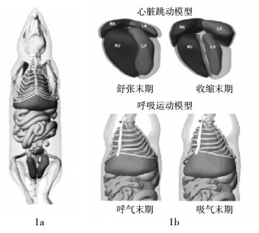
计算机仿真模型能够由基于方程的(程式化)数学函数、体素(数字)的容积阵列或混合方程-体素模型来定义[1],因而能够模拟与实际患者数据更加接近的医学影像数据。现有的电子化体模可分为两种普通类型:体素化体模和数学体模[2]。4D MOBY体模(图 1)和基于非均匀有理B样条(non-uniform rational B-splines, NURBS)曲线的四维心脏-躯干体模是Segars与其同事共同开发的数字体模[3-4]。基于NURBS曲线的四维心脏-躯干体模结合了体素化体模的逼真性和数学体模的灵活性,其概念设计是4D MOBY体模的基础。
在分子成像研究中,4D MOBY体模是一个现实而灵活的小鼠解剖和心脏呼吸运动的模型。MOBY体模用NURBS曲面来模拟器官形状(NURBS是基于三维计算机图形技术和建模的极好的工具,也能够用于人脑图像的模拟和分析[5-6]);用Duke中心的活体显微镜检查技术获得的高分辨三维磁共振显微镜数据作为形成NURBS曲面的基础;用正常小鼠门控黑血MRI数据集作为心脏模型呼吸门控MRI的基础;用已知的呼吸力学作为呼吸模型的基础,以模拟心脏和呼吸运动。在每一种情况下,由一组时间时变曲线定义三维曲面,以产生时间连续的动态或者四维NURBS曲面模型。因此,MOBY体模是基于高分辨门控磁共振显微镜而获得的数据和模拟器官形状的NURBS。4D MOBY体模基于最新型的计算机图形技术。在分子成像研究尤其是新成像仪器、图像采集策略以及图像处理和重建方式中,MOBY体模将发挥独特而重要的作用。
-
对ECAT EXACT HR+型扫描器几何形状而言,PET-SORTEO(是一种基于蒙特卡罗的模拟器)能快速产生真实的PET数据。为了解决对动物PET成像系统仿真模型日益增加的需要,以及为了适应和配置小动物PET这种仿真工具,尤其是广泛分布的用西门子临床解决方案(Siemens preclinical solutions)制造的微型PET R4和Focus 220系统,Lartizien等[7]提出一种能允许产生真实的啮齿类动物影像的仿真模型以便评价并优化采集和重建协议,并且在这个研究中,证实了用MOBY体模迅速产生真实的全身放射性分布的能力。
小动物生物动力学和剂量测定的研究先于临床放射性核素治疗。与在人体中的研究相同,对疗效进行可靠性评价是必要的,且必须基于精确的剂量测定,而精确的剂量测定又必须以实际的剂量测定模型为基础。为了评估解剖上使用更加逼真的小鼠体模时所得结果与以前的数学方式描述的体模所得结果之间的差异,以及评估EGS4和MCNPX2.6a(两种蒙特卡罗代码),Larsson等[8]用MOBY体模评估并优化小鼠活体成像,执行MOBY程序产生了一个基于体素的特定大小的三维体模,并改变体模将其用于输入吸收分数和S因子的蒙特卡罗模拟,用EGS4和MCNPX2.6a代码模拟辐射传递,用交互式数据语言程序计算18F、121I、131I、111In、177Lu和90Y等核素的放射剂量以及S因子,并与以前的模型所获得的结果相比较发现,MOBY体模和数学体模所获得的S因子在多数情况下比较相似,但对一些器官来说,数学体模获得的S因子被低估了。用MOBY体模研究MCNPX 2.6a和EGS4的差异时,发现在电子吸收份额中有一定的差异,其原因可能是由于代码之间的差异所致。因而笔者认为,作为一个放射剂量的测定模型,MOBY体模是一个逼真而且非常灵活的体模,是非常有用的。
目前,人们已经认识到小动物分子成像技术如PET、SPECT的重要性,所以已经加速优化了专门的小动物成像系统、相应的图像重建和校正方式,与此同时,也加速发展数据采集协定。为了整合MOBY体模、发射断层模拟系统(simulation system for emission tomography,SimSET)以及发射断层扫描Geant4应用程序(Geant4 application for tomographic emission,GATE)这3个公共领域软件工具,Chen等[9]用4D MOBY体模提供逼真的小鼠解剖和生理学模型,并结合了两个蒙特卡罗模拟包:SimSET和GATE,在直观且系统复杂的立体基阵的安装方面,保留了GATE的优点;在小鼠体模内模拟光子相互作用方面,为了克服相对较慢的Geant4蒙特卡罗代码的缺点,把GATE引进快速的SimSET光子历史生成器(photon history generator,PHG),从而开发了小动物PET和SPECT模拟工作流程。SimSET-PHG加速了蒙特卡罗模拟过程(加速约10倍),但没有牺牲精确性和多功能性。在小动物小孔SPECT成像流程中,GATE使复杂程度不同的单一的或多小孔准直和探测器几何形状的模拟更加方便,而仿真MOBY体模为不同类型的解剖和生理学模型提供了基础。因此,把MOBY体模与SimSET和GATE相结合,可成功建立一个新的、快速而逼真的多功能小动物分子成像模型。
为了开发和验证用小动物PET影像自动计算PET示踪剂生物分布的方式,Kesner等[10]用MOBY体模软件在三维图像上测量了用小动物PET扫描了的9只小鼠的长度,然后算出其平均长度和标准差,并在一系列机体尺寸内产生了9个体模;他们开发了一个软件(用一个小动物PET扫描的外观上可辨别的器官和其他标记物半自动配准数字小鼠体模)之后,在9只小鼠的9个模拟扫描和18个实际PET扫描中,软性改变体模,使其与相应的标记相符,自动计算反映全身和21个单个器官的22个感兴趣区的示踪剂浓度,用最小终端用户输入成功地产生了PET-地图集影像融合,能够迅速量化22个不同组织的感兴趣区,模拟扫描分析发现,用这种方式扫描轮廓尺寸和单个动物是稳健的。
对SPECT和PET系统来说,GATE模拟工具已经成为一个良好的验证工具,GATE的一个重要特征就是其能够完成时间依赖现象的建模。Sakellios等[11]用MOBY小鼠体模来模拟小鼠,并在GATE中建模,以试验性地评估两个小视角扫描仪。该研究模拟了两个扫描仪:第一个是小鼠大小的γ相机,另一个则是有4个探头的小动物PET,其安装了一个H8500型敏感的光电倍增管和一个像素化的硅酸钇镥闪烁晶体闪烁器,把在第一个扫描仪中引入的MOBY小鼠作为一个体素化原始资料,并实施了二维成像模拟。
NURBS曲线能用于计算机来进行形状模拟,目前,一些人类或动物的解剖结构模型已经在此基础上被开发出来了。图像处理技术的发展允许用来自CT、MRI或PET的数字图像产生的人体或者动物大脑的解剖和功能的模型和地图集[12]。Pushkin等[13]把PET和CT数据用于小鼠大脑的解剖结构和同位素吸收量的NURBS建模,用MOBY体模在Rhino三维开发框架(一个基于NURBS曲线和结构的绘制、动画制作和分析的工具)内构建和编辑所有的结构,结果用NURBS证明了小鼠大脑解剖结构数学建模的可行性。
为了通过用组合的光学-PET系统模拟生物荧光断层测量的图像形成过程来探讨其可行性,Alexandrakis等[14]用小鼠微型MRI图像容积MOBY体模来提供虚拟体模的解剖特征,为位于不同器官(如肝脏、肾脏和消化道等)的单点、两点和分布生物荧光源而模拟光学-PET度量,用期望值最大化代码恢复模拟源的强度和位置,并用MOBY体模提供不同组织的解剖定义。
Fahimian等[15]评估了一种均等斜面断层扫描的新的基于傅立叶的迭代采集和重建方式,并提出一系列定量图像质量和剂量减少的对照试验和模拟方式,用MOBY体模和Zubal全身人体体模,通过用均等斜面断层扫描的CT模拟并与滤过反投影相比,完成迭代代数法,用波状外形器官的信噪比和载噪比,调制传递函数和傅立叶环相关定量重建图像质量;用锥形束X线微型CT仪证实了模拟,制造了微流体分辨率体模和调制传递函数体模以定量验证影像的质量,因此,研究者认为,均等斜面断层扫描为CT中剂量的减少提供了一个有效的方式,并保证了进一步的研究。
-
在医学成像研究中,对描述、评估、优化医学成像体系来说,模拟是一个强有力的工具,其作用日益重要,已经成为理论推导、实验方式和临床研究的一个重要且不可或缺的补充。MOBY体模与基于NURBS曲线的四维心脏-躯干体模一样,为医学影像研究提供了一个很好的工具。可以预见,随着体模研究的进一步深入,将会出现更多新的数字体模,数字体模将会在医学领域中得到更广泛的应用。
四维小鼠全身体模及其在医学影像研究中的应用
The four-dimensional mouse whole-body phantoms and its application in medical imaging research
-
摘要: 医学成像模拟对描述、评估和优化医学成像设备而言是一个强有力的工具。模拟的关键是必须有研究对象解剖结构仿真体模或模型。四维小鼠全身体模为影像研究提供了小鼠解剖和生理的仿真模型。精确的模型与成像过程相结合,能提供研究对象在健康和疾病状态下不同的解剖和运动(心脏和呼吸)的大量逼真的成像数据。对研究解剖、生理、机体等因素对医学和小动物成像的影响来说,四维小鼠全身体模有巨大的潜力,其还可用于新仪器研究、图像采集策略、图像处理和重建方式、图像可视化和解释技术等。
-
关键词:
- 正电子发射断层显像术 /
- 体层摄影术,X线计算机 /
- 体模,显像术 /
- 计算机模拟
Abstract: Medical imaging simulation is a powerful tool for characterizing, evaluating, and optimizing medical imaging devices and techniques. A vital aspect of simulation is to have a realistic phantom or model of the subject's anatomy. Four-dimensional mouse whole-body phantoms provide realistic models of the mouse anatomy and physiology for imaging studies. When combined with accurate models for the imaging process, are capable of providing a wealth of realistic imaging data from subjects with various anatomies and motions(cardiac and respiratory)in health and disease. With this ability, the four-dimensional mouse whole-body phantoms have enormous potential to study the effects of anatomical, physiological and physical factors on medical and small animal imaging and to research new instrumentation, image acquisition strategies, image processing, reconstruction methods, image visualization and interpretation techniques. -
[1] Zaidi H, Xu XG. Computational anthropomorphic models of the human anatomy: the path to realistic Monte Carlo modeling in radiological sciences. Annu Rev Biomed Eng, 2007, 9: 471-500. doi: 10.1146/annurev.bioeng.9.060906.151934 [2] Segars WP, Tsui BM, Frey EC, et al. Development of a 4-D digital mouse phantom for molecular imaging research. Mol Imaging Biol, 2004, 6(3): 149-159. [3] Segars WP, Lalush DS, Tsui BMW. A realistic spline-based dynamic heart phantom. IEEE Trans Nucl Sci, 1999, 46(3): 503-506. doi: 10.1109/23.775570 [4] Segars WP. Development of a new dynamic NURBS-based cardiac-torso(NCAT) phantom. Chapel Hill: The University of North Carolina, 2001. [5] Liu L, Nowinski WL. A hybrid approach to shape-based interpolation of stereotactic atlases of the human brain. Neuroinformatics, 2006, 4(2): 177-198. doi: 10.1385/NI:4:2:177 [6] Anastasi G, Cutroneo G, Tomasello F, et al. In vivo basal ganglia volumetry through application of NURBS models to MR images. Neuroradiology, 2006, 48(5): 338-345. doi: 10.1007/s00234-005-0041-4 [7] Lartizien C, Kuntner C, Goertzen AL, et al. Validation of PET-SORTEO Monte Carlo simulations for the geometries of the MicroPET R4 and Focus 220 PET scanners. Phy Med Biol, 2007, 52(16): 4845-4862. doi: 10.1088/0031-9155/52/16/009 [8] Larsson E, Strand SE, Ljungberg M, et al. Mouse S-factors based on Monte Carlo simulations in the anatomical realistic Moby phantom for internal dosimetry. Cancer Biother Radiopharm, 2007, 22(3): 438-442. doi: 10.1089/cbr.2006.320 [9] Chen CL, Yang CC, Wang YC, et al. A novel workflow for fast, realistic, and versatile small animal molecular imaging simulations. J Nucl Med, 2007, 48 Supp12: 426P. [10] Kesner AL, Dahlbom M, Huang SC, et al. Semiautomated analysis of small-animal PET data. J Nucl Med, 2006, 47(7): 1181-1186. [11] Sakellios N, Rubio JL, Karakatsanis N, et al. GATE simulations for small animal SPECT/PET using voxelized phantoms and rotating-head detectors. Nuclear Science Symposium Conference Record, IEEE Xplore, 2006, 4: 2000-2003[2012-01-01]. http://ieeexplore.ieee.org/xpl/login.jsp?tp=&arnumber=4179419&url=http%3A%2F%2Fieeexplore.ieee.org%2Fxpls%2Fabs_all.jsp%3Farnumber%3D4179419. [12] Herholz K, Herscovitch P, Heiss WD. NeuroPET: positron emission tomography in neuroscience and clinical neurology. Berlin: Springer, 2004: 1-33. [13] Pushkin SV, Podoprigora GI, Comas L, et al. A computational model of rat cerebral blood flow using non-uniform rational B-splines. Conf Proc IEEE Eng Med Biol Soc, IEEE Xplore, 2007: 1098-1100[2012-01-01]. http://ieeexplore.ieee.org/xpl/articleDetails.jsp?arnumber=4352487. [14] Alexandrakis G, Rannou FR, Chatziioannou AF. Tomographic bioluminescence imaging by use of a combined optical-PET(OPET) system: a computer simulation feasibility study. Phys Med Biol, 2005, 50(17): 4225-4241. doi: 10.1088/0031-9155/50/17/021 [15] Fahimian B, Chatziioannou A, DeMarco J, et al. MO-D-332-06: dose reduction in CT using a novel fourier-based iterative reconstruction method. Med Phys, 2008, 35(6): 2870. -

 点击查看大图
图(1)
计量
- 文章访问数: 2015
- HTML全文浏览量: 882
- PDF下载量: 5