


 下载:
下载:
-
茎突综合征(styloid process syndrome,SPS)是因茎突的形态、长度和方位的异常以及茎突舌骨韧带骨化或茎突周围炎等原因刺激周围神经血管或其他组织而导致的以咽痛为主的征候群[1]。SPS在临床上并不少见,但由于其与慢性咽炎、咽异感症、眩晕症及颈椎病等具有相似的临床表现而容易被误诊。本研究回顾性分析了50例临床诊断为SPS患者的多层螺旋CT(multiple-detector spiral tomography,MDCT)和容积再现、表面遮盖显示(shaded surface display,SSD)重建、多平面重建(multi-planar reconstruction,MPR)及最大密度投影(maximum intensity projection,MIP)的重组资料,旨在探讨MDCT在诊断SPS中的价值,提高对该病的诊断水平。
-
选取2008年6月至2011年6月在我院就诊并经临床确诊为SPS的患者50例,其中,男性29例、女性21例,年龄28~72岁,平均39岁,病变为双侧者36例,病变为单侧者14例。
其诊断参考标准[2]:①临床症状;②X线平片及CT显示的茎突形态、长度及方位角;③用手指触诊扁桃体窝是否引起疼痛;④用质量分数为1%的利多卡因封闭扁桃体后部,检查症状是否暂时消失。确定诊断至少符合其中任意3条。
-
采用美国GE公司生产的Brightspeed 16层螺旋机,患者取仰卧位,保持左右对称,下颌抬高,头颅听眶线尽量与扫描床垂直,取头颅侧位片为定位片,扫描范围自听眶线上约1 cm起至第4颈椎下缘,扫描参数:120 kV、150 mA、层厚2.5 mm、重建层厚1.25 mm。使用美国GE公司的ADW 4.3CT工作站进行容积再现、MIP、SSD、MPR重组,剪切掉下颌骨等部位,充分暴露双侧茎突。
测量方法:本组资料对轴位CT薄层图像进行了多层面图像重建,在多层面重建图像上,通过双向角度的反复调整,可以清晰显示茎突根部,并尽可能将两侧茎突完整地显示在一幅图像上,测出茎突的长度。在容积再现正位图像上画一条与颅底水平的垂直线,测量此垂直线与茎突中心线的夹角,即为向内偏斜角。在容积再现侧位图像上画一条听眶线垂直线,测量此垂直线与茎突中心线的夹角,即为向前偏斜角。以茎突超过3 cm为过长;向前或向内偏斜角大于25°为方位角异常;以茎突超过2节为分节型茎突;以茎突中段不平直,呈现弯曲改变为中段弯曲;以茎突舌骨韧带处可见高密度钙化影为茎突舌骨韧带钙化。
-
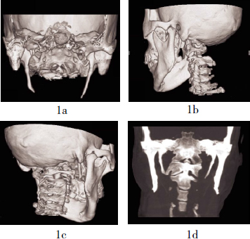
50例患者(86侧)均通过16层螺旋CT薄层螺旋扫描及三维重建,在测量的86侧茎突中,茎突过长者69侧,茎突平均长度为(38.12±5.66)mm,左侧为(37.67±5.15)mm,右侧为(38.76±5.49)mm,最长者为5.0 cm,检出率为80.2%。分节型茎突者45侧,检出率为52.3%。茎突中段弯曲者8侧,检出率为9.3%。茎突舌骨韧带钙化为6侧,检出率为7.0%。本组病例方位角异常中,以向内偏斜角 > 25°居多,占36侧,检出率达到41.9%,没有发现 > 40°或者 < 20°者;向前偏斜角则以 < 20°居多,发现 > 25°者仅5侧,检出率仅为5.8%(图 1)。

图 1 茎突综合征影像学图像
-
人类茎突由胚胎第二鳃弓的舌骨弓软骨即Reichert软骨发育而来,该软骨前下基部发育成舌骨,此基部的两端各有一条软骨链与每侧颞骨相连,软骨链分4段,鼓舌段出生前即开始骨化,1岁左右发展成茎突根部,并与颞骨的乳突、鼓骨、岩骨之间的骨质融合;茎舌段出生后才开始骨化,以后逐渐发展成为茎突体部;角舌段出生后发展成为茎突舌骨韧带;下舌段出生后发展为舌骨小角。茎突可因本身发育或茎突舌骨韧带钙化而导致茎突过长或者方位、形态异常,压迫血管、神经引起血管神经分布区疼痛或者其他异常感觉,这些症状可混合出现,极少数病例甚至以转头时反复眩晕、咽部异物感伴头晕、头痛、视物模糊以及牙痛等为主要症状,临床上称之为SPS,常见于成年人。
SPS起病缓慢,病史长短不一,常有扁桃体区、舌根区疼痛,多为单侧,但不剧烈,可放射到耳部或颈部,吞咽时加重。咽部常有异物感或梗阻感,多为一侧,吞咽时更为明显。当颈动脉受到压迫或磨擦时,疼痛可从一侧下颔角向上放射到头颈部或面部。目前,SPS确切的发病率还无法确定。一般认为SPS多出现在茎突软骨的骨化期间,即20至40岁之间[3]。
SPS病因复杂,主要包括:①茎突过长:茎突尖端伸向扁桃体窝内或其附近,无论扁桃体摘除与否,均可出现咽部异物感,如压迫神经末梢,可出现咽痛等症状,过长茎突压迫或磨擦颈部动脉,影响血液循环,可引起相应区域的疼痛;②茎突方位与形态异常:部分患者茎突长度在正常范围内,但其方位与形态异常,或颈动脉部位异常使两者相抵而引起头痛等症状;③扁桃体炎及扁桃体术后疤痕牵拉,也是发生SPS的原因之一;④舌咽神经炎与SPS有密切关系[4]。目前多数研究者认为,茎突呈细长圆锥形骨性突起,自根部向尖端逐渐变细,茎突后方有面神经出颅,尖端位于颈内、颈外动脉之间,接近颈外动脉终末分叉处,舌咽神经在茎突根部内侧下行,这是茎突过长引起症状的解剖基础[5]。
茎突根据发育情况分为4类:①完整型;②分节型;③发育不良型;④未发育型[6]。本组病例中,分节型茎突45侧,占52.3%,提示分节型茎突与SPS关系密切,而有关分节型茎突与SPS关系的文献报道较少[7]。
以往,X线诊断以超过2.5 cm为判断茎突过长的标准,从20世纪80年代开始,国内外学者对此提出了新的标准,但报道不一[5, 8-9]。邱大学等[10]观察研究458侧茎突的长度、方位及其周围结构的毗邻关系,测得茎突平均长度为(24.68±5.72)mm,左侧为(24.37±5.51)mm,右侧为(24.96±6.18)mm。肖轼之[11]认为,茎突长度在2.5~3.0 cm范围内为正常,超过3.0 cm为茎突过长。本组病例中,茎突长度超过3.0 cm者69侧,占80.2%,最长者为5.0 cm。但也有茎突过长而无症状者,国外文献报告,4%的人茎突过长,而过长者中只有4%的患者有临床症状[5],因此茎突过长并不是诊断SPS的唯一指标。
即使长度在正常范围,但茎突方位的异常同样可以压迫颈内外动脉以及神经,引起咽部异物感及疼痛,导致SPS。肖轼之[11]报道,正常茎突向前向内各偏斜25°, > 25°时可认为茎突方位角异常。兰宝森[12]则认为,正常茎突向内向前各偏斜30°, > 40°或 < 20°时可认为茎突方位角异常。张进华等[13]对10例成人行茎突CT三维重建测量茎突,发现前倾角为18.96°±5.12°,内倾角为23.67°±6.84°。我们观察到本组病例方位角异常中,以向内偏斜角>25°者居多,占36侧,没有发现>40°或者<20°者;向前偏斜角则以<20°居多,发现>25°者仅5侧。应该说,目前对于茎突偏斜异常角度尚未达成统一认识,尤其是向前偏斜角与发病的关系尚难定论,但借助容积再现图像的测量,并结合临床咽部指诊是否触及茎突,进而比较茎突角度的差异,就可大体了解茎突角度异常范围。
由于茎突的解剖位置复杂,易与周围复杂的骨性结构以及软组织影像重叠,因此,X线平片观察茎突效果较差,不能清晰显示茎突根部且有放大现象,使得茎突长度测量的准确性受到限制。曲面断层法摄片也存在着放大失真、测量偏斜角比较困难等缺点。常规CT冠状面薄层扫描虽然消除了重叠影像的干扰,失真也小,但受患者的茎突形态、方位角等因素的影响,茎突全景在一个层面上显示的机会较小,同时对偏斜角也无法测量。而MDCT的容积再现、SSD、MPR、MIP重组的应用弥补了以上缺陷,特别是对拟手术的患者尤为重要。
MDCT的优势之一就是强大的图像后处理功能。将扫描所获得的原始数据进行薄层重建后,传入微机工作站,可以重建出高质量的图像,根据需要可从不同角度、不同方位显示病变。MIP是以预设的角度对体积数据投射射线,将每束射线所遇到的强度最大值编成二维投影像显示出来,MIP灰阶反映相对的X射线衰减值,能描绘X射线衰减值的微小变化,MIP特别适合显示病灶与周围结构之间的三维关系。容积再现利用了全部的容积数据,是真正意义上的三维重建,它有很强的空间立体感,可任意角度旋转,它利用投影线通过容积数据,可将扫描容积内全部像素总合的投影以不同灰阶形式显示出来。SSD通过对不同结构的色彩编码和使用不同的透明度,可同时显示表浅或深在结构的影像,其伪彩技术的应用使图像质量更逼真,但缺点是不能显示肺内病变。MPR是利用容积扫描所获得的三维数据重建出冠状面、矢状面或任意斜面的二维图像,其操作简单,效果呈现快,可以从不同角度观察病灶的形态和相邻结构的解剖关系。三维容积再现重组图像及SSD重建图像看到的茎突是三维立体的全景图像,表面轮廓细腻逼真,能清晰地显示茎突的形态、偏斜角度以及与周围骨性组织的空间毗邻关系,再加上MPR、MIP图像对茎突长度的显示,极大地方便了茎突三维立体评估和测量,有着X线平片及常规冠状面CT图像无法比拟的优势[14-16]。
由于SPS的临床症状无特异性,因此该病常被忽略。临床诊断上要结合以下几个方面综合判断:临床症状、X线平片及MDCT、手指触诊扁桃体窝是否引起疼痛,以及1%的利多卡因封闭扁桃体后部是否使症状暂时消失等;此外,还应与下咽部肿瘤、舌咽神经痛、咽部异物感、三叉神经痛、眩晕症、颈椎病等相鉴别;其他疾病如扁桃体炎及摘除术后、茎突附着的肌肉、韧带的病变、风湿性茎突炎、茎突骨折等,也具有相应的临床表现和体征,针对性检查不难与SPS相鉴别[17-18]。
目前,茎突截短术是治疗SPS的主要方法,手术方式一般采取经口内或颈部入路。由于茎突的形态、长度和方位角变异较大,因此MDCT的容积再现、SSD、MPR、MIP技术对于术前了解茎突的情况、选择适当的手术入路就显得较为重要。一般来说,对于咽部指诊可触及茎突而MDCT的容积再现、SSD、MPR、MIP技术显示茎突过长、向内和向前偏斜角过大者,选择口内进路较为适合;而对于咽部指诊不易触及茎突者,MDCT的容积再现、SSD、MPR、MIP技术显示茎突过长、纤细、分节而偏斜角正常的患者,手术入路则应视病例的具体情况慎重选择。个别患者术后症状有复发,可能为茎突截短的长度不够或术后咽部瘢痕挛缩刺激茎突残根所致[17]。
多层螺旋CT在诊断茎突综合征中的应用价值
Multiple-detector spiral CT in diagnosis of the application value of styloid process syndrome
-
摘要:
目的 探讨16层螺旋CT容积再现、表面遮盖法重建、多平面重建技术及最大密度投影技术在成人茎突测量中的应用及对茎突综合征(SPS)诊断的价值。 方法 回顾性分析50例86侧临床诊断为SPS患者的16层螺旋CT及容积再现、表面遮盖法重建、多平面重建、最大密度投影重组资料,并测量茎突的长度及方位角。 结果 茎突长度大于3 cm者69侧(80.2%),分节型茎突者45侧(52.3%),茎突向内偏斜角 > 25°者36侧(41.9%),向前偏斜角 > 25°者5侧(5.8%)。 结论 茎突过长、茎突发育呈分节型和方位角异常是诊断SPS的重要依据。多层螺旋CT的容积再现、表面遮盖法重建、多平面重建、最大密度投影重组可为诊断SPS提供重要依据。 Abstract:Objective To evaluate the value of styloid process measurement using 16-detector row spiral CT with volume rendering, shaded surface display, multi-planar reconstruction and maximum intensity projection reconstruction techniques in the diagnosis of adult styloid process syndrome (SPS). Methods 16-detector row spiral CT with volume rendering, shaded surface display, multi-planar reconstruction and maximum intensity projection reconstruction techniques were used in 50 patients (86 sides) with clinically suspected SPS, the length and deviated angle of styloid process were measured. Results In these 86 sides of styloid process, 69 sides showed the length longer than 3 cm (80.2%), 45 sides showed segmented styloid process (52.3%), the oblique angle of styloid process was deviated internally more than 25° in 36 sides (41.9%), and deviated anteriorly angle more than 25° in 5 sides (5.8%). Conclusion Over-length, segmentation andabnormal deviation of the oblique angle of styloid process are important criteria in the diagnosis of SPS, helpful information could be provided by multiple-detector spiral CT with volume rendering, shaded surface display, multi-planar reconstruction and maximum intensity projection reconstruction techniques. -
Key words:
- Stem axon syndrome /
- Image processing /
- computer-assisted /
- Tomography /
- spiral
-
[1] 乐杰.妇产科学. 6版.北京:人民卫生出版社, 2005: 110.
[2] 谭旭民, 杨桦, 邓安春.茎突综合征的临床分析.中国医师杂志, 2004, 6(7): 953-954. doi: 10.3760/cma.j.issn.1008-1372.2004.07.041
[3] Karam C, Koussa S. Eagle syndrome: the role of CT scan with 3D reconstructions. J Neuroradiol, 2007, 34(5): 344-345. [4] 董学武, 郭英杰, 王志彬.临床症状不同、诊断结果一致病案两例--(1)转头时反复出现眩晕2年多; (2)咽部异物感, 偶伴头晕、头痛、视物模糊6年.中国医刊, 2005, 40(9): 60. doi: 10.3969/j.issn.1008-1070.2005.09.029
[5] Thot B, Revel S, Mohandas R, et al. Eagle' syndrome. Anatomy of the styloid process. Indian J Dent Res, 2000, 11(2): 65-70. [6] 徐秀娥, 耿永志.牙痛为首发症状的茎突综合征1例报告.临床口腔医学杂志, 2002, 18(6): 466. doi: 10.3969/j.issn.1003-1634.2002.06.038
[7] 黄连庆, 陆武, 周胜利, 等.螺旋CT三维重建SSD和多平面重建MPR对成人茎突测量的研究.实用放射学杂志, 2004, 20(11): 975-978. doi: 10.3969/j.issn.1002-1671.2004.11.005
[8] 童远明, 邓士俊, 李济安.茎突综合征28例临床与X线表现分析.临床耳鼻咽喉科杂志, 2008, 7(1): 48-50.
[9] 王志斌, 刘秋润, 崔永华, 等.茎突X线平片与CT扫描三维成像的临床评价.临床耳鼻喉科杂志, 2006, 20(2): 60-63.
[10] 邱大学, 施建辉, 曹文建, 等.颞骨茎突的测量及其临床意义.解剖学杂志, 2002, 25(1): 76-78. doi: 10.3969/j.issn.1001-1633.2002.01.019
[11] 肖轼之.耳鼻咽喉科学.北京:人民卫生出版社, 1985: 269-270.
[12] 兰宝森.中华影像医学:头颈部卷.北京:人民卫生出版社, 2002: 219-220.
[13] 张进华, 周义成, 肖明.茎突CT三维重建检查技术及测量方法探讨.放射学实践, 2001, 16(4): 224-225. doi: 10.3969/j.issn.1000-0313.2001.04.005
[14] 顾卫根.茎突CT薄层冠状扫描方法及临床应用.实用放射学杂志, 2001, 17(6): 424-427. doi: 10.3969/j.issn.1002-1671.2001.06.031
[15] 彭俊红, 袁德华, 王仁法. MSCT VR与MIP诊断成人茎突综合征的临床价值.放射学实践, 2008, 23(1): 23-25. doi: 10.3969/j.issn.1000-0313.2008.01.008
[16] 陈春美, 秦将均. 64排螺旋CT重建枝术在茎突综合征中的诊断价值.海南医学院学报, 2009, 15(7): 799-801.
[17] 林达, 邱乾德, 许加俊, 等.螺旋CT MPR重建在诊断茎突综合征中的应用价值.医学影像学杂志, 2008, 18(11): 1229-1232. doi: 10.3969/j.issn.1006-9011.2008.11.005
[18] 王霞, 盛蕾, 刘军, 等.多层螺旋CT对成人茎突测量与茎突综合征的诊断.医学影像学杂志, 2006, 16(5): 452-454. doi: 10.3969/j.issn.1006-9011.2006.05.007
-

 点击查看大图
图(1)
计量
- 文章访问数: 2075
- HTML全文浏览量: 841
- PDF下载量: 2