


-
电离辐射会对DNA产生一系列直接或间接的辐射损伤,在显微镜下可以表现为多种类型的染色体畸变,染色体畸变率和辐射剂量在一定范围内呈正相关,被称为生物剂量计,其中,染色体双着丝粒的畸变率是相对成熟的生物剂量计。目前,核医学的一个重要研究内容是应用开放性核素进行组织内照射治疗,但其剂量估算比较困难,本研究尝试应用G显带的染色体双着丝粒畸变率评估32P胶体对培养中的淋巴细胞的辐射剂量。
-
采用瑞典ELEKTA公司直线加速器,美国Thermo公司CO2细胞培养箱,台湾Baso公司姬姆萨染液,日本OLYMPUS公司显微镜,北京麦思奇高科技有限公司核型自动分析系统,北京原子高科股份有限公司32P-磷酸铬胶体和美国CAPINTEC公司CRC-15R型活度计。
-
外周血采集:正常志愿者,男性,无辐射接触史,静脉采血3 ml,采用肝素抗凝。
染色体制片:将采集的外周血接种到5 ml的淋巴细胞培养液中,培养72 h,以离心半径为20 cm、2000 r/min离心15 min,收集淋巴细胞;细胞经低渗处理、染色体预固定、固定和再固定处理后制片;采用气干法制片,玻片干燥后置于37 ℃水浴中的胰酶(0.06%)中消化2 min,立即放入新配置的吉姆萨染液中染色4 min,取出后用清水漂洗, 自然干燥后于显微镜核型自动分析系统下读片分析。
-
淋巴细胞接种到培养液后1 h,置于直线加速器下,采用4 MV的X射线进行垂直照射,球靶距100 cm,剂量率3.8 Gy/min,一次性照射剂量分别为0.5 Gy、1 Gy、2 Gy、4 Gy。
-
用活度计量取7.4 MBq的32P胶体加入10 ml的生理盐水中,充分混匀后,取0.1 ml(即74 kBq)的32P胶体加入到接种淋巴细胞1 h后的培养液中,72 h后,以离心半径为20 cm、2000 r/min离心15 min,进行染色体制片。
-
先用低倍镜按照从上到下、从左到右的“之”字形顺序观察,发现合适的中期细胞后,再转到高倍镜下进行核型分析。应用染色体核型自动分析系统进行核型自动配对排列后,由两名有经验的工作者进行修正和确认。
-
观察100个染色体分散良好、形态清晰的细胞核型,统计经照射后染色体双着丝粒的畸变数,并计算其畸变率,建立外照射剂量和染色体双着丝粒畸变率的剂量-效应曲线,并通过该剂量-效应曲线计算74 kBq的32P胶体在5 ml培养液中72 h的累积辐射剂量。
-
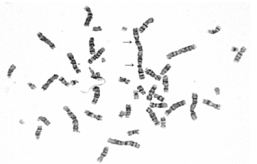
通过淋巴细胞培养和染色体制片可以进行细胞染色体的核型分析。应用G显带技术可以使染色体呈现深浅相间的条纹,相对常规的非显带技术,可以更加准确地识别染色体畸变。图 1为经X射线照射后的染色体G显带核型,可以清晰地识别双着丝粒畸变。

图 1 1 Gy X射线照射后的淋巴细胞染色体G显带核型图
-
应用X射线和32P胶体对培养的淋巴细胞进行照射,不同照射剂量下的染色体双着丝粒畸变率见表 1。
照射剂量 染色体双着丝粒畸变率 X射线(Gy) 0.5 0 1 15 2 26 4 86 32P胶体(kBq) 74 18 表 1 不同剂量X射线及32P胶体照射后的双着丝粒染色体畸变率(%)
-
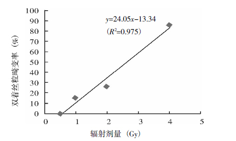
根据X射线外照射0.5 Gy、1 Gy、2 Gy、4 Gy剂量和相应的染色体双着丝粒畸变率,建立辐射剂量-双着丝粒畸变率拟合剂量-效应曲线(图 2),该曲线为:y=24.05x-13.34(R2=0.975),其中,x为辐射剂量,y为双着丝粒畸变率,R2为决定系数,并根据曲线推算74 kBq的32P胶体的辐射剂量。32P胶体照射后的染色体双着丝粒的畸变率为18%,则74 kBq的32P胶体在5 ml的培养液中72 h的累积辐射剂量约为1.3 Gy。

图 2 不同剂量X射线照射后辐射剂量-双着丝粒畸变率的拟合剂量-效应曲线
-
染色体畸变分析作为一种生物剂量测量方法已经运用了50余年,是公认的比较成熟的技术。通过在国内外多次辐射事故中的实际应用,其临床应用价值已得到了充分的肯定[1-2]。
目前报道较多的染色体畸变分析是应用非显带的染色体技术分析双着丝粒畸变[3-4],本研究采用G显带技术进行染色体分析。G显带技术可以使染色体呈现深浅相间的带纹,较非显带的染色体均匀着色,有着很强的识别性,在对双着丝粒的判断上准确率更高[5]。如图 1所示,G显带技术不仅可以清楚地识别双着丝粒染色体,而且可以判断该畸变是由1号和3号染色体的易位造成的,较非显带技术有明显优势。
本研究通过建立4 MV的X射线照射后的辐射剂量-双着丝粒畸变率的剂量-效应曲线,计算得出74 kBq的32P胶体在5 ml的培养液中72 h的累积辐射剂量为1.3 Gy。笔者在另一项研究中采用胶片显色的方法测量,将辐射显色胶片加入培养瓶中,72 h后对照外照射建立的辐射剂量-显色曲线,得出74 kBq的32P胶体72 h内在5 ml的培养液中的累积辐射剂量为2.46 Gy[6]。根据国际医学内辐射剂量委员会1999年对32P进行的体素水平的剂量估算,即假设1 MBq的32P在6 mm3的体素(质量为0.216 g的立方体)内,能量没有溢出,则其剂量率为0.537 mGy·MBq-1·s-1,计算得出74 kBq的32P在5 g培养液中72 h的累积辐射剂量为0.45 Gy[7-8]。
本研究应用生物剂量方法评估放射性核素的辐射剂量,能客观、全面、真实地反映辐射的生物效应,但也存在较大的误差,包括:①应用单次高剂量率辐射建立的曲线来评估持续低剂量率辐射的情况,由于辐射方式不同,可能存在一定的剂量估算偏差;②32P活度的测量误差;③通过稀释法加样可能造成一些误差;④计数染色体形成良好的细胞核型为100个,考虑到辐射对染色体损伤的随机性,辐射剂量也只能是估算。胶片显色法应该是测量辐射剂量较准确的方法,但在测量中也需要应用外照射建立胶片辐射剂量-显色曲线,再对测量胶片进行对应的分析,在操作过程中也存在一些误差。理论计算法是根据国际医学内辐射剂量委员会提供的基于体素S因子的计算方法,计算过程需要假设较多的条件,其计算结果必须与实际测量值进行相互验证。
总之,生物剂量方法、胶片显色法和理论计算法的结果存在一些差异,还需进一步研究探讨,然而,这三种方法是可以互相补充、互相参考的。笔者认为,从辐射剂量估算的角度考虑,染色体畸变率在一定范围内是可以有效评估放射性核素的内照射剂量的。
染色体畸变率估算32P的辐射剂量
The quantitative radiation dose of 32P by chromosomal aberration
-
摘要:
目的 应用G显带的染色体双着丝粒畸变率评估32P的辐射剂量。 方法 应用4 MV的X射线对离体人血液进行0.5 Gy、1 Gy、2 Gy和4 Gy的照射,建立染色体双着丝粒畸变率和X射线的辐射剂量的剂量-效应曲线。将74 kBq的32P胶体加入淋巴细胞的培养液后72 h,进行G显带染色体分析,通过X射线的辐射剂量和染色体双着丝粒畸变率的剂量-效应曲线来评估32P的辐射剂量。 结果 4 MV的X射线的辐射剂量和染色体的双着丝粒畸变率呈线性正相关,剂量-效应曲线为y=24.05x-13.34(R2=0.975)。74 kBq的32P胶体产生的染色体双着丝粒畸变率为18%,即74 kBq的32P胶体在5 ml淋巴细胞培养液中72 h约产生1.3 Gy的辐射剂量。 结论 应用染色体畸变率可以有效地评估放射性核素的内照射剂量。 Abstract:Objective To quantify the radiation dose of 32P by the G-banding chromosomal aberration rate. Methods The human blood was irradiated byX-ray of 0.5, 1, 2, 4 Gy respectively. The dose-response curve between the radiation dose and the dicentrics aberration rate was assayed. 74 kBq 32P colloid was put into culture solution and, after 72 h, the dicentrics aberration rate was observed. The radiation dose of 32P was assayed by the dose-response curve. Results The dose-response curve between the radiation dose and the dicentrics aberration rate was y=24.05x-13.34(R2=0.975). The dicentrics aberration rate of 74 kBq 32P was 18% and the radiation dose of 32P in 5 ml culture solution during 72 h was 1.3 Gy. Conclusion The radiation dose of radionuclide can be estimated by chromosomal aberration rate. -
Key words:
- Chromosome aberrations /
- Dose-response relationship /
- radiation /
- Phosphorus radioisotopes /
- X-rays
-
表 1 不同剂量X射线及32P胶体照射后的双着丝粒染色体畸变率(%)
照射剂量 染色体双着丝粒畸变率 X射线(Gy) 0.5 0 1 15 2 26 4 86 32P胶体(kBq) 74 18  下载: 导出CSV
-
[1] 李秀芹, 于瑞敏, 赵进沛, 等.染色体畸变研究方法及其在生物剂量估算中的应用.中国辐射卫生, 2006, 15(1): 116-117. doi: 10.3969/j.issn.1004-714X.2006.01.070
[2] 闵锐.电离辐射生物剂量研究现状.国外医学放射医学核医学分册, 2004, 28(3): 121-127.
[3] 李世珍.深圳市部分放射作业人员染色体畸变分析.职业与健康, 2011, 27(13): 1473-1474.
[4] 杜杰, 陈英, 闫学昆, 等.18. 8 MeV质子与60Co-γ射线诱发人淋巴细胞染色体畸变比较. 2009, 29(3): 13-17.
[5] García Sagredo JM, Vallcorba I, López-Yarto, et al. Chromosome painting in biological dosimetry: assessment of the ability to score stable chromosome aberrations using different pairs of paint probes. Environ Health Perspect, 1996, 104Suppl3: 475-477. [6] 施常备, 梁晓燕, 袁彬, 等. 32P持续低剂量率辐射敷贴治疗的胶片剂量验证.现代肿瘤医学, 2009, 17(9): 1772-1774. doi: 10.3969/j.issn.1672-4992.2009.09.061
[7] Samei E, Kearfott KJ, Gillespie TJ, et al. An atlas of selected beta-ray spectra and depth-dose distributions in lithium fluoride and soft tissue generated by a fast Monte Carlo-based sampling method. Radiat Phys Chem, 1996, 6(48): 719-725. [8] Bolch WE, Bouchet LG, Robertson JS, et al. MIRD pamphlet No. 17: the dosimetry of nonuniform activity distributions-radionuclide S values at the voxel level. J Nucl Med, 1999, 40(1): 11s-36s. -

 点击查看大图
图(2)表(1)
计量
- 文章访问数: 1950
- HTML全文浏览量: 850
- PDF下载量: 3