


 下载:
下载:
-
神经母细胞瘤是一种儿童常见的实质性肿瘤,其恶性程度极高,起病隐匿,易早期发生多部位转移,往往因转移症状就诊而确诊[1-2]。全身骨显像是诊断神经母细胞瘤骨转移的最常用方法。儿童正常骨骺及干骺端即可摄取较多的显像剂,而干骺端因血流丰富,是肿瘤转移最常见的部位,因此,鉴别正常与异常干骺端是提高诊断准确率的关键。为了评价全身骨显像半定量分析在神经母细胞瘤骨转移中的应用价值,本研究对45例病理确诊的神经母细胞瘤患儿行全身骨显像,并进行半定量分析和视觉分析的比较。
-
经病理证实的神经母细胞瘤患儿45例,其中,男性33例、女性12例,年龄2个月~14岁,平均5.6±3.9岁。肿瘤起源于肾上腺髓质者28例、腹膜后者15例、上纵隔者1例、未找到原发灶者1例。45例患儿共90个股骨远端干骺端,根据骨髓涂片、CT、X线片、MRI或临床随访结果将其分为转移组及无转移组。转移组21个部位,其中,双侧病变8例、单侧病变5例;无转移组69个部位,其中,双侧无病变32例、单侧无病变5例。所有收治患儿均经其家属知情并签署同意书。
-
全身骨显像在术前或术后完成。99Mo-99Tcm发生器由北京原子高科股份有限公司提供,MDP由北京师宏药物有限公司提供,采用常规标记法,放化纯 > 95%。显像剂99Tcm-MDP的注射剂量按9.25 MBq/kg(250 μCi/kg)计算,最小总剂量不能低于74 MBq(2 mCi)。显像仪器为GE公司Hawkeye-VG5型双探头SPECT仪,配低能通用型准直器,能峰为140 keV,能窗为20%。静脉注射显像剂后4 h显像,患者取仰卧位,常规行全身前后位及后前位显像,扫描速度为15 cm/min。
-
取全身后前位图像,分别框取双侧股骨远端干骺端及股骨骨干中部的ROI,计算同侧干骺端/骨干的平均放射性计数比值(用x ± s表示)。以无转移组干骺端/骨干的平均放射性计数比值(x±2s)作为判断标准,高于x±2s者诊断为阳性,低于x±2s者诊断为阴性。
-
由两名核医学医师共同阅片,以①双侧股骨远端干骺端放射性分布不对称,②出现干骺端放射性局灶性或弥漫性异常浓集或稀疏、缺损区,③股骨远端骨骺板线状结构变厚、模糊者诊断为阳性,其余诊断为阴性[3]。
-
采用SPSS10.0软件进行统计学分析,应用两独立样本的t检验对半定量分析法结果进行分析,应用χ2检验对两种方法的诊断结果进行分析,P < 0.05为差异有统计学意义。
-
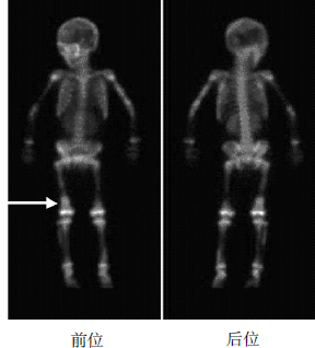
股骨远端干骺端转移组干骺端/骨干的放射性计数比值为3.66±0.71,无转移组为1.83±0.68,2组间差异有统计学意义(t=8.334,P < 0.01)(图 1)。半定量分析法诊断股骨远端干骺端转移组真阳性19个、假阴性2个,无转移组真阴性66个、假阳性3个。半定量分析法判断股骨远端干骺端转移的灵敏度、特异度、准确率、阳性预测值及阴性预测值分别为90.5%(19/21)、95.7%(66/69)、94.4%(85/90)、86.4%(19/22)及97.1%(66/68)。

图 1 神经母细胞瘤股骨远端干骺端骨转移99Tcm-MDPSPECT全身骨显像图
-
视觉分析法诊断股骨远端干骺端转移组真阳性17个、假阴性4个,无转移组真阴性69个、假阳性0个。视觉分析法判断股骨远端干骺端转移的灵敏度、特异度、准确率、阳性预测值及阴性预测值分别为81%(17/21)、100%(69/69)、95.6%(86/90)、100%(17/17)及94.5%(69/73)。
两种方法诊断结果的差异无统计学意义(χ2=0.68,P > 0.05)。
-
神经母细胞瘤是儿童常见的实质性肿瘤,其恶性程度极高,是最容易发生早期多部位转移的肿瘤之一[1]。神经母细胞瘤的发病较隐匿,原发病变缺乏特异性的症状及体征,往往是在出现转移症状时才确诊[2]。由神经母细胞瘤的国际分期可知,不同的转移部位与临床分期密切相关,进而影响到临床治疗方案的选择。因此,明确有无骨转移的发生对于神经母细胞瘤的分期、治疗方案的选择和预后具有重要的意义。
骨髓及骨骼是神经母细胞瘤早期最易转移的部位,骨转移中以四肢长骨的转移率最高[4]。长骨受累的发生率高于其他部位,原因是儿童长骨的干骺端血运丰富,经血循环的肿瘤细胞较易侵犯这些部位[5]。10岁以下的正常儿童属于骨质生长的活跃时期,全身骨骼影像普遍增浓,尤其是骨骺及干骺端。因此,如何区分正常的干骺端与有肿瘤转移的干骺端具有极其重要的意义。
神经母细胞瘤股骨远端干骺端转移在临床上以视觉分析法的使用最为广泛。当双侧干骺端放射性分布不对称时,视觉分析法较易诊断,如本研究21个四肢受累部位中,5个表现为单侧骨放射性分布异常浓集,8个表现为双侧骨放射性分布呈对称性异常浓集。
ROI技术是对图像进行半定量分析。原则上讲,ROI放射性计数为所选区域内的平均像素,放射性分布越浓集则计数越高,故血流丰富处的ROI放射性计数高于其他部位。但这种方法存在假阳性率及假阴性率,如本研究中半定量分析法诊断股骨远端干骺端转移结果中,真阳性19个、假阴性2个、真阴性66个、假阳性3个。因转移病灶有溶骨性与成骨性两种,而溶骨性病灶周围一般都会有成骨反应,可能会表现为放射性分布浓集而非放射性分布稀疏缺损,此时所选ROI若溶骨性占大部分,则会造成假阴性的发生。另外,儿童尿液少量污染衣物或皮肤时,会造成假阳性的发生。
本研究结果显示,半定量分析法与视觉分析法对神经母细胞瘤股骨远端干骺端转移的诊断价值差异无统计学意义,两种方法均可较正确地诊断出转移灶,但其均存在一定的假阳性率及假阴性率。因此,可将两种方法联合使用以相互弥补不足,从而提高诊断的准确率。
神经母细胞瘤股骨远端干骺端骨转移全身骨显像分析
Application of semiquantitative analysis of whole body bone imaging on distal femoral metaphysis osseous metastasis of neuroblastoma
-
摘要:
目的 评价全身骨显像半定量分析在神经母细胞瘤股骨远端干骺端骨转移中的应用价值。 方法 45例经病理证实的神经母细胞瘤患儿,按骨髓涂片、X线片、CT、MRI或临床随访结果等将其分为股骨远端干骺端转移组和无转移组,术前或术后行99Tcm-MDP SPECT全身骨显像,以两种方法对其进行分析:①半定量分析法:对每个患儿双侧股骨远端干骺端及股骨骨干中段勾画ROI,并测定其平均计数,计算每个患儿干骺端/骨干的放射性比值;②视觉分析法:根据骨显像视觉分析诊断患儿是否存在股骨远端干骺端转移。比较这两种方法的诊断结果有无差异。 结果 半定量分析法:转移组及无转移组干骺端/骨干的放射性比值差异有统计学意义(t=8.334,P < 0.01)。半定量分析法诊断股骨远端干骺端转移的灵敏度、特异度、准确率、阳性预测值及阴性预测值分别为90.5%、95.7%、94.4%、86.4%及97.1%;视觉分析法分别为81%、100%、95.6%、100%及94.5%。两种方法诊断结果的差异无统计学意义(χ2=0.68,P > 0.05)。 结论 放射性核素全身骨显像对神经母细胞瘤骨转移的诊断具有重要的价值,结合视觉分析法及半定量分析法可提高诊断的准确率。 Abstract:Objective To evaluate the value of semiquantitative analysis of whole body bone imag-ing on distal femoral metaphysis osseous metastasis of neuroblastoma. Methods Twenty-nine patients with confirmed neuroblastoma by pathological reports were divided into group of metastasis and group of no metastasis by bone marrow slides, X-ray, CT, MRI or clinical follow-up. Whole body bone imaging was performed pre-or postoperation. All cases were analysed by two methods: ①Semi-quantitative analysis: Regions of interest on bilateral distal femoral metaphysic and middle of femoral were drawn, and their average counts were measured. The ratio of radioactivity of distal femoral metaphysic to middle of femoral was calculated; ②Visual analysis: Bilateral distal femoral metaphysic metastasis were diagnosed by visual analysis according to whole body bone imaging. The differences between this two= Methods were compared. Results There were differences of the ratio of radioactivity of distal femoral metaphysic to middle of femoral between group of metastasis and group of no metastasis(t=8.334, P < 0.01), and there was no significant difference betweent the two methods(χ2=0.68, P > 0.05). The sensitivivy, specificity, accuracy, positive predictive value and negative predictive value of semiquantitative analysis in detecting osseous metastasis were 90.5%, 95.7%, 94.4%, 86.4% and 97.1%, while visual analysis were 81%, 100%, 95.6%, 100% and 94.5%. Conclusions Radionuclide whole body bone imaging was of great importance in diagnosis of osseous metastasis of neuroblastoma. The diagnostic accuracy was improved by combination of visual analysis and semi-quantitative analysis. -
Key words:
- Neuroblastoma /
- Femur /
- Neoplasm metastasis /
- Tomography /
- emission-computed /
- single-photon
-
[1] Berthold F, Hero B, Breu H, et al. The recurrence patterns of stageⅠ, Ⅱ and Ⅲ neuroblastoma: experience with 77 relapsing patients. Ann Oncol, 1996, 7(2) : 183-187. doi: 10.1093/oxfordjournals.annonc.a010547 [2] 李正, 王慧贞, 吉士俊.实用小儿外科学(下册).北京: 人民卫生出版社, 2001: 1298-1304.
[3] 邵虹, 施美华, 周莺, 等.骨显像和CT联合应用对儿童神经母细胞瘤临床分期的价值.中华核医学杂志, 2004, 24(4): 227-228. doi: 10.3760/cma.j.issn.2095-2848.2004.04.012
[4] 仲卿雯, 邵虹, 杜隽, 等. CT、超声和骨显像的联合应用对儿童神经母细胞瘤临床分期的研究.中国医学计算机成像杂志, 2008, 14(2): 150-153. doi: 10.3969/j.issn.1006-5741.2008.02.013
[5] Treves ST, Connolly LP, Kirkpatrick JA, et al. Bone. 2nd ed. New York: Springer-Verlag, 1995: 674-675. -

 点击查看大图
图(1)
计量
- 文章访问数: 2128
- HTML全文浏览量: 1002
- PDF下载量: 6