


-
实体肿瘤普遍存在乏氧, 是肿瘤的特性之一。试验表明: 肿瘤乏氧是导致肿瘤局部出现放疗不敏感和产生局部复发及远位转移的重要原因。同时, 近期研究表明: 肿瘤乏氧存在明显的异质性, 不同病理类型、同一类型的不同个体以及同一肿瘤的内部各部分之间, 其乏氧状态也不尽相同[1]。以往人们尝试用各种方法来评价肿瘤的乏氧特性, 以期指导放疗和评价预后, 但是这些方法或者因其具有创伤性, 临床难以推广应用或者只能提供瘤体某一部分的乏氧水平, 难以从整体评价和预测放疗效果, 使其临床应用受到极大限制。99Tcm-4, 9-二氮-2, 3, 10, 10-四甲基十二烷-2, 11-二酮肟(99Tcm-4, 9-diaza-2, 3, 10, 10-tetramethyldodecan-2, 11-dioxime, 99Tcm-HL91)是一种新近开发的乏氧组织显像剂, 细胞培养和动物实验结果显示: 肿瘤摄取水平与其乏氧程度呈良好的正相关[2-5]。利用99Tcm-HL91显像对病灶乏氧进行定性和定量检测, 可动态检测肿瘤再氧合及预测放疗疗效, 具有较其他乏氧检测技术更为突出的优势。研究表明, 肿瘤18F-FDG符合显像也可以很好的评价肿瘤的治疗效果及判断预后。本研究用99Tcm-HL91作为乏氧显像剂, 应用18F-FDG符合显像作为对照, 探讨肿瘤放射治疗前、后的乏氧状态与放疗疗效之间的关系, 以期为临床应用99Tcm-HL91提供实验依据, 从而可以更好地帮助临床评价疗效, 指导治疗方案。
-
(1) 动物: 无特定病原体(SPF)级小鼠由西安交通大学医学院实验动物中心提供并饲养, 选取20只6周龄健康雄性小鼠供实验用, 体质量为(27±3)g, 采用抽签方法随机分为2组, 每组10只, 另备5只用于腹水模型。
(2) 细胞: S180腹水瘤细胞株由西安交通大学医学院第一附属医院分子生物中心提供, 复苏后分别接种于5只小鼠腹腔, 待腹水形成后, 抽取1ml稀释至活瘤细胞计数大于2×10⁶个/ml瘤细胞悬液, 于每只小鼠右后肢皮下接种0.2 ml。待瘤体长至1~1.5 cm时, 用于实验。
-
99Tcm-HL91由广东希埃核医药中心提供。通过小鼠尾静脉注射99Tcm-HL91 37 MBq, 4h后分别于放疗前、放疗后1h、2 d和10 d个时间点进行显像。显像仪器采用美国GE Hawkeye VG SPECT系统, 配以低能通用准直器, 矩阵128×128, 采集时间150 s, 放大1.3倍。
-
18F-FDG由广东希埃核医药中心提供, 分别于放疗前和放疗后11d显像。显像前12 h禁食, 小鼠尾静脉注射18F-FDG 11.5MBq, 30 min后腹腔注射0.1%戊巴比妥钠麻醉。显像仪器采用美国GE Hawkeye VG SPECT系统, 配以高能符合线路准直器, 矩阵128×128, 采集时间150 s, 放大1.3倍。
-
第一次99Tcm-HL91和18F-FDG显像结束后, 对照组和实验组的小鼠分别给予0 Gy(假照射)和8Gy的X线加速器照射。
-
于图像上肿瘤部位和小鼠肺野设等大ROI, 获得显像中T/NT的摄取比值(uptakingratio, UR)。
-
数据处理采用SPSS13.0软件, 两样本数据用均数±标准差(x±s)表示, 组间比较采用t检验, P < 0.05为差异有统计学意义。
-
(1) 放疗前和放疗后1 h、2 d、10 d99Tcm-HL91的UR见表 1, 显像结果见图 1。结果表明: 放疗后不同时相的UR表现为依次递减趋势, 而对照组各时相的UR无明显递减趋势, 甚至在2 d时段出现UR增高。
放疗前 放疗后 1h 2d 10d 放疗组 3.53±1.62 3.41±1.42 2.55±1.57 ** 1.26±0.03*# 对照组 3.62±1.65 3.02±1.94 4.10±1.48 2.96±2.02 t值 1.231 0.513 2.272 8.475 注:表中,*: 与放疗前比较,P < 0.05; #: 与对照组比较,P < 0.05。 表 1 99Tcm-HL91显像的摄取比值(n=10)

图 1 S180腹水瘤细胞小鼠放疗前和放疗后不同时间99Tcm-HL91显像结果(箭头所指为瘤体部位)
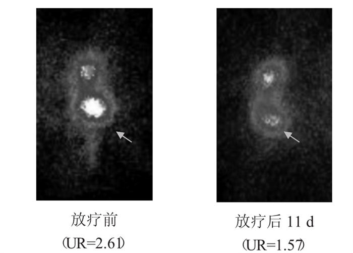
(2) 放疗前和放疗后11d18F-FDG显像的UR见表 2, 显像结果见图 2。结果表明, UR亦呈现依次递减趋势, 与99Tcm-HL91显像一致。而对照组的UR虽然也出现减低, 但变化不明显, 而且大部分实验鼠在显像前死亡。
放疗前 放疗后 t值 放疗组 2.49 ± 1.29 1.49 ±0.56* 2.249 对照组 2.22 ±0.45 1.89 ±0.08 2.083 注:表中,*: 与放疗前比较,P < 0.05。 表 2 放疗前和放疗后18F-FDG显像的摄取比值(n=10)

图 2 S180腹水瘤细胞小鼠放疗前和放疗后11d的18FFDG显像结果(箭头所指为瘤体部位)
-
肿瘤乏氧是导致肿瘤局部出现放疗不敏感和产生局部复发及远位转移的重要原因。而肿瘤乏氧存在明显的异质性, 不同病理类型、同一类型的不同个体以及同一肿瘤的内部各部分之间, 其乏氧状态也不尽相同[6-7]。正是由于肿瘤细胞的这些特点, 导致不同个体间对放疗和化疗的反应及预后出现明显差异。所以, 在活体水平上寻找一种简便、无创的评价肿瘤乏氧方法, 成为临床研究的热点之一。研究证实: 99Tcm-HL91在乏氧组织中有高摄取, 其T/NT可达10倍之多[8-9], 非常有利于临床应用。
本研究中, 放疗前和放疗后不同时相的UR表现为依次递减趋势, 表明放疗后肿瘤出现再氧合, 乏氧状态依次减低, 而在有氧存在的条件下, 射线对肿瘤的辐射生物学效应会增强, 肿瘤细胞对放射治疗会更敏感, 有利于肿瘤的治疗。对照组各时相的UR无明显递减趋势, 说明肿瘤乏氧状态无明显变化, 甚至在2 d时段还出现乏氧加重, 提示在未进行放射治疗的前提下, 肿瘤持续进展。同时, 放疗前和放疗后18F-FDG的葡萄糖代谢显像的UR亦呈现依次递减的趋势, 与99Tcm-HL91显像高度一致, 表明放疗后除肿瘤的乏氧水平减低外, 肿瘤的葡萄糖代谢活性也减低, 提示放射治疗有效地降低了肿瘤的代谢和增殖水平。虽然对照组18F-FDG显像的UR也出现减低趋势, 但变化不明显, 无统计学意义; 而且, 大部分小鼠在显像前死亡, 表明肿瘤进展。
以上这些结果提示, 99Tcm-HL91显像的UR能够较好地评价放疗前后肿瘤乏氧水平变化, 这为活体上评价肿瘤的乏氧状态与放疗疗效之间的关系提供了一种潜在性的新方法, 有很好的临床应用价值。
99Tcm-HL91和18F-FDG显像与放疗关系的实验研究
Experimental study on the relationship between radiotherapy effect and 99Tcm-HL91 and 18F-FDG imaging
-
摘要:
目的 用99Tcm-4,9-二氮-2,3,10,10-四甲基十二烷-2,11-二酮肟(99Tcm-HL91)和18FFDG为显像剂,探讨肿瘤放疗前、后的乏氧状态与放疗疗效之间的关系。 方法 ①动物:选取20只昆明种、清洁级健康成年雄性小鼠供实验用(另备5只用于腹水模型),采用完全随机方法分为2组(对照组和实验组),每组10只。②细胞:将S180腹水瘤细胞株复苏后分别接种于5只小鼠腹腔,待腹水形成后,抽取1 ml稀释至活瘤细胞计数大于2×10⁶个/ml瘤细胞悬液,于每只小鼠右后肢皮下接种0.2 ml。待瘤体长至1~1.5 cm时用于实验。③99Tcm-HL91显像:通过小鼠尾静脉注射99Tcm-HL91 37 MBq,4 h后分别于放疗前、放疗后1 h、2 d和10 d进行显像。④18F-FDG显像:分别于放疗前和放疗后11 d进行18F-FDG显像。显像前12 h禁食,小鼠尾静脉注射18F-FDG 11.5 MBq,30 min后腹腔注射0.1%戊巴比妥钠麻醉。⑤放射治疗:第1次99Tcm-HL91和18F-FDG显像结束后,2组小鼠分别给予0 Gy和8 Gy的X线照射。⑥图像处理和半定量分析:于图像上肿瘤部位和小鼠肺野设等大感兴趣区(ROI),获得显像中瘤/非瘤(T/NT)摄取比值(UR)。 结果 ①放疗前和放疗后1 h、2 d、10 d99Tcm-HL91的UR分别为3.53±1.62、3.41±1.42、2.55±1.57和1.26±0.03,表明放疗后肿瘤出现再氧合,乏氧状态依次减低。对照组各时相的UR值分别为3.62±1.65、3.02±1.94、4.10±1.48和2.96±2.02,UR无明显递减趋势,表明肿瘤乏氧状态未见减低,甚至在2 d时段出现加重。②放疗前和放疗后11 d18F-FDG显像的UR分别为2.49±1.29和1.49±0.56,UR亦呈现依次递减趋势,与99Tcm-HL91显像一致。表明放疗后肿瘤葡萄糖代谢活性减低。对照组的UR分别为2.22±0.45和1.89±0.08,UR也出现减低,但变化不明显,而且大部分小鼠在显像前死亡。 结论 放疗前后99Tcm-HL91显像的UR能够较好地评价肿瘤乏氧水平,并可初步评价疗效,为在活体评价放疗后肿瘤再氧合过程提供了一种新方法。 -
关键词:
- 放射疗法 /
- 氟脱氧葡萄糖F18 /
- 体层摄影术, 发射型计算机, 单光子 /
- HL-91
Abstract:Objective To investigate the relationship between radiotherapy effect and 99Tcm-4, 9-diaza-2, 3, 10, 10-tetramethyldodecan-2, 11-dioxime(99Tcm-HL91)and18F-FDG imaging in S180 mouse. Methods ①Animals: twenty male Kunming mice were randomly divided into two groups of radiotherapy and non-radiotherapy control group.②Cells: S180 cell lines were injected into peritoneal cavity of the other 5 mice.When the S180 tumor developed, 1ml liquid were dripped and diluted to the suspension solution of 2×10⁶cells.Then, 0.2 ml cells solution were was injected into the hippo of right rear leg of mice.The mouse model was used to experiment while the tumor dimension developed to 1-1.5 cm.③99Tcm-HL91 imaging: 37 MBq99Tcm-HL91 was injected into mouse models by tail vein.After 4 h, SPECT were performed before and at 1 h, 2 d and 10 d after radiotherapy.④18F-FDG imaging: 11.5 MBq 18F-FDG was injected into mouse models by tail vein.After 30 min, SPECT were perfored before and after radiotherapy at the time of 11 d.⑤Radiotherapy: two groups of mice were irradiated to 0 Gy and 8 Gy X-ray after the first 99Tcm-HL91 and 18FFDG imaging.⑥Images analysis: the region of interest(ROI)region, in tumor and lung site, was drawed to calculate the uptake ratio(UR). Results Before and at 1 h, 2 d, and 10 d after radiotherapy, the UR in 99Tcm-HL91 imaging were 3.53±1.62, 3.41±1.42, 2.55±1.57 and 1.26±0.03, respectively, while the UR were 3.62±1.65, 3.02±1.94, 4.10±1.48 and 2.96±2.02 in control group.This revealed that tumors hypoxic level was decreased after radiation and suggested that tumors developed reoxygenation.Before and at 11d after radiotherapy, the UR in 18F-FDG imaging were 2.49±1.29 and 1.49±0.56, while the UR were 2.22±0.45 and 1.89±0.08 in control group, which suggested a coincident trend with 99Tcm-HL91 imaging.The tumor glucose metabolism rate was also decreased which suggested a good radiotherapy outcome. Conclusions 99Tcm-HL91 UR can effectively be used to evaluate the tumor hypoxic level in vivo.This affords a new useful method to assess the whole volume of tumor hypoxic level in clinic. -
表 1 99Tcm-HL91显像的摄取比值(n=10)
放疗前 放疗后 1h 2d 10d 放疗组 3.53±1.62 3.41±1.42 2.55±1.57 ** 1.26±0.03*# 对照组 3.62±1.65 3.02±1.94 4.10±1.48 2.96±2.02 t值 1.231 0.513 2.272 8.475 注:表中,*: 与放疗前比较,P < 0.05; #: 与对照组比较,P < 0.05。  下载: 导出CSV
表 2 放疗前和放疗后18F-FDG显像的摄取比值(n=10)
放疗前 放疗后 t值 放疗组 2.49 ± 1.29 1.49 ±0.56* 2.249 对照组 2.22 ±0.45 1.89 ±0.08 2.083 注:表中,*: 与放疗前比较,P < 0.05。
下载: 导出CSV
-
[1] Iyer RV, Haynes PT, Schneider RF, et al. Marking hypoxia in rat prostate carcinomas with beta-D-[125I]azomycin galactopyranoside and[Tc-99m]HL-91: Correlation with microelectrode measurements. J Nucl Med, 2001, 42(2): 337-344. [2] Siim BG, Laux WT, Rutland MD, et al. Scintigraphic imaging of the hypoxia marker(99m)technetium-labeled 2, 2′-(1, 4-diaminobutane)bis(2-methyl-3-butanone)dioxime(99mTc-labeled HL-91; Prognox): noninvasive detection of tumor response to the antivas-cular agent 5, 6-dimethylxanthenone-4-acetic acid. Cancer Res, 2000, 60(16): 4582-4588. [3] Zhang X, Melo T, Ballinger JR, et al. Studies of 99mTc-BnAO(HL-91): a non-nitroaromatic compound for hypoxic cell detection. Int J Radiat Oncol Biol Phys, 1998, 42(4): 737-740. doi: 10.1016/S0360-3016(98)00301-0 [4] Yutani K, Kusuoka H, Fukuchi K, et al. Applicability of 99mTc-HL91, a putative hypoxic tracer, to detection of tumor hypoxia. J Nucl Med, 1999, 40(5): 854-861. [5] Ballinger JR. Imaging hypoxia in tumors. Semin Nucl Med, 2001, 31(4): 321-329. doi: 10.1053/snuc.2001.26191 [6] Vaupel P, Kallinowski F, Okunieff P. Blood flow, oxygen and nutrient supply and metabolic microenvironment of human tumors. Cancer Res, 1989, 49(23): 6449-6465. [7] 高远红, 杨伟志, 徐国镇. 肿瘤乏氧问题的研究现状. 国外医学肿瘤学分册, 1999, 26(5): 270-273.
[8] Okada, RD, Johnson, G, Nguyen, KN, et al. HL-91-technetium-99m: a new marker of viability in ischemic myocardium. J Nucl Cardiol, 1999, 6(3): 306-315. doi: 10.1016/S1071-3581(99)90043-0 [9] Honess DJ, Hill SA, Collingridge DR, et al. Preclinical evaluation of the novel hypoxic marker 99mTc-HL91(prognox)in murine and xenograft system in vivo. Int J Radiat Oncol Biol Phys, 1998, 42(4): 731. doi: 10.1016/S0360-3016(98)00300-9 -

 点击查看大图

图(2)表(2)
计量
- 文章访问数: 1748
- HTML全文浏览量: 545
- PDF下载量: 3