


 下载:
下载:
-
肺栓塞在美国心血管疾病中的发病率居第三位, 仅次于冠心病和高血压, 误诊漏诊率达70%~90%, 病死率高达20%~30%, 仅次于肿瘤和心肌梗死。但若经及时诊断和合理治疗, 该病病死率可下降至8%左右[1]。多层螺旋CT肺血管造影(CT pulmonary angiography, CTPA)是一种非侵袭性方法, 尤其是64排螺旋CT, 其空间分辨率和时间分辨率明显提高, Z轴达到了各向同性, 成为临床诊断肺栓塞的首选影像学检查方法[2]。
-
选取2006年8月至2010年8月在我院经64层螺旋CT诊断为肺栓塞的患者63例, 其中, 男性45例、女性18例, 年龄35~78岁, 平均61岁。临床表现主要为胸闷、呼吸困难, 其中伴下肢深静脉血栓者34例, 全部病例均有D-二聚体增高。
-
采用德国siemens sensation 64层螺旋CT仪进行CTPA检查, 扫描参数为: 管电压120 kV, 管电流140 mAs, 准直0.6 mm, 扫描层厚3 mm, 矩阵512×512。患者仰卧, 用高压双筒注射器经前臂肘静脉以4.0~5.0 ml/s的速度注入非离子型造影剂碘普罗胺(370 mg/ml, 拜耳医药保健有限公司广州分公司生产)60 ml和0.9%生理盐水30 ml, 延迟扫描时间采用智能示踪自动团注触发技术, 测试点放在分叉前的主肺动脉, 当感兴趣区肺动脉内造影剂浓度达到120 HU时, 自动触发扫描, 扫描范围从膈顶至肺尖。
对原始数据以1 mm层厚进行多维平面重建、最大强度投影和容积重建, 采用横轴位及冠状位图像可更好地确定血栓的范围和边界。由两位主治以上的CT医师独立阅片, 观察肺动脉内直接征象和肺野、心脏及胸腔等的间接征象。
-
64层螺旋CTPA对63例患者肺动脉各级管腔内的栓子均明确显示, 共累及肺动脉303支。其中, 主肺动脉12支, 右肺动脉28支, 右下叶肺动脉37支, 右上叶肺动脉26支, 右中间段动脉12支, 右上叶前段动脉10支, 右上叶尖后段动脉13支, 右中叶内侧段动脉2支, 右中叶外侧段动脉4支, 右下叶前、后、外、内基底段动脉各2、6、12、3支, 左肺动脉17支, 左下肺动脉38支, 左舌叶肺动脉27支, 左上、下舌叶肺动脉各8、4支, 左下叶前、后、外、内基底段动脉各1、23、16、2支。
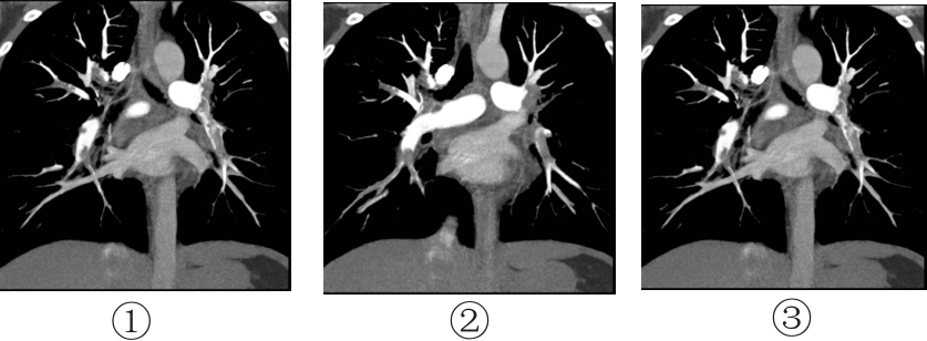
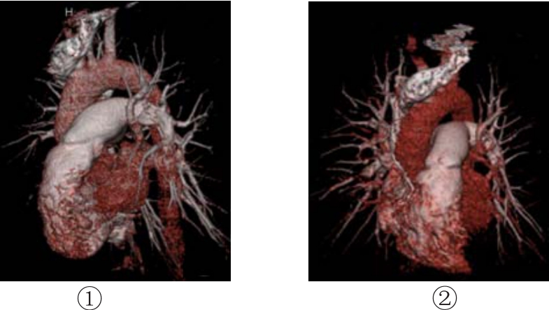
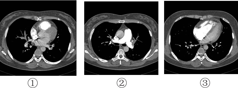
直接征象: 栓子在轴位显示为管腔部分或完全性低密度充盈缺损, 周围有高密度对比剂(图 1), 在冠状位多维平面重建与最大强度投影显示轨道样充盈缺损(图 2)。容积重建显示周围肺动脉分支明显减少(图 3)。
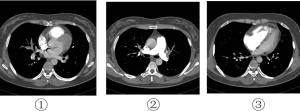
图 1 肺栓塞多层螺旋CT肺血管造影轴位图 示两侧肺叶、段、亚段肺动脉栓塞,管腔部分或完全性低密度充盈缺损。

图 2 肺栓塞多层螺旋CT肺血管造影多维平面重建图 示两侧肺叶、段、肺动脉栓塞,血管内轨道样充盈缺损。
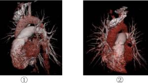
图 3 肺栓塞多层螺旋CT肺血管造影容积重建图 示左侧肺动脉周围血管分支明显减少。
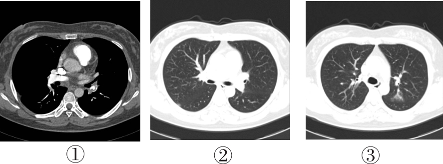
间接征象: 栓塞合并肺内渗出病变者3例; 伴胸腔积液者17例(图 4-①、②), 累及双侧者8例, 右侧者6例, 左侧者3例; 主肺动脉增宽(直径 > 30 mm)者36例; 肺梗死者3例, 肺窗显示楔形高密度影(图 4-③); 支气管动脉扩张者12例。
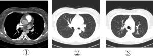
图 4 肺栓塞多层螺旋CT肺血管造影图 图中,①②示双侧叶间裂外侧少量积液,②③为肺窗图;③示左肺上叶楔形渗出性改变。
治疗后复查: 1例患者未经治疗死亡, 为主肺动脉明显栓塞合并右心房、室内栓子形成及右心功能衰竭; 其余62例溶栓治疗后复查, 栓子完全消失者47例, 栓子明显缩小者11例, 溶栓治疗效果差者3例, 考虑慢性肺栓塞。
-
肺栓塞是临床常见的急诊疾病, 下肢深静脉血栓形成是其最常见病因, 其他病因包括手术后、产后或长期卧床形成的脂肪、空气栓子及肿瘤的瘤栓等。肺栓塞的临床表现及实验室检查缺乏特异性, 常被误诊为心脑血管或者肺部疾病, 也有不少病例被漏诊。肺动脉造影被认为是评价肺栓塞的金标准, 但因其检查手段有创伤性、价格贵、并发症多、死亡率较高(达0.5%)等, 多年来临床上一直不能广泛应用。因此, 肺栓塞的有效、方便的影像学检查手段一直倍受关注[3]。
彩色多普勒超声可以检出肺动脉高压及右心室、右心房的受损, 并能检查出少部分主肺动脉及左右叶肺动脉内的栓塞, 但不能观察到叶动脉以上的栓子[4]。过去, 肺栓塞的确诊主要依靠核素肺通气/灌注(ventilation/perfusion, V/Q)显像, 该法具有无创性、灵敏度高等特点, 多年来一直作为重要的诊断手段。与多层螺旋CT相比, 核医学V/Q显像的空间分辨率及时间分辨率较低, 小范围的灌注缺损不能被识别, 特异性差, 显示肺动脉供血区域存在重叠, 可以通过侧支循环向相应肺段供血, 另外, 任何引起肺血流受损的因素, 如肺部肿瘤、慢性阻塞性肺部疾病、大动脉炎等均可造成局部血流降低。该法对大约70%的患者可以做出确切的诊断, 30%不能确切诊断。国内近期的研究结果显示: 肺V/Q显像与螺旋CTPA对叶及段肺动脉栓塞的诊断敏感性基本相同; 对亚段肺动脉栓塞, 肺V/Q显像的敏感性略高; 对肺血管轻度栓塞, 螺旋CTPA的敏感性略高[5]。
螺旋CTPA的空间分辨率和时间分辨率高, 注入对比剂后可显示血管及栓子, 并能清晰显示叶、段、甚至部分亚段肺动脉分支, 敏感性高。另外, 螺旋CTPA能够显示引起患者类似症状的其它非栓塞性疾病, 如渗出、实变等, 提高了肺栓塞诊断的特异性, 多层螺旋CTPA目前已经成为肺栓塞患者诊断的首选方法, 可以清晰显示肺动脉5~8级分支[6-7]。本研究中64排螺旋CTPA清晰显示叶、段、亚段肺动脉及分支栓子, 排除了引起患者类似症状的其它非栓塞性疾病, 明显提高了肺栓塞的诊断率, 减少了误诊及漏诊率。
多层螺旋CT对肺栓塞治疗后的随访具有重要意义, 本研究中, 通过多层螺旋CT对肺栓塞各级肺动脉栓子治疗前后的情况比较, 可以确定栓子大小、位置的改变, 明确治疗效果, 指导临床医师及时调整溶栓抗凝方案, 使大部分患者在急性期得到有效治疗并痊愈, 明显提高了患者的治愈率, 降低了病死率, 也提高了患者的生存质量。
总之, 成像时间短、薄层扫描、广泛的覆盖以及良好的重建图像质量是多层螺旋CT用于肺栓塞诊断的主要优势, 可以确诊肺栓塞, 但仍有少部分周围型、亚段以下轻度病变可能被漏诊。目前, 双能量CT肺血管及灌注成像在显示肺血管解剖分支的同时, 还可以显示碘对比剂在肺实质内的分布情况, 确定肺实质有无灌注缺损, 诊断肺栓塞及亚段级肺栓塞, 并且对肺栓塞的疗效观察有重要的指导意义[8]。
64层螺旋CT肺血管造影在肺栓塞诊疗中的应用价值
Assessment of 64-slice spiral CT pulmonary angiography in diagnosing of pulmonary embolism
-
摘要:
目的 探讨64层螺旋CT在肺栓塞诊断及治疗中的指导作用。 方法 63例患者均行64层螺旋CT肺血管造影(CTPA),并进行多种形式的图像重建结合轴位图像分析。 结果 64层螺旋CTPA对63例患者肺动脉各级管腔内的栓子均明确显示,共累及肺动脉303支;图像分析结果全部显示肺栓塞的直接征象为:主肺动脉和(或)左右肺叶、段、亚段血管腔内充盈缺损和血管阻塞;部分显示肺栓塞的间接征象为:马赛克征、右心房及右心室肥厚及扩张、肺动脉扩张、胸腔积液、肺不张及实变(肺梗死)等。62例患者经溶栓治疗后复查CTPA,其中,栓子完全消失者47例,栓子明显缩小者11例,溶栓治疗效果差者3例,考虑为慢性肺栓塞。 结论 64层螺旋CTPA是临床最有效的诊断肺栓塞及溶栓后疗效评价的无创性方法之一。 -
关键词:
- 肺栓塞 /
- 体层摄影术, 螺旋计算机
Abstract:Objective To assess the clinical usefulness of 64-slice spiral CT pulmonary angiography (CTPA)in diagnosing of pulmonary embolism. Methods Sixty-three patients with clinically known pulmonary embolism underwent 64-slice spiral CTPA.The source images of all the patients were reconstructed with the 3D techniques including multiplanarreconstruction, maximum intensity projection and volume rendering, CT axial images features were analyzed. Results Three hundreds and three embolus were found in 63 cases patients.The direct diagnostic findings of pulmonary embolism including: 64-slice spiral CTPA clearly showed the sites, extensions and stenosed lumens of pulmonary embolism in all patients.Pulmonary embolism lesions appeared as intra-luminal irregular-plaque shape and adhered-mural filling defects.The indirect diagnostic findings of pulmonary embolism including: dmosaic signs, dilation of the right ventricle and atrium, pulmonary artery enlargement, pleural effusion, pulmonary atelectasis and pulmonary consolidation(pulmonary infarction).There are 47 patients whose thrombosis disappeared after therapy in 62 patients, 11 became smaller and 3 were chronic pulmonary embolism. Conclusion 64-slice spiral CTPA is one of the most effective and non-invasive imaging modalities in the diagnosis before and aftertherapy of pulmonary embolism. -
Key words:
- Pulmonary embolism /
- Tomography, spiral computed
-
[1] 王乐民, 魏林. 肺栓塞与深静脉血栓形成. 北京: 人民卫生出版社, 2001: 32-39.
[2] 马大庆. 多排螺旋CT对于肺栓塞的诊断. 中国介入影像与治疗学, 2005, 2(3): 161-165. doi: 10.3969/j.issn.1672-8475.2005.03.002
[3] Remy-Jardin M, Pistolesi M, Goodman LR, et al. Management of suspected acute pulmonary embolism in the era of CT angiography: a statement from the Fleischner Society. Radiology. 2007, 245(2): 315-329. doi: 10.1148/radiol.2452070397 [4] 赵博文, 应可净, 陈丽英, 等. 彩色多普勒超声检查在急性肺栓塞诊断与治疗中的价值. 中华急诊医学杂志, 2006, 15(4): 342-344. doi: 10.3760/j.issn:1671-0282.2006.04.014
[5] 谭业颖, 田嘉禾. 肺灌注/通气显像与螺旋CT肺动脉造影诊断肺栓塞的对比研究. 医学影像学杂志, 2007, 17(2): 137-139. doi: 10.3969/j.issn.1006-9011.2007.02.010
[6] Schoepf UJ, Costello P. CT angiography for diagnosis of pulmonary embolism: state of the art. Radiology, 2004, 230(2): 329-337. doi: 10.1148/radiol.2302021489 [7] 王辰. 肺栓塞. 北京: 人民卫生出版社, 2003: 1-279.
[8] 黄小勇, 濮欣, 张兆琪, 等. 双能量CT肺灌注及血管成像与核素肺通气灌注成像诊断肺栓塞的对照研究. 中华放射学杂志, 2010, 44(9): 926-930. doi: 10.3760/cma.j.issn.1005-1201.2010.09.009
-

 点击查看大图
图(4)
计量
- 文章访问数: 898
- HTML全文浏览量: 413
- PDF下载量: 1