


 下载:
下载:

-
PET-CT是将PET和CT两个设备有机地结合在一起, 使PET与CT优势互补, 达到了1+1 > 2的效果, 明显提高了诊断的准确性[1-2]。随着PET-CT的广泛应用, 其对肿瘤定位定性诊断的优越性已得到临床医师的肯定, 本研究回顾性分析了70例经病理确诊的周围型肺癌病例的18F-FDGPET-CT结果。
-
2005年7月至2009年7月经在我院手术病理确诊的周围型肺癌患者70例, 其中男性56例、女性14例, 年龄28~81岁, 平均年龄62.4岁。70例患者中, 鳞癌患者32例, 腺癌患者25例, 小细胞癌患者8例, 腺鳞癌患者3例, 大细胞癌患者2例。其中2例分别于3个月和6个月后复查。
-
使用美国GE公司生产的Discovery-ST PET-CT扫描仪, 采用Xeleris工作站进行图像后处理。检查前患者禁食4~6 h, 并确保血糖控制在7.0 mmol/L以内。患者静息、平卧15 min后注射示踪剂18FFDG, 注射剂量为3.7~5.6 mBq/kg, 静卧45 min后行全身PET-CT。扫描范围从颅顶至股骨上段, 先行CT(8排螺旋CT), 层厚3.75 mm, 然后进行PET采集, 使用三维扫描, 每个床位采集2 min, 共采集6~7个床位。采集结束后融合图像并重建, 分别得到全身或局部的冠状、矢状、横断面CT、PET以及PET-CT的融合图像。
-
CT及PET-CT图像由3名有多年工作经验的医师共同阅片。使用PET分析软件对病变部位选用合适感兴趣区, 测量病灶的平均标准化摄取值(standardized uptake value, SUV)。平均SUV≥2.5诊断为恶性。
-
用SPSS13.0软件包处理数据, 行配对t检验。以P < 0.05表示差异有统计学意义。
-
70例周围型肺癌患者中, 16例病灶为孤立性肺结节(其中1 cm以下者2例, 1.1~2.9 cm者14例), 54例呈团块状改变; 68例病灶出现异常放射性浓聚, 2例未见异常放射性浓聚(术后病理检查为高分化鳞癌1例, 肺泡细胞癌1例, 病灶均小于1 cm)。
70例周围型肺癌患者的平均SUV=4.94±1.53, 其中66例SUV≥2.5, 阳性率为94.3%, 4例SUV < 2.5, 阴性率为5.7%。4例假阴性的患者中, 病灶 < 1.0 cm者2例, 1.1~2.0 cm者1例, 2.1~3.0 cm者1例, 病灶均出现在肺部孤立结节中, 阴性患者数占病灶为肺部孤立结节患者数的25%。假阴性患者中有2例因具有典型肺癌CT形态学改变(分叶、毛刺)而明确诊断, 另2例CT结果无特征性, 但在随后3个月和6个月的PET-CT复查中发现, 患者病灶较前增大、代谢增高, 因而诊断为肺癌。
将病灶按大小分为 < 1.0 cm、1.1~2.0 cm、2.1~3.0 cm、3.1~5.0 cm和 > 5.0 cm5组, 则各组SUV分别为1.81±0.73、2.68±1.21、4.88±1.51、7.72±1.86和8.43±2.92。 < 1.0 cm组与1.1~2.0 cm组比较, 差异无统计学意义(t=1.83, P > 0.05);1.1~2.0 cm组与其余3组比较, 差异均有统计学意义(t=2.82, t= 2.56, t=2.47, P均 < 0.05);2.1~3.0 cm组与3.1~5.0 cm和 > 5.0 cm组比较, 差异均有统计学意义(t=2.76, t=2.64, P均 < 0.05);3.1~5 cm组和 > 5 cm组比较, 差异无统计学意义(t=1.12, P > 0.05)。54例病灶≥3 cm的患者, 其病灶的SUV均≥2.5, 阳性数占周围型肺癌团块影患者数的100%。
-
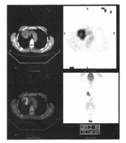
66例SUV≥2.5的阳性病灶中, 12例呈结节状, 54例呈团块状且异常放射性浓聚, 放射性分布均匀、致密, 边界清楚。另外, 有32例病灶边缘放射性浓聚, 其中29例病灶 > 3 cm; 12例病灶内中央放射性缺损, 缺损影周围肿瘤组织呈厚薄不一、非均匀性放射性浓聚, 浓聚部分厚度均 > 5 mm, SUV > 2.5(图 1)。4例阴性病灶中, 2例无明显异常放射性浓聚, 另2例为片状轻度放射性浓聚, SUV > 2.0(图 2)。

图 1 肺鳞癌PET-CT融合图像 患者为男性,62岁,右上肺鳞癌,PET示肿块呈团块状异常放射性浓聚影,中央呈放射性缺损;CT示肿块中央液化、坏死。

图 2 肺腺癌PET-CT融合图像 患者为女性,58岁,左下肺背段腺癌,PET示结节影呈轻度放射性浓聚影;CT示结节影内见小空泡征。
-
经过多年的研究, 特别是最近十多年影像技术的迅速发展, 肺癌的影像诊断已积累了丰富的临床经验。在PET-CT出现以前, 一般认为CT是评价肺癌形态和密度较好的影像方法, 对其征象的研究也比较深入, 周围型肺癌的CT征象包括分叶、毛刺、空泡征、棘状突起、血管集束征、胸膜凹陷等。但肺癌的CT征象复杂多变, 定性诊断有时较困难, 特别是对肺部单发结节的定性[3], CT的诊断价值有限, 常规CT的灵敏度只有12%~63%, 增强螺旋CT虽然可以通过对肿块的强化提高诊断灵敏度(可达98%), 但特异度只有58%。PET对良恶性病变的鉴别诊断主要是通过观察病灶对18F-FDG的代谢情况, 通过目测比较肿块与纵隔血池对放射性摄取程度的差异判断肿块的代谢活性; 另外, 半定量分析的SUV也是判断病灶代谢活性的方法。虽然PET定位不精确, 组织分辨力也明显低于CT, 但是据文献报道, PET的特异性明显高于CT[4]。Präuer等[5]发现, CT的灵敏度即使达到100%, 特异度却只有52%, 而PET虽然灵敏度略低(90%), 特异度却可以达到83%。因此只有将二者结合起来, 才能同时弥补CT定性困难和PET定位不准确的缺陷。
本组病例以病灶SUV≥2.5作为判断为恶性的标准, 结果显示阳性率为94.3%, 阴性率为5.7%, 这对周围型肺癌的诊断有较高的临床价值。另外, 将本组病例按病灶大小进行分组, 结果提示病灶大者其放射性浓聚程度也较高, SUV与肺癌大小之间存在显著差异, 肿块≥3 cm者, 其阳性率达100%;而部分周围性小肺癌病灶SUV低。这可能为部分容积效应所致, 也可能与病变早期肿瘤细胞数目较少、增殖速度较慢、恶性程度较低有关。
目前关于PET-CT在肺癌诊断中的临床应用方面的报道较多, 而有关肺癌形态学改变方面的报道较少, 本研究结果显示, 虽然PET对病灶组织结构的分辨能力低于CT, 但大多数肺癌仍有较特殊的影像学表现。主要表现有: ①病灶异常放射性高浓聚, 边界清楚, SUV≥2.5;②病灶主要呈结节状、团块状改变; ③病灶大多呈较均匀性浓聚, 但约17%的病灶中有放射性缺损(可能为局部组织坏死所致), 缺损灶周边浓聚部分常厚薄不一、浓聚程度不均匀, SUV > 2.5, 这与CT所见肺癌厚壁不规则空洞相一致; ④病灶边缘呈分叶状为诊断肺癌的依据之一, 本组病例中45.7%的肺癌病灶有此征象, 但由于病灶都较大(≥3 cm), 结节病灶可能因PET的组织分辨能力有限而无法清楚显示。
据文献报道, PET-CT的假阴性率小于5%[6], 这与本研究结果相一致, 造成假阴性的原因是有些低度恶性的肿瘤或较小病灶的18F-FDG代谢活性不一定增高所致, 如分化程度较高的恶性肿瘤、细支气管肺泡癌、类癌等; 另一方面, 对于体积小的肿瘤, 尤其是 < 7 mm的小病灶, 由于PET分辨力和部分容积效应的限制可导致假阴性结果。对这类患者应仔细观察其CT表现及密切进行随访复查, 及时诊断。本组4例阴性患者中, 2例因CT图像具有恶性征象而确诊为肺癌, 故必须重视诊断性CT的应用, 以减少假阴性的结果; 对于PET和CT结果均无特征的阴性患者, 建议积极定期复查, 密切关注其动态变化, 本组中另2例阴性患者其病灶大小和代谢活性在复查前后均有明显变化, 故确诊为肺癌, 因此动态观察对周围型肺癌的诊断亦有非常重要的临床价值。
以SUV作为鉴别良恶性的指标, 其临界值目前尚无统一标准, 如果以病灶SUV≥2.0作为诊断肺癌的指标, 可大幅提高肺癌诊断的敏感性, 但假阳性率必然会明显升高。Gould等[7]认为, 诊断标准的选择应倾向于提高诊断的敏感性而不是特异性, 也就是应尽可能减少假阴性的发生, 因为假阴性比假阳性对患者的影响更加重大。因此, 如何既提高敏感性又减少假阳性, 有待今后进一步研究。
18F-FDG PET-CT对周围型肺癌的诊断价值研究
The diagnostic value of PET-CT on peripheral lung cancer
-
摘要:
目的 探讨PET-CT对周围型肺癌的诊断价值。 方法 回顾性分析70例经病理证实的周围型肺癌患者的PET-CT影像资料,分析病灶的标准化摄取值(SUV)及其形态学表现。 结果 70例周围型肺癌患者中,鳞癌患者32例、腺癌患者25例、小细胞肺癌患者8例、腺鳞癌患者3例、大细胞肺癌2例。平均SUV=4.94±1.53,其中,66例患者的PET影像结果可见18F-FDG高浓聚,SUV≥2.5;另外4例的SUV < 2.5。SUV与肿块大小呈显著相关性。 结论 周围型肺癌大多具有较特殊的PET-CT影像学表现。PET-CT对周围型肺癌的诊断及鉴别诊断有较好的效能。SUV结合形态学分析有助于病灶的鉴别诊断。 -
关键词:
- 肺肿瘤 /
- 正电子发射断层显像术 /
- 体层摄影术, X线计算机 /
- 氟脱氧葡萄糖F18
Abstract:Objective To evaluate the value of PET-CT in the diagnosis of peripheral lung cancer. Methods The morphologic characteristics and standardized uptake value (SUV) of 70 patients with lung cancer proved by pathology were analyzed retrospectively. Results Of the 70 cases, 32 cases were squamous carcinoma, 25 cases were adenocarcinoma, 8 cases were small cell lung cancer, 3 cases were adenosquamous carcinoma and 2 cases were megacell lung cancer.The average SUV of the lung cancer was 4.94±1.53.In the group of lung cancer, hypermetabolic lesions were found in 66 cases and the SUV was more than 2.5 while the SUV was less than 2.5 in 4 cases.Positive correlation was showed in the SUV and the size of tumors. Conclusions The peripheral lung cancer has its special imaging appearances of PET-CT.PET-CT is an excellent modality in the diagnosis and differential diagnosis of peripheral lung cancer.The SUV combining with morphological findings sometimes may be helpful for the differential diagnosis. -
[1] 屈婉莹. 正电子发射计算机体层摄影-CT及其在肿瘤学中的临床应用. 中华放射学杂志, 2005, 39(11): 1127-1129.
[2] 屈婉莹, 郑建国, 林嘉滨. PET/CT临床应用优化选择的思考. 中华核医学杂志, 2006, 26(6): 327-329.
[3] Gaeta M, Barone M, Russi EG. et al. Carcinomatous solitary pulmonary nodules: evaluation of the tumor-bronchi relationship with thin-section CT. Radiology, 1993, 187(2): 535-539. [4] Shon IH, O'doherty MJ, Maisey MN, et al. Positron emission tomography in lung cancer. Semin Nucl Med, 2002, 32(4): 240-271. [5] Präuer HW, Weber WA. Römer W, et al. Controlled prospective study of positron emission tomography using the glucose analogue [18F] fluorodeoxyglucose in the evaluation of pulmonary nodules. Br J Surg, 1998, 85(11): 1506-1511. [6] Bousson V, Moretti JL, Weinmann P, et al. Assessment of malignancy in pulmonary lesions: FDG dual-head coincidence gamma camera imaging in association with serum tumor marker measurement. J Nucl Med, 2000, 41(11): 1801-1807. [7] Gould MK, Maclean CC, Kuschner WG, et al. Accuracy of positron emission tomography for diagnosis of pulmonary nodules and mass lesions: a meta-analysis. JAMA, 2001, 285(7): 914-924. -

 点击查看大图
图(2)
计量
- 文章访问数: 1826
- HTML全文浏览量: 521
- PDF下载量: 2