


-
X线摄影是诊断骨折的主要影像学手段,它可以观察骨折的部位、类型及移位情况,指导临床治疗[1]。但在实际工作中我们注意到,当骨折的侧方移位和成角移位方向不平行于冠状或矢状面时,常规X线摄影(正侧位)所显示的移位情况就不够准确,从而会误导临床治疗。因此,本研究结合X线摄影的几何原理设计了骨折移位的校正方法并通过CT给予验证。
-
本研究中所用的材料为一枚肱骨标本,做成其中段骨折并牢固固定在一木板之上,使骨折之两段保持空间位置不变。本研究所用CT机为西门子公司Emotion 16层螺旋CT,采用的扫描参数为:8 mm×1.5 mm,螺距:1.0,电压:110 kV。
-
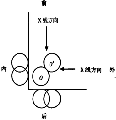
常规四肢X线摄影,采用相互垂直的正侧位摄影(以下简称正侧位摄影),其中心线分别垂直于人体冠状面和矢状面,当骨折远折端的移位方向不平行于冠状或矢状面(不垂直于X线的中心线)时,正侧位图像上均表现骨折的移位,如图 1所示,圆O代表近折端横断面,圆O'代表远折端横断面,远折端向外前方移位,两断端完全错位,经相互垂直的X线投照,投影在正侧位片上的对位情况和实际存在较大的误差,均表现为部分重叠,移位方向和X线夹角越小(0°~90°),投影的误差越大,夹角越大则误差越小,移位方向垂直于X线中心线时X线表现的移位和实际一致。

图 1 骨折移位X线摄影原理示意图
-
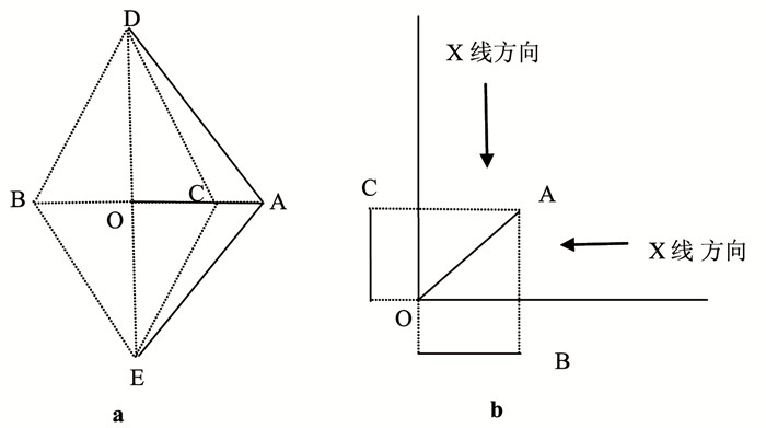
骨折成角移位与侧方移位相似,当成角的方向和X线垂直时,X线投影成角和实际一致,否则就和实际产生不同程度误差,如图 2a,AD和AE分别代表骨折的两折段,∠DAE为骨折成角,DAE三点构成三角形,OA为底边DE上的高,无论正位还是侧位投照,骨两端的连线DE不变;当成角平面不平行于人体冠状面或矢状面时,如图 2b,OA在正侧两位置上分别投影为OB和OC,根据三角函数原理,OB、OC小于OA,因投影三角形底边上的高OB、OC均小于OA,所以骨折成角的投影成角无论正侧位必大于实际成角,也就是骨折成角平面不平行于冠状面或矢状面时,X线投影图像上表现的骨折投影角大于实际成角。

图 2 骨折成角移位的投影示意图
-
首先从正侧位图像上分别测量出远折端向前(或后)、内(或外)移位的距离,如图 3根据移位的方向分别在坐标轴上找到相应距离的点B、C,经过此两点向坐标内引垂线,两垂线交点A和O点的连线OA就是远折端移位的实际距离,该连线所指的方向就是侧方移位的确切方向。
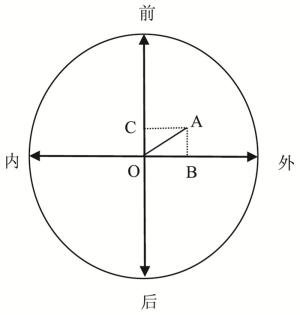
图 3 骨折成角移位的校正示意图
-
在正侧位图像上分别做出骨折近段、远段的中轴线,如图 2a中的DB、BE,DC、CE,分别交于B、C两点,连接DE,分别在正侧位图像上构成两个三角形,做出三角形DE边上的高OB、OC,测量其长度,将其带入图 3中的相应坐标轴上,过相应点向相应象限内引垂线,两垂线交于A点,连接OA,即为骨折实际成角移位所构成三角形两骨端连线DE边上的高。在正位或侧位图像上,如图 2a延长投影三角形DE边上的高至OA长度,连接AD、AE,∠DAE就是骨折的实际成角。图 3中OA所指的方向就是骨折成角的确切方向。
-
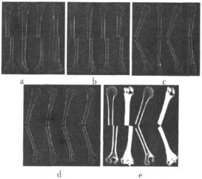
(1) 取肱骨标本一枚,做成其中段骨折,两断端构成侧方移位,牢固固定在一木板之上,使骨折之两段保持空间位置不变。经X线拍摄(本研究采用正侧位定位扫描图像模拟X线结果),取得图像结果,然后对标本进行前后(正位)、左右(侧位)方向定位像扫描和整个骨标本的容积扫描,然后将标本整体旋转15°、30°、45°,分别对其进行容积扫描并将数据进行多维平面重建(multiplanar reconstrution,MPR)及表面遮盖显示(shaded surface display,SSD)。
(2) 用同样的方法使骨折构成一定的成角移位,对其进行扫描、重建。所得图像分别如图 4所示。对不同旋转角度X线图像进行测量(本研究采用正侧位定位扫描图像模拟X线结果)。分别应用侧方移位和成角移位的校正方法进行校正处理,获得侧方移位的距离及成角移位的角度。将上述结果与CT数据MPR、SSD结果及实际设定移位情况进行比较,分析其可靠性。

图 4 骨折成角移位的扫描、重建图像
-
应用侧方移位和成角移位的校正方法求得的校正数据、MPR及SSD后所得数据与标本实际设置侧方移位、成角移位程度误差较小,结果见表 1、表 2。证明常规X线摄影骨折移位的情况经坐标图测量校正的方法真实可靠,可明显提高X线诊断骨折移位程度的准确性。
旋转角度(°) X线正位图像侧方移位 X线侧位图像侧方移位 X线图像校正后侧方移位 MPR图像侧方移位 SSD图像侧方移位 实际侧方移位 0 22 0 22.0 21 21 21 15 20 6 21.0 21 21 21 30 18 14 22.5 21 21 21 45 17 15.5 22.5 21 21 21 注:表中,MPR: 多维平面重建;SSD:表面遮盖重建;X线结果为CT正侧位定位扫描图像模拟X线图像所得数据。 表 1 不同旋转角度下骨折X线数据、校正数据、MPR及SSD所得侧方移位比较(mm)
旋转角度 X线正位图像成角移位 X线侧位图像成角移位 X线图像校正后成角移位 MPR图像成角移位 SSD图像成角移位 实际成角移位 0 127.3 180 127.3 127.8 128 128 15 128.5 166 128.0 127.8 128 128 30 137.0 150 126.5 127.8 128 128 45 142.5 142 127.5 127.8 128 128 注:表中,MPR: 多维平面重建;SSD:表面遮盖重建; X线结果为CT正侧位定位扫描图像模拟X线图像所得数据。 表 2 不同旋转角度下骨折X线数据、校正数据、MPR及SSD所得侧方成角移位比较(°)
-
正确的骨折复位是骨折愈合顺利进行的必要条件,侧方移位、成角移位受移位方向和X线投照位置的影响较大,其移位的方向与X线成45°角时误差最大,本来完全错位的骨折,在正侧位图像上可表现为约1/3的对位,造成X线表现明显好于实际的错觉,易误导临床医师延误治疗,给患者带来日后难以恢复的功能障碍。
CT定位图像是通过床的运动对被照射物进行定位扫描[2],正、侧位定位扫描与常规X线摄影的正侧位投照是一致的,两角度是相互垂直的,通过放置在CT检查床上的标本整体旋转来完成模拟X线正侧位投照,其实验结果是可靠的。
骨折侧方移位的复位标准在不同的部位要求不一[1],越接近解剖复位功能恢复越好,解剖复位无论X线从任何角度投照,得到的图像和实际一致,不存在误差。只有当正侧位图像的其中一个位置表现为侧方移位时,其移位的距离和方向才和实际一致;当正侧两位置上均表现侧方移位时,两位置图像上的移位程度和实际移位均存在差距,移位明显的一侧相对接近实际。
骨折成角移位只有在成角方向与X线垂直时才是骨折实际成角的真实表现,因为常规X线检查采用相互垂直的正侧位投照,所以当骨折端只在正位或侧位一个位置上表现成角而另一位置不表现成角时,成角一侧图像上的角度才是可信的,否则两位置上均表现成角时,两投影角的角度都大于实际成角,成角小的一侧接近实际成角。骨折成角移位的方向是以骨纵轴为中心的任意方位,用前、后、内、外四个方位对其描述尚不够,当成角方向指向内前、外前、外后、内后时,应明确成角方向与人体冠状面或矢状面的夹角。
骨折侧方移位、成角移位的程度只有在移位方向与X线投影垂直时才是准确的,因此在透视下旋转患肢,X线表现移位程度最大时摄影所得的移位数据是相对准确的,不过需要患者重复检查且无记录、辐射量较大、危重患者不适合。多层螺旋CT实现了图像的各向同性[3],且CT的MPR及SSD能够较准确的显示骨折移位情况[1],但检查辐射量大、费用高、患者难以接受。
总之,常规X线摄影当相互垂直的正侧位图像上都表现侧方移位或成角移位时,两位置上的移位程度均和实际存在不同程度的差距,侧方移位明显或成角移位小的一侧相对接近实际,经坐标图校正的移位结果接近实际,可明显提高骨折移位程度X线诊断的可靠性。骨折移位X线检查是建立在稳定或相对稳定骨折的基础上,即不受体位的改变而变化,否则测量的移位程度不准确,故此校正方法对于临床医师评价闭合整复后外固定及手术切开内固定的骨折具有积极意义。
长管状骨骨折移位X线表现的校正方法探讨
The study on the X-ray correction method of long bone fracture displacement
-
摘要:
目的 探讨常规X线摄影(正、侧位)时骨折移位影像表现的校正方法,并用CT验证。 方法 根据X线摄影几何学原理设计出移位的校正方法。取一枚中段骨折的肱骨标本构成一定程度的侧方移位和成角移位后,将标本沿横轴多角度旋转并分别行前后(正位)、左右(侧位)方向X线拍摄,然后利用CT进行整个标本的容积扫描,并将容积扫描数据进行多维平面重建(MPR)及表面遮盖显示(SSD)。对X线图像的移位程度及校正数据、MPR及SSD后的移位程度、标本实际移位程度分别进行比较分析。 结果 经X线图像求得的校正数据、MPR及SSD后所得的数据与标本实际设置移位方向、程度误差较小,位置差异 < 1.5 mm,角度差异 < 1.5°。 结论 常规X线摄影显示的骨折移位的影像表现经坐标图测量校正,方法真实可靠,可以显著提高X线诊断骨折移位程度的准确性。 -
关键词:
- 体层摄影术,X线计算机 /
- 图像处理,计算机辅助 /
- 肱骨骨折 /
- 骨折移位
Abstract:Objective To explore the image correction of fracture displacement by conventional X-ray photography(orthotropic and lateral) and test by computed tomgraphy(CT). Methods The correction method of fracture displacement was designed according to geometry principles of X-ray photography. Selected one midhumeral fracture specimen which designed with lateral shift and angular displacement, and scanned from anteroposterior and lateral position respectively, and also volume scanned using CT, the data obtained from volume scan were processed using multiplanar reconstruction (MPR) and shaded surface display(SSD). The displacement data relied on X-ray image, CT with MPR and SSD processing, actual design of specimens were compared respectively. Result The direction and degree of displacement among correction data of X-ray im-ages and the data from MPR and SSD, actual design of specimen were little difference, location difference < 1.5 mm, degree difference < 1.5°. Conclusion It is really reliable for fracture displacement by conventional X-ray photography with coordinate correction, and it is helpful to obviously improve the diagnostic accuracy of the degree of fracture displacement. -
表 1 不同旋转角度下骨折X线数据、校正数据、MPR及SSD所得侧方移位比较(mm)
旋转角度(°) X线正位图像侧方移位 X线侧位图像侧方移位 X线图像校正后侧方移位 MPR图像侧方移位 SSD图像侧方移位 实际侧方移位 0 22 0 22.0 21 21 21 15 20 6 21.0 21 21 21 30 18 14 22.5 21 21 21 45 17 15.5 22.5 21 21 21 注:表中,MPR: 多维平面重建;SSD:表面遮盖重建;X线结果为CT正侧位定位扫描图像模拟X线图像所得数据。  下载: 导出CSV
表 2 不同旋转角度下骨折X线数据、校正数据、MPR及SSD所得侧方成角移位比较(°)
旋转角度 X线正位图像成角移位 X线侧位图像成角移位 X线图像校正后成角移位 MPR图像成角移位 SSD图像成角移位 实际成角移位 0 127.3 180 127.3 127.8 128 128 15 128.5 166 128.0 127.8 128 128 30 137.0 150 126.5 127.8 128 128 45 142.5 142 127.5 127.8 128 128 注:表中,MPR: 多维平面重建;SSD:表面遮盖重建; X线结果为CT正侧位定位扫描图像模拟X线图像所得数据。
下载: 导出CSV
-
[1] 吴恩惠, 白人驹, 刘望彭, 等. 医学影像诊断学. 北京: 人民卫生出版社, 2001: 292-295.
[2] 李果珍, 戴建平, 王仪生, 等. 临床CT诊断学. 北京: 中国科学技术出版社, 1994: 9-10.
[3] 李松柏, 徐克. 多层螺旋CT临床诊断实践图谱. 北京: 人民军医出版社, 2004: 35-38.
-

 点击查看大图
图(4)表(2)
计量
- 文章访问数: 1032
- HTML全文浏览量: 457
- PDF下载量: 1