


-
磁共振弥散加权成像(diffusion weighted imaging, DWI)作为分子水平上观察组织结构的无创性检查新技术, 已逐渐应用于骨骼系统, 为骨髓病变的早期异常征象提供了新的影像学方法。目前, 国内外已有DWI应用于骨关节的报道, 但关于股骨头无菌性坏死(femoral head necrosis, FHN)的DWI研究报道尚少。本文对我院诊断的FHN患者及股骨头正常者的髋关节DWI影像学特征进行了分析总结, 以期为FHN的诊断提供依据。
-
2007年6月至2009年6月在我院行双髋关节MRI检查的64例受检者, 均对其行常规MRI及DWI, 其中股骨头正常者22名(男性13名、女性9名), 平均年龄为(44.8±7.21)岁, FHN患者42例(男性28例、女性14例), 平均年龄为(47.7±9.54)岁。42例FHN患者中, 27例有服用激素史, 6例有骨外伤史。
-
使用美国通用公司的GE SIGNA EXCITE 1.5T超导型磁共振扫描仪。所有受检者均使用8通道体部线圈。每位受验者在做DWI前均行常规扫描轴位、冠状位快速自旋回波序列T1加权像、快速恢复快速自旋回波/压脂序列T2加权像。DWI序列参数为: 采用自旋回波-回波平面成像行轴位扫描, 扩散梯度因子b=800 s/mm2, 重复时间/回波时间为6000/81.3 ms, 层厚为6.0 mm, 层距为1.0 mm, 矩阵为128mm×128mm, 激励次数为2, 视野为40 cm× 23 cm。
-
在表观弥散系数(apparent diffusion coefficient, ADC)图上直接测量数值。FHN患者选择病变层面的病灶中心为ROI, 尽量避开周围伪影的影响。正常受检者数据测量在股骨头关节面下2 cm处, ROI大小约为100 ram2。使用SPSS13.0统计软件进行统计分析, 三组间的均数比较采用方差分析, 两两间均数比较采用q检验, P < 0.05为差异有统计学意义。
-
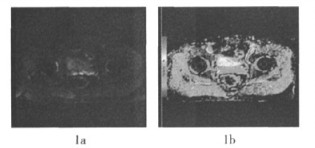
股骨头正常者与FHN患者股骨头的磁共振信号分布见表 1。股骨头正常者的股骨头T1加权像及T2加权像呈均匀高信号, 与股骨颈信号相一致, DWI为均匀低信号(图 1)。FHN患者的T1加权像呈低信号37例(图 2a), 稍低信号5例; T2加权像压脂像呈不均匀高信号37例(图 2b), 稍高信号5例; DWI呈不均匀高信号改变, 边缘不清, 病变范围与常规扫描显示相吻合(图 2c), ADC伪彩图显示病灶边缘较清晰(图 2d、2e)。在股骨头坏死的病变中央区及周围区DWI可见稍高信号影, 通过对应的ADC伪彩图测量的ADC显示病变范围较平扫更大, 且数值有显著差别(图 3、图 4)。
股骨头正常者(n=22) 股骨头无茼性坏死患者(n=42) 低信号 稍低信号 等信号 稍高信号 高信号 低信号 稍低倍号 等信号 稍高 高信号 T1加权像 0 0 0 0 22 37 5 0 0 0 T2加权像 0 0 0 0 22 0 0 0 5 37 DWI 22 0 0 0 0 0 0 2 7 33 注: 表中,DWI: 磁共振弥散加权成像。 表 1 股骨头正常者与股骨头无菌性坏死患者的股骨头磁共振信号分布表(例数)

图 1 股骨头正常者的双侧股骨头磁共振弥散加权图及表观弥散系数图 1a:磁共振弥散加权图;1b:表观弥散系数图。
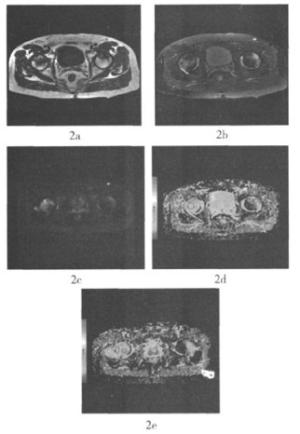
图 2 双侧股骨头无菌性坏死患者的MRI、磁共振弥散加权图及表观弥散系数图 2a:T1加权像显示不规则的低信号改变;2b:T2加权像显示不规则的高信号改变;2c:磁共振弥散加权图显示不均匀的高信号改变;2d:表现弥散系数图显示病变中央及周围区域的表观弥散系数分别为0.79×10⁻⁵mm2/s、(1.0~1.01)×10-5mm2/s;2e:同一患者的双侧股骨头稍下方的表观弥散系数图,显示病变水肿区域的表观弥散系数为1.07×10-5mm2/s。
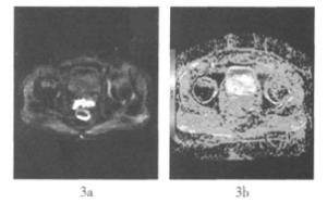
图 3 双侧股骨头无菌性坏死患者的磁共振弥散加权图及表观弥散系数图 3a:磁共振弥散加权图;3b:表观弥散系数图;病变以左侧较重。
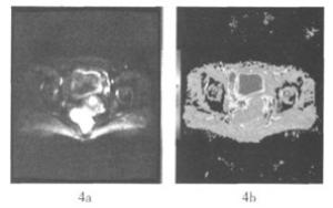
图 4 左侧股骨头无菌性坏死患者的磁共振弥散加权图及表观弥散系数图 4a:磁共振弥散加权图;4b:表观弥散系数图;左侧股骨头弥散加权图显示稍高异常信号改变,测量表观弥散系数为0.731×10⁻³mm2/s,诊断无菌坏死早期表现。
-
股骨头正常者及FHN患者的ADC见表 2。其中, 股骨头正常者的股骨头ADC为(0.000~0.036)×10-3mm2/s, FHN中央坏死水肿区及病变周围区的ADC则分别(0.845~1.397)×10-3mm2/s和(0.111~0.311)×10-3mm2/s。正常人、股骨头坏死患者的病变区与病变周围区三者的ADC之间差异有统计学意义(F=1384.28, P < 0.05)。
x±s x±1.96s 95%信赖区间 股骨头正常者 0.018±0.009 0.000±0.036 0.016±0.020 股骨头无菌性 坏死患者 病变周围区 0.211±0.050 0.111±0.311 0.213±0.229 病变区 1.121±0.141 0.845±1.397 1.099±1.143 注: 表中,三组数据间比较,F= 1384.28、P < 0.001;两两比较使用q检验,正常受枪者与患者病变周围区比较,q=10.9775、正常受检者与患者病变区比较, q=62.7366,患者病变周围区与病变区比较: q=62.4239,P均 < 0.01。 表 2 股骨头正常者及股骨头无菌性坏死患者的表观弥散系数(×10-3mm2/s)
-
目前, DWI是唯一能检测活体组织内水分子扩散运动的无创性影像技术, 能通过检测活体组织内水分子的微观扩散运动状态, 反映机体组织结构的生理、病理特点, 是不同于常规T1加权像、T2加权像的一项新的磁共振成像技术。该技术在脑部疾病特别是在超早期脑梗死诊断中已经得到肯定, 但对骨关节系统的应用价值尚处于探索阶段[1-2]。DWI可以在分子水平检测骨髓病变, 通过检测组织内水分子运动来反映组织的结构特点与生物学行为, 还可通过ADC对骨髓病变进行定量分析, 为骨髓病变的检出提供客观的诊断依据[3-4]。
FHN是一种临床常见疾病, 无论什么原因引起的FHN, 其病理改变都是相似的[2-4]: 早期表现为细胞坏死, 中、晚期表现为细胞坏死与修复共存。股骨头缺血坏死包括骨髓坏死和骨组织坏死。骨髓内各种细胞成分对缺血的耐受性不同, 最敏感的是骨髓造血细胞, 在缺血6~12 h开始坏死, 12~48 h后骨细胞发生坏死, 2~5 d后脂肪细胞坏死。
股骨头缺血坏死的MRI表现: 早期股骨头缺血坏死表现为“双线征”, 晚期可出现典型的股骨头形态变扁、关节面的塌陷等。股骨头骨松质在急性或超急性缺血期, 病变区的骨髓水肿的MRI表现为T1加权像呈广泛的低信号, T2加权像呈高信号。Koo等[5]研究表明, 其与患者的临床疼痛症状显著相关。随着骨髓压力增大, 骨髓细胞外液增多, 使局部缺血区骨组织自由水减少, 细胞内水分子扩散受限, ADC显著下降, 因而在DWI上表现为高信号区, 而在ADC图上表现为低信号, 该区域形态与缺血区一致, 表面为小片状、带状、地图状或其他不规则状改变。刘斯润等[6]对髌软骨的DWI研究进行了报道。Ward等[7]对35例正常膝关节骨髓和15例骨髓创伤者进行了常规MRI和DWI研究(扩散梯度因子b=980s/mm2), 结果显示, 创伤者骨髓的平均ADC为(0.8×10⁻³mm2/s), 明显高于正常骨髓(0.1×10⁻³mm2/s)。
本研究结果显示, 股骨头正常者、FHN患者中央坏死水肿区与病变周围区的ADC分别为(0.000~0.036)×10-3mm2/s, (0.845~1.397)×10-3mm2/s和(0.111~0.311)×10-3mm2/s。FHN患者的ADC明显高于股骨头正常者, 且病变周围区域可进一步提示病变的进展及预后状态。FHN及骨挫伤的ADC增高均是由骨髓充血水肿引起骨髓内自由水增加, 导致细胞外容积增加, 进而使病变内水分子运动增加引起的。骨髓DWI能为股骨头无菌性坏死提供早期的影像学表现, 还可通过ADC的测量对骨髓病变进行量化分析。
DWI新技术是活体上研究细胞内水肿的唯一方法, 能更准确指导临床分期和治疗。但现有的骨髓DWI方法尚不统一。有关序列基本是从颅脑DWI序列修改而来, 有必要研究和开发适于FHN的专用检查序列。
股骨头无菌坏死弥散加权成像的临床应用
Clinical application of diffusion-weighted imaging in the femoral head necrosis patients
-
摘要:
目的 对股骨头无菌性坏死(FHN)患者的磁共振弥散加权成像(DWI)的影像学特征进行总结分析,为FHN的诊断提供依据。 方法 对22名股骨头正常者及42例FHN患者的磁共振信号分布及表观弥散系数(ADC)进行分析比较。 结果 股骨头正常者T1加权像及T2加权像呈均匀高信号,DWI为低信号;FHN患者的T1加权像呈低信号和稍低信号,T2加权像呈不均匀高信号和稍高信号,DWI呈不均匀高信号。股骨头正常者、FHN患者病变周围区和中央区的ADC分别为(0.000~0.036)×10-3mm2/s、(0.111~0.311)×10-3mm2/s和(0.845~1.397)×10-3mm2/s,三者间的差异具有统计学意义(F=1384.28,P < 0.05)。 结论 FHN患者的DWI信号分布及ADC与股骨头正常者相比,具有明显的特异性,可用于对FHN患者的明确诊断,并可根据ADC的测量对骨髓病变进行量化分析。 Abstract:Objective To summarize and analyze the imaging features of diffusion-weighted imaging (DWI)in femoral head necrosis(FHN)patients and provide the basis for the diagnosis of FHN. Methods The magnetic resonance signal distribution and apparent diffusion coefficient(ADC)in 22 normal person and 42 FHN patients were analyzed and compared. Results The homogeneous high signal was showed in T1WI and T2WI imaging for normal person and low signal in DWI.The slightly low and low signal in T1WI, the high signal and slightly inhomogeneous signal in T2WI and the heterogeneous high signal in DWI were showed for FHN patients.The ADC normal value of normal person, the around area and central area of FHN patients were(0.000-0.036)×10-3mm2/s, (0.111-0.311)×10-3mm2/s and(0.845-1.397)×10-3mm2/s respectively, there was statistically significant difference among three areas(F=1384.28, P < 0.05). Conclusion There is specificity in DWI signal distribution and ADC normal value in FHN patients compared with normal person.DWI can be used for the diagnosis of FHN patients and the ADC value can be used for quantify analysis of bone marrow lesions. -
Key words:
- Femur head necrosis /
- Diffusion magnetic resonance imaging
-
图 2 双侧股骨头无菌性坏死患者的MRI、磁共振弥散加权图及表观弥散系数图 2a:T1加权像显示不规则的低信号改变;2b:T2加权像显示不规则的高信号改变;2c:磁共振弥散加权图显示不均匀的高信号改变;2d:表现弥散系数图显示病变中央及周围区域的表观弥散系数分别为0.79×10⁻⁵mm2/s、(1.0~1.01)×10-5mm2/s;2e:同一患者的双侧股骨头稍下方的表观弥散系数图,显示病变水肿区域的表观弥散系数为1.07×10-5mm2/s。
图 4 左侧股骨头无菌性坏死患者的磁共振弥散加权图及表观弥散系数图 4a:磁共振弥散加权图;4b:表观弥散系数图;左侧股骨头弥散加权图显示稍高异常信号改变,测量表观弥散系数为0.731×10⁻³mm2/s,诊断无菌坏死早期表现。
表 1 股骨头正常者与股骨头无菌性坏死患者的股骨头磁共振信号分布表(例数)
股骨头正常者(n=22) 股骨头无茼性坏死患者(n=42) 低信号 稍低信号 等信号 稍高信号 高信号 低信号 稍低倍号 等信号 稍高 高信号 T1加权像 0 0 0 0 22 37 5 0 0 0 T2加权像 0 0 0 0 22 0 0 0 5 37 DWI 22 0 0 0 0 0 0 2 7 33 注: 表中,DWI: 磁共振弥散加权成像。  下载: 导出CSV
表 2 股骨头正常者及股骨头无菌性坏死患者的表观弥散系数(×10-3mm2/s)
x±s x±1.96s 95%信赖区间 股骨头正常者 0.018±0.009 0.000±0.036 0.016±0.020 股骨头无菌性 坏死患者 病变周围区 0.211±0.050 0.111±0.311 0.213±0.229 病变区 1.121±0.141 0.845±1.397 1.099±1.143 注: 表中,三组数据间比较,F= 1384.28、P < 0.001;两两比较使用q检验,正常受枪者与患者病变周围区比较,q=10.9775、正常受检者与患者病变区比较, q=62.7366,患者病变周围区与病变区比较: q=62.4239,P均 < 0.01。
下载: 导出CSV
-
[1] Nonomura Y, Yagumoto M, Yoshimura R, et al. Relationship between bone marrow cellularity and apparent diffusion coefficient. J Magn. Reson. Imaging, 2001, 13(5): 757-760. doi: 10.1002/jmri.1105 [2] Raya JG, Dietrich O, Reiser MF, et al. Techniques for diffusion-weighted imaging of bone marrow. Eur J Radiol, 2005, 55(1): 64-73. doi: 10.1016/j.ejrad.2005.01.014 [3] Herneth AM, Friedrich K, Weidekamm C, el a1. Diffusion weighted imaging of bone marrow pathologies. Eur J Radiol, 2005, 55(1): 74-83. doi: 10.1016/j.ejrad.2005.03.031 [4] Raya JG, Dietrich O, Reiser MF, et al. Methods and applications of diffusion imaging of vertebral bone marrow. J Magn Reson Imaging, 2006, 24(6): 1207-1220. doi: 10.1002/jmri.20748 [5] Koo KH, Kim R. Quantifying the extent of osteonecrosis of the femoral head. A new method using MRI. J Bone Joint Surg Br, 1995, 77(6): 875-880. [6] 刘斯润, 朱天缘, 陈汉方, 等. MR扩散加权成像诊断膝关节骨关节病髌骨软骨病变的价值. 中华放射学杂志. 2006, 40(10): 1098-1101.
[7] Ward R, Caruthers S, Yablon C, el a1. Analysis of diffusion changes in posttraumatic bone marrow using navigator-corrected diffusion gradients. AJR Am J Roentgenol, 2000, 174(3): 731-734. doi: 10.2214/ajr.174.3.1740731 -

 点击查看大图
图(4)表(2)
计量
- 文章访问数: 1685
- HTML全文浏览量: 494
- PDF下载量: 2