


 下载:
下载:

-
McCune-Albright综合征(McCune-Albright syndrome,MAS)是一种罕见且复杂的疾病。影像检查可通过骨纤维结构不良(fibrous dysplasia of bone,FDB)的检出辅助MAS的临床诊断。全身骨显像是诊断FDB的一种重要的影像方法,一次显像可灵敏、直观地评价全身骨骼病变的部位、范围、程度。本文报道了1例行 99Tcm-MDP全身骨显像的MAS病例,最终经组织病理学检查结果确诊,并通过文献复习其临床和影像特征,以期提高对该病的认识和诊断。
-
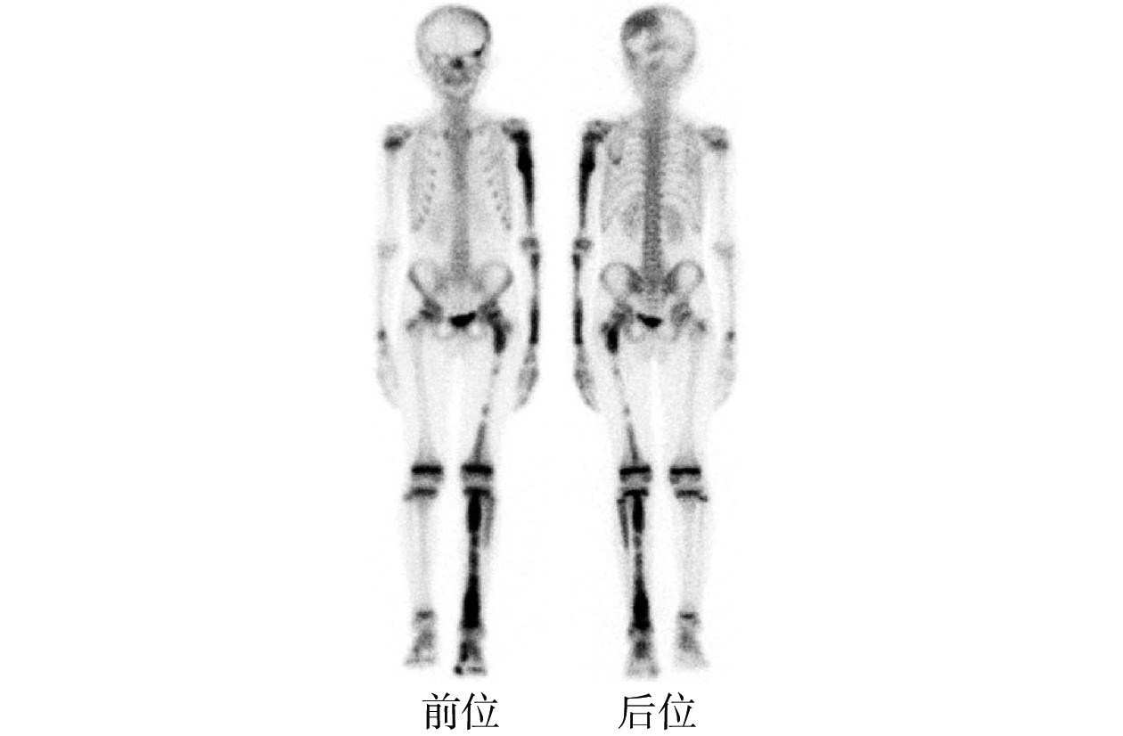
患儿男性,9岁,因“左侧小腿疼痛1周”就诊。家属诉患儿1周前无明显诱因出现左小腿疼痛,行走后疼痛加重,无负重时疼痛缓解。体格检查:腿部皮肤可见牛奶咖啡斑。实验室检查:碱性磷酸酶水平为295 U/L(正常参考值范围:20~200 U/L),其余指标正常。X线成像示:左侧尺桡骨局部髓腔膨大,密度不均匀,呈“磨玻璃”样改变,左侧桡骨中下段可见环形高密度影,内见结节样致密影;左手指骨局部膨大,髓腔呈“磨玻璃”样密度增高(图1A)。CT成像示:颅骨多发骨质膨胀,呈“磨玻璃”样改变。左侧胫骨矢状面CT成像示:左侧胫骨髓腔内可见团片、结节状高密度影,骨皮质连续(图1B、1C)。MRI成像示:左侧胫骨髓腔内可见多发团片状长T1、稍长T2信号影,信号不均匀(图1D、1E)。99Tcm-MDP全身骨显像示:左侧顶骨、枕骨、左侧肱骨、左侧尺骨、左手部分掌骨及指骨、左侧股骨、左侧胫腓骨及左侧足骨可见显像剂摄取异常增高灶,结合临床及影像综合诊断,考虑为多发性FDB(图2)。患儿在局部麻醉下通过CT引导行左侧胫骨病变穿刺活检术,随后行左侧胫骨病骨刮除+植骨术。组织病理学检查结果示:左胫骨FDB(图3)。结合病史和临床表现,综合诊断为MAS。
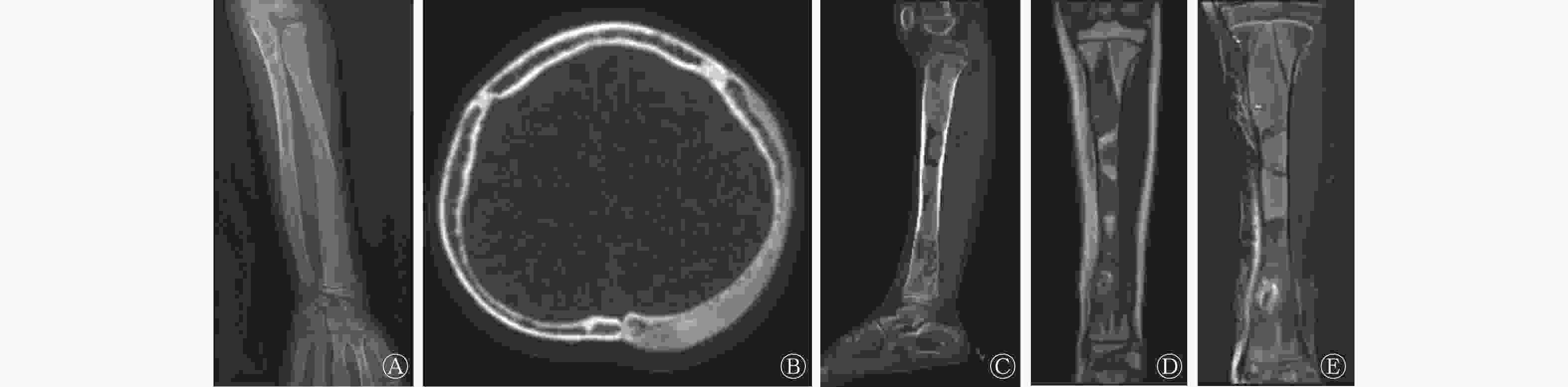
图 1 McCune-Albright综合征患儿(男性,9岁)的X线、CT和MRI图
Figure 1. X-ray, CT and MRI imagings of a child (male, 9 years old) with McCune-Albright syndrome

图 2 McCune-Albright综合征患儿(男性,9岁)的99Tcm-MDP全身骨显像图
Figure 2. 99Tcm-MDP whole body bone imaging of a child (male, 9 years old) with McCune-Albright syndrome

图 3 McCune-Albright综合征患儿(男性,9岁)左侧胫骨病灶的组织病理学检查图 (苏木精-伊红染色,×40)
Figure 3. Histopathological imaging of the left tibial bone in a child (male, 9 years old) with McCune-Albright syndrome (hematoxylin eosin staining, ×40)
-
MAS是一种以FDB、牛奶咖啡斑和青春期性早熟为主要特征的罕见的复杂疾病[1],至少包括上述两个以上临床特征才可确诊。MAS的发生是由于鸟苷酸结合蛋白α亚基激活多肽1(GNAS)基因突变导致Gs信号蛋白的α-亚基基因突变,属于非遗传性疾病[2]。FDB亦称骨纤维异常增殖症,是一种罕见的良性骨病,其特征是正常骨和骨髓组织被纤维组织取代,从而导致骨痛、骨骼畸形、骨折和运动功能障碍[3]。绝大多数FDB患者病情进展缓慢,但也有不到1%的患者具有向恶性转化的可能[4-5]。FDB可单独发生或合并其他骨骼疾病,可表现为单骨或多骨受累[2],颅骨、股骨近端和骨盆是其最常见的受累部位。FDB的病因及发病机制目前尚未明确[6],研究人员认为其与基因突变、染色体异常、骨发育异常及内分泌异常等有关。
影像检查的目的是通过检出FDB辅助临床医师诊断MAS,FDB的影像特征根据年龄不同而不同。FDB的X线、CT、MRI具有特征性影像表现,CT和MRI可以清晰地显示骨病变部位细微的解剖结构,对于FDB的诊断具有重要价值。X线和CT成像表现为:(1)骨骼“磨玻璃”样改变;(2)骨骼囊状改变;(3)骨骼“丝瓜瓤”样改变;(4)“虫蚀”样骨破坏;(5)骨骼硬化改变[7]。MRI的影像特征取决于FDB的组织病理学改变,其与病变组织中纤维的增生程度、病灶的含水量、代谢功能有关,其成像以长T2信号为主,伴片状低信号影[8]。本例患儿X线及CT成像表现为“磨玻璃”样改变,MRI为长T1、稍长T2信号影,影像特征与文献[7-8]报道一致。X射线、CT、MRI对多发FDB病变的诊断能力有限,加之辐射剂量及经济负担等因素,X线、CT、MRI对FDB全身病变的评估不全面,易对无症状病变漏诊。
99Tcm-MDP全身骨显像是功能影像,可早期、灵敏地显示FDB病变,通过一次扫描可获得全身骨骼的影像,评估全身骨损伤的部位、范围和程度,病灶多表现为显像剂摄取增高,但其特异性较低[5]。不同部位FDB病变的全身骨显像具有不同的特征[7]:(1)四肢长骨呈节段样分布的显像剂摄取增高灶;(2)股骨病灶为典型的“牧羊拐杖”征;(3)肋骨呈沿肋骨长轴分布的显像剂摄取增高灶,病变肋骨呈连续分布征象;(4)颅骨、脊柱和骨盆呈块状显像剂摄取增高灶;(5)大部分病灶骨的形态改变不显著,有单侧受累的趋势。本例患儿99Tcm-MDP全身骨显像表现为左侧骨骼多发FDB病变,主要累及颅骨和四肢骨,影像表现典型。有学者指出,全身骨显像有助于更好地描述FDB病变,并在指导治疗和随访中具有重要作用[2]。然而,FDB的影像诊断不能仅依靠全身骨显像,还需要结合X线、CT、MRI检查。
FDB需与畸形性骨炎、骨转移瘤及多发性骨髓瘤等鉴别[7]。组织病理学检查是诊断FDB的“金标准”,患者的临床资料、病史和相关影像检查可辅助诊断和综合评估。本例患儿因左小腿疼痛就诊,腿部皮肤可见牛奶咖啡斑,结合影像评估,经组织病理学检查确诊为FDB。本例患儿无明显的青春期性早熟的临床表现,但临床上MAS男患儿出现青春期早熟的比例较女患儿低得多,因此该患儿最终诊断为MAS。
99Tcm-MDP全身骨显像是诊断FDB的一种重要的影像检查方法,一次显像可灵敏、直观地评价全身骨骼病变的部位、范围及程度。同时具有方便、经济的优点,且不会对患者产生过大的辐射,因而在FDB的筛查、诊断和长期随访中具有独特优势。
利益冲突 所有作者声明无利益冲突
作者贡献声明 王茸、刘自强负责文献的收集、论文的撰写;王海军、王晓平负责论文的修订与审阅;王永乐、陈得魁负责病例资料的收集与整理
McCune-Albright综合征99Tcm-MDP全身骨显像1例
99Tcm-MDP whole body bone imaging of McCune-Albright syndrome: a case report
-
摘要: 笔者报道了1例McCune-Albright综合征(MAS)99Tcm-亚甲基二膦酸盐(MDP)全身骨显像的病例,显像结果表现为全身多处显像剂摄取异常增高灶,并通过X线、CT和MRI影像全面评估,经组织病理学检查结果证实为多发性骨纤维结构不良(FDB)。MAS临床上少见,笔者分别从临床特点及影像表现等方面分析了该病的特点,并结合文献复习了不同部位FDB的99Tcm-MDP全身骨显像特征,以为其诊断提供更多参考。Abstract: The authors reported a case of 99Tcm-methylenediphosphonate(MDP) whole-body bone imaging in a child with McCune-Albright syndrome (MAS). The imaging showed multiple abnormal hyperactive and elevated bone metabolism, and were comprehensively evaluated by X-ray, CT and MRI imaging. Pathological examination confirmed the diagnosis of multiple fibrous dysplasia of bone (FDB). MAS is rare in clinical, the authors analyzed the characteristics of MAS from the clinical characteristics and imaging manifestations respectively, and combined with the literature review of 99Tcm-MDP whole body bone imaging features of FDB in different parts, which to provide more references for its diagnosis.
-
Key words:
-
图 1 McCune-Albright综合征患儿(男性,9岁)的X线、CT和MRI图
Figure 1. X-ray, CT and MRI imagings of a child (male, 9 years old) with McCune-Albright syndrome
图 2 McCune-Albright综合征患儿(男性,9岁)的99Tcm-MDP全身骨显像图
Figure 2. 99Tcm-MDP whole body bone imaging of a child (male, 9 years old) with McCune-Albright syndrome
-
[1] Nicolaides NC, Kontou M, Vasilakis IA, et al. McCune-Albright syndrome: a case report and review of literature[J/OL]. Int J Mol Sci, 2023, 24(10): 8464[2023-02-19]. https://www.mdpi.com/1422-0067/24/10/8464. DOI: 10.3390/ijms24108464. [2] Jreige M, Hall N, Becce F, et al. A novel approach for fibrous dysplasia assessment using combined planar and quantitative SPECT/CT analysis of Tc-99m-diphosphonate bone scan in correlation with biological bone turnover markers of disease activity[J/OL]. Front Med (Lausanne), 2022, 9: 1050854[2023-02-19]. https://www.frontiersin.org/articles/10.3389/fmed.2022.1050854/full. DOI: 10.3389/fmed.2022.1050854. [3] Zhang K, Qu P, Wang B, et al. Management of the temporal bone fibrous dysplasia with external auditory canal stenosis and secondary cholesteatoma in an Asian population: a 11-case series[J]. Ear Nose Throat J, 2021, 100(10): NP469−NP474. DOI: 10.1177/0145561320927922. [4] Ju HJ, Paycha F. Osteoblastic and hyperostotic craniofacial lesion detected by 99mTc-labeled methylene diphosphonate bone scintigraphy and single-photon emission computed tomography/computed tomography: a pictorial essay[J]. Nucl Med Commun, 2021, 42(2): 117−126. DOI: 10.1097/MNM.0000000000001318. [5] Zhang LQ, He Q, Li W, et al. The value of 99mTc-methylene diphosphonate single photon emission computed tomography/computed tomography in diagnosis of fibrous dysplasia[J/OL]. BMC Med Imaging, 2017, 17(1): 46[2023-02-19]. https://bmcmedimaging.biomedcentral.com/articles/10.1186/s12880-017-0218-4. DOI: 10.1186/s12880-017-0218-4. [6] Lianou AD, Martini T, Tsimos K, et al. Fibrous dysplasia of the temporal bone: a demanding entity for radiologists and ENT surgeons[J]. Maedica (Bucur), 2022, 17(2): 524−527. DOI: 10.26574/maedica.2022.17.2.524. [7] 冯瑾, 张连娜, 高璇, 等. 骨纤维异常增殖症全身骨显像影像特征分析[J]. 标记免疫分析与临床, 2021, 28(9): 1452−1456, 1463. DOI: 10.11748/bjmy.issn.1006-1703.2021.09.003.
Feng J, Zhang LN, Gao X, et al. An analysis of features of the whole-body bone imaging in osteofibrous dysplasia[J]. Labeled Immunoassays Clin Med, 2021, 28(9): 1452−1456, 1463. DOI: 10.11748/bjmy.issn.1006-1703.2021.09.003.[8] 赵娜, 董江宁, 高飞, 等. 单骨局灶性骨纤维结构不良磁共振DWI及增强表现特征及病理学基础[J]. 临床放射学杂志, 2020, 39(4): 741−745. DOI: 10.13437/j.cnki.jcr.2020.04.025.
Zhao N, Dong JN, Gao F, et al. MRI DWI and enhancement features and pathological basis of focal fibrous dysplasia of single bone[J]. J Clin Radiol, 2020, 39(4): 741−745. DOI: 10.13437/j.cnki.jcr.2020.04.025. -

 点击查看大图

图(3)
计量
- 文章访问数: 2628
- HTML全文浏览量: 2112
- PDF下载量: 19