


 下载:
下载:
-
结节性筋膜炎(nodular fasciitis,NF)是一种相对少见的纤维组织增生性良性病变,组织病理学上具有假肉瘤的特征,该病常突然出现,生长迅速,易被误诊为恶性肿瘤[1]。NF的临床及影像学表现缺乏特异性,误诊后导致的手术等过度治疗会给患者造成不必要的伤害。笔者报道了1例误诊为霍奇金淋巴瘤(Hodgkin lymphoma,HL)复发的NF病例。
-
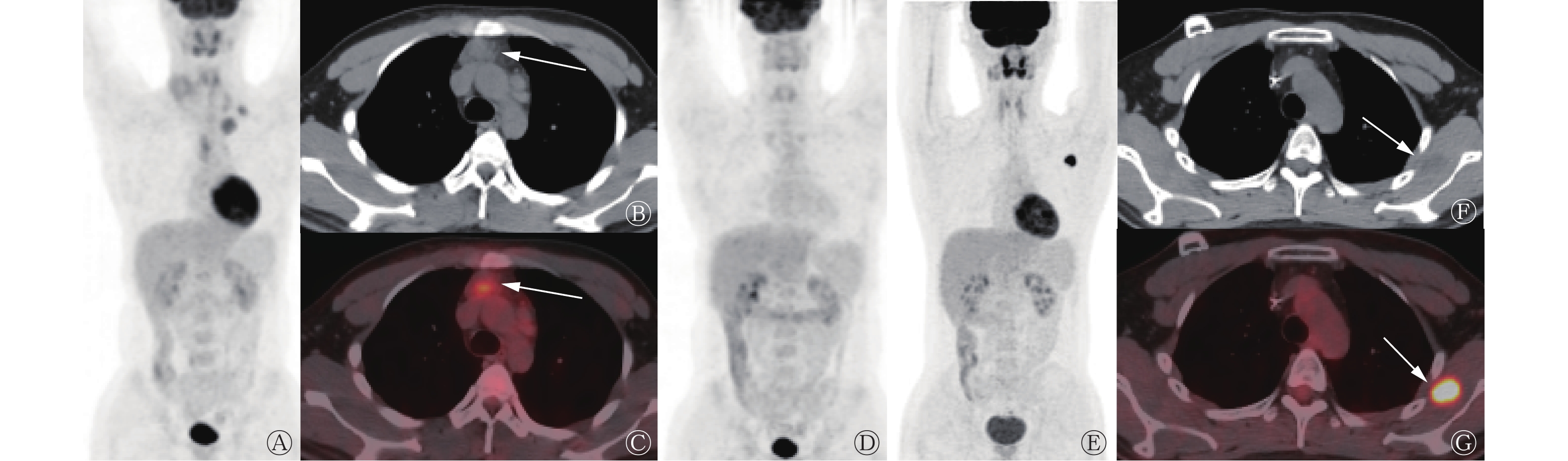
患者男性,25岁,2018年9月初无意间触及右侧颈部有一肿物,质地硬,活动度尚可,无明显压痛,2018年9月14日于东部战区总医院口腔科行右侧颈部肿物摘除术,术后经组织病理学检查结果确诊为结节硬化型经典型霍奇金淋巴瘤(nodular sclerosis classical Hodgkin lymphoma,NSCHL)。2018年9月18日于东部战区总医院核医学科行18F-FDG PET/CT检查,结果显示:左侧颈部、左侧锁骨区、左上纵隔、前纵隔胸腺区、主动脉弓旁可见多发肿大淋巴结,18F-FDG代谢增高,SUVmax=4.7,考虑为淋巴瘤浸润(图1A~C)。骨髓形态学+活体组织病理学检查结果提示正常。结合病史及检查结果,诊断为NSCHL[Ann Arbor分期为ⅡA期,淋巴瘤国际预后指数(IPI)评分0分,年龄调整后的淋巴瘤国际预后指数(aaIPI)评分0分,低危组 ]。对患者行ABVD方案治疗:在第1天和第15天,分别静脉滴注多柔比星脂质体30 mg+博来霉素15 mg +长春瑞宾10 mg+达卡巴嗪0.6 mg,1个周期为21 d,化疗2个周期后行18F-FDG PET/CT复查,结果显示,原发病灶明显缩小,18F-FDG代谢程度明显降低。化疗4个周期后,再次行18F-FDG PET/CT复查,结果显示,原发病灶均未见18F-FDG代谢增高灶,评估病情为完全缓解(图1D)。

图 1 结节性筋膜炎患者(男性,25岁)在霍奇金淋巴瘤治疗过程中的3次18F-FDG PET/CT显像图
Figure 1. Three 18F-FDG PET/CT imaging results of a patient with nodular fasciitis (male, 25 years old) during the treatment of Hodgkin's lymphoma
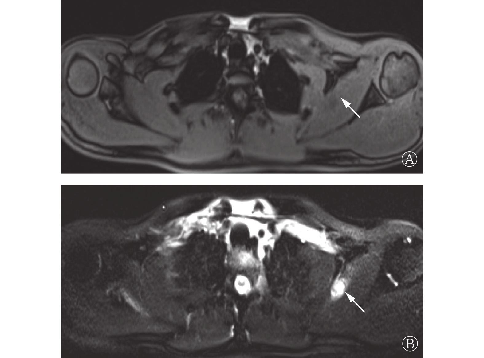
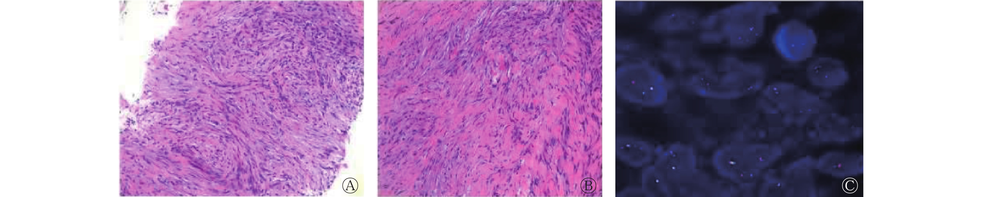
2021年3月,患者出现左侧肩部疼痛,于东部战区总医院行MRI检查,结果显示左侧肩胛下肌有异常信号(图2),2021年5月复查MRI,结果提示病灶增大,结合病史,考虑可能为淋巴瘤浸润。2021年7月19日,患者行18F-FDG PET/CT检查,结果显示:左侧肩胛下肌处有类圆形低密度灶,18F-FDG代谢明显增高,SUVmax=13.06,考虑可能为淋巴瘤浸润(图1E~G)。2021年7月21日,患者行经皮穿刺左肩胛下肌结节活体组织病理学检查,结果提示:梭形细胞增生性病变,伴黏液样变性及炎细胞浸润(图3);结合影像表现(肌内肿块长径约2 cm)及免疫组织化学、分子生物学方法检测结果,考虑为NF。

图 2 结节性筋膜炎患者(男性,25岁)肩部的MRI图
Figure 2. MRI images of shoulder in a patient with nodular fasciitis (male, 25 years old)

图 3 结节性筋膜炎患者(男性,25岁)的组织病理学检查图(苏木精-伊红染色,×100,×200)及基因检测图(荧光原位杂交法,×1000)
Figure 3. Histopathological examination (hematoxylin eosin staining, ×100, ×200) and genetic testing (fluorescence in situ hybridization, ×1000) results of a patient with nodular fasciitis (male, 25 years old)
-
NF是由纤维母细胞和肌纤维母细胞增生形成的假肉瘤性病变,一般被认为是一种自限性、反应性、增生性的肿瘤样病变,通常具有生长迅速、细胞丰富、分裂活跃等特点,它最常见于上肢、躯干、头颈部的皮下组织,发病年龄从2岁到50岁不等,无性别差异[2-3]。第5版WHO软组织肿瘤分类将其归于成纤维细胞和(或)肌成纤维细胞性良性肿瘤[4]。NF按照部位一般可分为3种亚型:皮下型、肌内型、筋膜(肌间)型;按照病理类型可分为黏液型、细胞型、纤维型。NF的MRI表现与其组织病理学特点及生长方式有关,MRI上大多呈 T1WI等信号、均匀或不均匀T2WI高信号(病灶内的黏液成分多,则呈明显高信号;纤维和细胞成分多,则信号减低)[2, 5-7]。本病例MRI表现为T1WI低信号,T2WI脂肪抑制高信号,符合文献报道中对NF的归纳与描述。
18F-FDG PET/CT被广泛应用于肿瘤代谢显像,在淋巴瘤的诊断、临床分期、疗效评估中也发挥着重要作用[8]。NF的病理基础为纤维母细胞和肌纤维母细胞的增生,其糖代谢也处于较高水平,所以此类病变在行18F-FDG PET/CT显像时也可表现为高摄取,因此在诊断上具有一定迷惑性,国外也有相关个案报道[3, 9-10]。18F-FDG PET/CT检查的灵敏度高,但特异度相对较低。同样,在进行MRI诊断时也会面临这一困难。肿瘤转移的MRI信号特征通常由原发灶的性质确定,可呈现为多样性,因此仅凭影像学表现很难做出准确判断,针对本病例,如果没有组织病理学检查的进一步诊断,很容易将左侧肩胛下肌的高摄取灶误判为NSCHL复发,从而造成过度治疗,给患者的身心带来损伤。近年来,随着核医学技术的不断发展,68Ga 标记的成纤维细胞活化蛋白抑制剂 (fibroblast activating protein inhibitor,FAPI)现已作为一种新型的正电子肿瘤显像剂逐渐被应用于临床,当机体存在强烈的纤维增生反应、患有以间质组织活化为特征的疾病或有伤口处于愈合期,成纤维活化蛋白呈现为高表达状态[11],68Ga-FAPI已作为PET显像剂被应用于实体肿瘤、感染及炎症的诊断,未来我们或可应用68Ga-FAPI作为18F-FDG的补充显像剂,提高对该类炎性病变的诊断及鉴别诊断能力,其临床价值有待进一步探索。
NF的发病机制不详,目前多被认为可能与外伤或感染性因素有关。追溯本病例的病史后,我们发现患者在NSCHL治疗过程中一直有网球运动的习惯,或与其NF发病有一定关联。18F-FDG PET/CT对于葡萄糖高代谢的病变具有较高的检出率,但是其鉴别诊断效果有待提高。回顾分析本病例可发现,患者的治疗效果一直很好,随访过程中进行的18F-FDG PET/CT检查也从未有复发迹象,此次左侧肩胛下肌病灶的SUV明显高于其原发NSCHL病灶,或可提示它们并非同源。因此,在日常影像诊断工作中,不断增加对病种的积累、学会对比各项影像检查的表现、结合患者整个病史过程考虑病情发展的可能性等都显得十分重要。
利益冲突 所有作者声明无利益冲突
作者贡献声明 姜思远负责数据的采集与分析、论文的撰写;周晓蝶负责论文的审阅;吉蘅山负责论文的审阅与指导;吴江负责论文的修订与指导
结节性筋膜炎18F-FDG PET/CT误诊为霍奇金淋巴瘤复发1例
18F-FDG PET/CT misdiagnosed nodular fasciitis as recurrence of Hodgkin's lymphoma: a case report
-
摘要: 笔者报道了1例结节性筋膜炎(NF)误诊为霍奇金淋巴瘤(HL)复发的病例。患者2年多前患HL,本次发病表现为左侧肩部有疼痛性肿块,行MRI、PET/CT检查均提示可能为HL复发,最终经穿刺组织病理学活检结果确诊为NF。笔者分析了NF的影像学及病理学特点,并进行了相关文献复习,以期分享18F-FDG PET/CT在诊断该类型病例方面的相关经验。Abstract: The authors reported a case of nodular fasciitis (NF) misdiagnosed as recurrence of Hodgkin's lymphoma (HL). The patient developed HL more than 2 years ago. This time the disease presents as a painful mass on the left shoulder, MRI and PET/CT examinations indicated the possibility of HL recurrence, and the pathological result of puncture confirmed NF. The authors analyzed the imaging and pathological features of NF and reviewed the relevant literature in order to share the experience in 18F-FDG PET/CT diagnosis of this type of cases.
-
Key words:
-
图 1 结节性筋膜炎患者(男性,25岁)在霍奇金淋巴瘤治疗过程中的3次18F-FDG PET/CT显像图
Figure 1. Three 18F-FDG PET/CT imaging results of a patient with nodular fasciitis (male, 25 years old) during the treatment of Hodgkin's lymphoma
图 2 结节性筋膜炎患者(男性,25岁)肩部的MRI图
Figure 2. MRI images of shoulder in a patient with nodular fasciitis (male, 25 years old)
-
[1] Luna A, Molinari L, Bollea Garlatti LA, et al. Nodular fasciitis, a forgotten entity[J]. Int J Dermatol, 2019, 58(2): 190−193. DOI: 10.1111/ijd.14219. [2] 徐贵平, 强永乾, 曹培龙. 结节性筋膜炎的临床及影像学表现[J]. 实用放射学杂志, 2020, 36(1): 91−94. DOI: 10.3969/j.issn.1002-1671.2020.01.023.
Xu GP, Qiang YQ, Cao PL. Clinical and imaging features of nodular fasciitis[J]. J Pract Radiol, 2020, 36(1): 91−94. DOI: 10.3969/j.issn.1002-1671.2020.01.023.[3] Choi BW, Kim HW, Jeon JC, et al. Nodular fasciitis mimicking malignant soft tissue tumor in an infant, with 18F-FDG PET/CT findings: a case report[J]. Nuklearmedizin, 2018, 57(3): N29−N31. DOI: 10.3413/Nukmed-0930-17-09. [4] Sbaraglia M, Bellan E, Tos APD. The 2020 WHO classification of soft tissue tumours: news and perspectives[J]. Pathologica, 2021, 113(2): 70−84. DOI: 10.32074/1591-951X-213. [5] Lin WL, Bao LY. Nodular fasciitis of the breast: the report of three cases[J/OL]. BMC Womens Health, 2022, 22(1): 54[2022-06-27]. https://bmcwomenshealth.biomedcentral.com/articles/10.1186/s12905-022-01631-2. DOI: 10.1186/s12905-022-01631-2. [6] 刘壮盛, 黄云海, 王建明, 等. MRI筋膜尾征诊断结节性筋膜炎的价值[J]. 中华放射学杂志, 2015, 49(7): 531−534. DOI: 10.3760/cma.j.issn.1005-1201.2015.07.012.
Liu ZS, Huang YH, Wang JM, et al. Application value of fascial tail sign at MRI in the detection of nodular fasciitis: a retrospective study[J]. Chin J Radiol, 2015, 49(7): 531−534. DOI: 10.3760/cma.j.issn.1005-1201.2015.07.012.[7] 杨利, 薛川, 李方海, 等. 结节性筋膜炎临床表现及MRI诊断探讨[J]. 社区医学杂志, 2023, 21(11): 584−588. DOI: 10.19790/j.cnki.JCM.2023.11.08.
Yang L, Xue C, Li FH, et al. Findings of MRI and clinical features of nodular fasciitis[J]. J Commun Med, 2023, 21(11): 584−588. DOI: 10.19790/j.cnki.JCM.2023.11.08.[8] 乔文礼, 牛家华, 金文雅, 等. 自体干细胞移植前或后18F-FDG PET/CT显像及相关因素对经典霍奇金淋巴瘤预后的评估价值[J]. 中华核医学与分子影像杂志, 2020, 40(3): 147−152. DOI: 10.3760/cma.j.cn321828-20190729-00149.
Qiao WL, Niu JH, Jin WY, et al. Prognostic value of 18F-FDG PET/CT imaging and related factors for patients with classic Hodgkin lymphoma before or after autologous stem cell transplantation[J]. Chin J Nucl Med Mol Imaging, 2020, 40(3): 147−152. DOI: 10.3760/cma.j.cn321828-20190729-00149.[9] Kim JY, Park J, Choi YY, et al. Nodular fasciitis mimicking soft tissue metastasis on 18F-FDG PET/CT during surveillance[J]. Clin Nucl Med, 2015, 40(2): 172−174. DOI: 10.1097/rlu.0000000000000559. [10] Seo M, Kim M, Kim ES, et al. Diagnostic clue of nodular fasciitis mimicking metastasis in papillary thyroid cancer, mismatching findings on 18F-FDG PET/CT and 123I whole body scan: a case report[J]. Oncol Lett, 2017, 14(1): 1167−1171. DOI: 10.3892/ol.2017.6181. [11] Chen HJ, Zhao L, Ruan D, et al. 68Ga-FAPI PET/CT improves therapeutic strategy by detecting a second primary malignancy in a patient with rectal cancer[J]. Clin Nucl Med, 2020, 45(6): 468−470. DOI: 10.1097/rlu.0000000000003000. -

 点击查看大图
图(3)
计量
- 文章访问数: 1391
- HTML全文浏览量: 928
- PDF下载量: 12