


 下载:
下载:


-
卵巢甲状腺肿(struma ovarii,SO)起源于卵巢的生殖细胞,是指肿瘤全部由甲状腺组织构成或肿瘤中甲状腺组织占50%以上[1]。目前对该病的认识较局限,而且该病的临床表现缺乏特异性,易被误诊为卵巢癌,确诊需术后组织病理学检查。笔者报道了1例SO伴腹膜转移患者的18F-FDG PET/CT 显像结果,结合其临床资料及影像学检查,并复习相关文献,分析其误诊原因,从而提高影像医师对该病诊断的准确率。
-
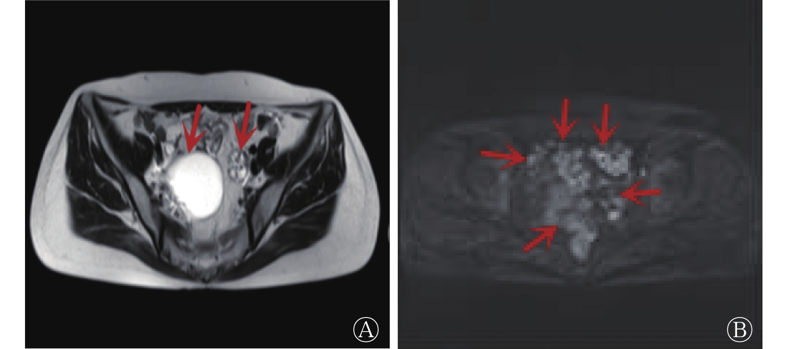
患者女性,52岁,自述5个月前无明显诱因感觉腹胀,因腹胀加重于2022年4月6日来我院就诊。患者自发病以来,无发热、寒战,无心慌、乏力,无下肢肿胀,无明显消瘦。患者精神尚可,饮食、睡眠佳,大小便正常。既往因子宫肌瘤、子宫腺肌症行子宫全切术,无高血压、冠心病及糖尿病等病史。妇科检查示:盆腔右侧可触及囊性肿物,活动度差。实验室检查结果示:血常规及肝功能未见明显异常,CA125水平为 79.2(0~23.0) U/ml、甲状腺球蛋白(Tg)水平<0.9 (0~4.0)IU/ml、FT3 水平为3.5(2.3~4.2) pg/ml、FT4 水平为0.89(0.89~1.76) ng/dl、TSH水平为 1.33(0.55~4.78) μIU/ml、CA199水平为13.2(0~36.0) U/ml、癌胚抗原(carcinoembryonic antigen,CEA)水平为0.6(0~4.5) ng/ml、甲胎蛋白(AFP)水平为2.8(0~7.0) ng/ml。超声检查结果示:右侧附件区可见囊性包块,其内可见中等回声;左侧附件区、盆腔可见多处不均质回声包块,血流信号丰富。MRI检查结果示:T2WI示双侧附件可见类圆形高信号,其内可见不规则低信号,病灶边界清晰(图1A),DWI示右侧附件肿块边缘稍高信号及盆腔多发高信号结节影(图1B)。结肠脾曲T2WI可见不规则等长信号,病灶与周围肠管分界欠清。PET/CT检查示:子宫全切术后,右侧附件区可见囊实性密度影,实性部分18F-FDG摄取略增高,SUVmax=3.4(图2A),延迟显像18F-FDG摄取较前期增高,SUVmax=5.4(图2B);左侧附件区可见不规则稍高密度肿块影,18F-FDG摄取略增高,SUVmax=3.1(图2A),延迟显像18F-FDG摄取较前期增高,SUVmax=3.9(图2B);左侧中腹部可见不规则软组织密度影,18F-FDG摄取稍增高,SUVmax=4.0,延迟显像18F-FDG摄取较前期变化不明显(图2C);盆腹腔内肠间隙可见多发大小不等稍高密度结节影, 18F-FDG摄取略增高,SUVmax=3.5(图2D),延迟显像18F-FDG摄取较前期增高,SUVmax=4.8(图2E)。PET/CT显像诊断结果为右侧附件区卵巢癌,左侧附件区考虑恶性可能,盆腹腔内肠间隙多发结节考虑转移,左侧中腹部考虑转移灶或肠道间质瘤。外科医师认为不能排除肠道间质瘤破裂,造成腹腔广泛播散的可能。患者于2022年4月14日行“腹腔镜下双侧附件切除术+大网膜切除术+经腹结肠部分切除术+肠吻合术+肠系膜肿物剥除术”。术后组织病理学检查结果:双侧附件、腹膜、肠系膜、大网膜多发增生性SO(图3)。患者术后恢复良好,于2022年4月23日出院。

图 1 卵巢甲状腺肿伴腹膜转移患者(女性,52岁)腹部MRI图
Figure 1. MRI images of abdomen in a patient (female, 52 years old) with struma ovarii with peritoneal metastasis

图 2 卵巢甲状腺肿伴腹膜转移患者(女性,52岁)的PET/CT显像图
Figure 2. PET/CT images of a patient (female, 52 years old) with struma ovarii with peritoneal metastasis

图 3 卵巢甲状腺肿伴腹膜转移患者(女性,52岁)组织病理学检查图(苏木精-伊红染色,×40)
Figure 3. Histopathological examination images of a patient (female, 52 years old) with struma ovarii with peritoneal metastasis (hematoxylin eosin staining, ×40)
-
SO是一种罕见的卵巢畸胎瘤,约占卵巢肿瘤的0.3%~1%[1]。SO患者的临床表现缺少特异性,往往因下腹痛、腹胀及腹部肿块等非特异性症状而就诊,或在妊娠、盆腔检查中偶然发现[2-5]。患者的临床表现各不相同,有的患者可出现甲亢的症状,切除SO后症状逐渐消退[6];有的患者还会出现胸腔积液和腹水,切除SO后症状缓解 [7];有的患者Tg水平升高,在接受手术和131I治疗后,Tg水平逐渐恢复正常[6]。患者的CA125水平升高似乎较为多见,最高可达正常值的数10倍 [7]。
SO的影像学表现多样,其MRI表现以囊性或囊实性为主,肿瘤在T1WI中表现为高、等、低信号,在T2WI中表现为不均匀高、低信号;CT表现为囊实性,有些病灶可见沿壁钙化灶[4, 7-9]。陈凤英等[10]发现SO的实性成分在T2WI中呈高信号,T1WI中呈中等信号,增强扫描实性部分呈明显强化;囊性成分随着黏液的黏性增加,MRI信号会降低,特征性表现为“黑珍珠”样低信号颗粒。由此可见,SO的MRI及CT表现多样,缺乏特异性,但实性成分及分隔呈明显强化类似甲状腺组织,这一表现可以作为SO诊断与鉴别诊断的依据[11]。Savelli等[12]回顾性分析了21例SO患者的超声影像学表现,结果显示,多数超声表现为囊性低回声,彩色多普勒血流显像(CDFI)示血流信号从无到丰富不等,特异性表现为“甲状腺肿珠”,即光滑的圆形实性球体位于囊肿内,“甲状腺肿珠”可以作为SO的一个诊断依据。与18F-FDG PET/CT不摄取或低摄取不同的是, 131I闪烁扫描可以显示SO呈高摄取,多种影像学表现可以为该病提供更多的诊断信息[6-7]。
SO通常以良性肿瘤为主,其确诊还需依靠组织病理学检查。但是Shaco-Levy等[13]收集了88例SO患者并进行了长期随访,结果表明,医师不能仅仅根据组织病理学检查结果的良恶性来预测SO的临床结果,他们提出无论组织病理学结果如何,仅需要满足以下1项指标即符合生物学恶性:(1)就诊时已有卵巢外扩散;(2)卵巢浆膜上有肿瘤;(3)手术后SO复发。本例患者就符合这一现象,属于组织病理学良性、生物学恶性的肿瘤,因为其不仅发生在双侧附件、结肠脾曲,还发生了腹膜种植。
本例患者因持续性腹胀而就诊,其Tg、TSH、FT3、FT4水平以及其他肿瘤标志物水平均在正常范围内,PET/CT显示甲状腺的形态、密度、代谢均无异常。虽然该患者的CA125水平升高程度较典型卵巢癌略低,但PET/CT显示其双侧附件囊实性18F-FDG代谢增高影伴腹盆腔多发稍高代谢结节,诊断结果为卵巢癌伴腹膜种植。最终该患者的术后组织病理学检查结果为增生性SO。因此,仅仅依靠影像学诊断SO较为困难,SO组织病理学良性、生物学恶性的特点使此病与卵巢恶性肿瘤伴多发转移难以鉴别。SO的MRI或CT表现多为囊实性,18F-FDG PET/CT不摄取或摄取较低,这些并不特异的影像学表现使得此病在术前诊断较为困难。肿瘤的实性部分在CT与MRI上强化明显,在PET/CT低摄取时或许可以为诊断提供帮助。正常甲状腺组织葡萄糖代谢较低且均匀,只有恶性甲状腺组织失去摄碘能力后,才会出现高代谢的情况[14],因此,当131I呈现高摄取,而PET/CT显示低摄取,在排除其他肿瘤后,要考虑是否为甲状腺组织。
SO需要与其他疾病相鉴别。(1)成熟囊性畸胎瘤。其是卵巢肿瘤中最常见的良性肿瘤,肿瘤表面光滑,瘤体内可发现毛发、牙齿、骨骼等。CT表现为肿瘤边界清晰、呈高低混杂密度影;MRI表现为肿瘤呈混杂信号,脂肪的T1WI、T2WI均为高信号,压脂序列上脂肪信号明显降低,牙齿和骨骼的T1WI、T2WI均为低信号;PET/CT通常无18F-FDG摄取。(2)卵巢囊腺癌。其主要来自卵巢的上皮成分,其中黏液性囊腺癌常为多房表现,早期临床表现不明显,常见的表现有腹水、腹痛、腹部包块,患者就诊时常常已为晚期。CT表现多为囊实性,体积较大,分隔薄厚不均;MRI实性成分在T1WI中呈等信号,T2WI呈中高信号,囊性部分在浆液性囊腺癌T1WI中呈低信号,T2WI呈高信号,在黏液性囊腺癌T1WI、T2WI中均呈高信号;PET/CT呈18F-FDG高摄取。PET/CT联合解剖显像与功能显像,能够早期、准确地诊断肿瘤,结合CA125水平升高,可以大大提高卵巢癌的诊断率[15]。(3)肠道间质瘤。其是最常见的间叶来源肿瘤。CT表现为肿瘤类圆形,多呈腔外生长,肿瘤内可有钙化灶形成,增强扫描动脉期呈均匀强化,门脉期呈延迟强化。PET/CT大多呈18F-FDG高摄取。
综上所述,SO在临床较为罕见,诊断较为困难,我们应该尽可能完善相关临床资料及影像学检查。即使有类似恶性病变的间接征象,如PET/CT延迟显像附件区、腹膜多发结节代谢较早期扫描稍增强,也不可轻易地诊断为恶性病变,要将每一处不符合典型病变的疑点进行逐一排查,得到较为客观的结论,从而为临床提供早期诊断的依据。
利益冲突 所有作者声明无利益冲突
作者贡献声明 暴丽洁负责文献的检索与分析、论文的撰写;赵子龙负责研究命题的提出、论文的修订;张计负责文献的检索与分析;杨慧、武建飞负责图像的分析;周茂荣负责研究命题的设计、论文的审阅
卵巢甲状腺肿伴腹膜转移18F-FDG PET/CT显像误诊1例
Misdiagnosis of struma ovarii with peritoneal metastasis by 18F-FDG PET/CT imaging: a case report
-
摘要: 卵巢甲状腺肿是一种罕见的卵巢肿瘤,肿瘤全部由甲状腺组织构成,或甲状腺组织占全部肿瘤的50%以上。笔者报道了1例因持续性腹胀就诊的卵巢甲状腺肿伴腹膜转移患者,根据患者的临床表现、实验室检查及影像学表现分析该病例的特点,并结合相关文献进一步复习卵巢甲状腺肿的影像表现及误诊原因,从而为临床提供早期诊断依据。Abstract: Struma ovarii is a rare disease in which the tumor is composed entirely of thyroid tissue, or thyroid tissue accounts for more than 50% of the total tumor. The author reported a case of a patient with struma ovarii with peritoneal metastasis who received medical treatment due to persistent abdominal distension. The characteristics of the case were analyzed according to the clinical manifestations, laboratory examination and imaging findings of the patient. Combined with relevant literatures, the imaging manifestations and causes of misdiagnosis of struma ovarii were further reviewed, so as to provide clinical basis for early diagnosis.
-
Key words:
-
图 1 卵巢甲状腺肿伴腹膜转移患者(女性,52岁)腹部MRI图
Figure 1. MRI images of abdomen in a patient (female, 52 years old) with struma ovarii with peritoneal metastasis
图 2 卵巢甲状腺肿伴腹膜转移患者(女性,52岁)的PET/CT显像图
Figure 2. PET/CT images of a patient (female, 52 years old) with struma ovarii with peritoneal metastasis
-
[1] 钟桂棉, 陈海玉, 向之明. 卵巢甲状腺肿的CT误诊分析[J]. 中国CT和MRI杂志, 2022, 20(8): 130−132. DOI: 10.3969/j.issn.1672-5131.2022.08.043.
Zhong GM, Chen HY, Xiang ZM. CT image misdiagnosis of struma ovarii[J]. Chin J CT MRI, 2022, 20(8): 130−132. DOI: 10.3969/j.issn.1672-5131.2022.08.043.[2] Ayhan S, Kilic F, Ersak B, et al. Malignant struma ovarii: from case to analysis[J]. J Obstet Gynaecol Res, 2021, 47(9): 3339−3351. DOI: 10.1111/JOG.14902. [3] Li Z, Wang JX, Chen Q. Struma ovarii and peritoneal strumosis during pregnancy[J]. BMC Pregnancy Childbirth, 2021, 21(1): 347. DOI: 10.1186/s12884-021-03815-4. [4] Wang SY, He XQ, Yang HJ, et al. Struma ovarii associated with ascites and elevated CA125: two case reports and review of the literature[J/OL]. Int J Womens Health, 2022, 14: 1291−1296[2022-06-13]. https://www.dovepress.com/struma-ovarii-associated-with-ascites-and-elevated-ca125-two-case-repo-peer-reviewed-fulltext-article-IJWH. DOI: 10.2147/IJWH.S379128. [5] Donato S, Simões H, Leite V. Malignant struma ovarii with concurrent thyroid cancer: outcomes during and after pregnancy[J]. Eur Thyroid J, 2021, 10(6): 523−527. DOI: 10.1159/000512735. [6] Wolff EF, Hughes M, Merino MJ, et al. Expression of benign and malignant thyroid tissue in ovarian teratomas and the importance of multimodal management as illustrated by a BRAF-positive follicular variant of papillary thyroid cancer[J]. Thyroid, 2010, 20(9): 981−987. DOI: 10.1089/thy.2009.0458. [7] Fujiwara S, Tsuyoshi H, Nishimura T, et al. Precise preoperative diagnosis of struma ovarii with pseudo-Meigs' syndrome mimicking ovarian cancer with the combination of 131I scintigraphy and 18F-FDG PET: case report and review of the literature[J/OL]. J Ovarian Res, 2018, 11(1): 11[2022-06-13]. https://ovarianresearch.biomedcentral.com/articles/10.1186/s13048-018-0383-2. DOI: 10.1186/s13048-018-0383-2. [8] 魏伟, 刘向一, 何庄贞, 等. 卵巢甲状腺肿的MR表现与病理对照分析[J]. 现代医用影像学, 2022, 31(6): 1040−1043. DOI: 10.3969/j.issn.1006-7035.2022.06.011.
Wei W, Liu XY, He ZZ, et al. Struma ovarii: comparison of MR features and pathological findings[J]. Mod Med Imageol, 2022, 31(6): 1040−1043. DOI: 10.3969/j.issn.1006-7035.2022.06.011.[9] 白杨. 卵巢甲状腺肿的影像学表现与误诊分析[J]. 现代医用影像学, 2019, 28(12): 2660−2661.
Bai Y. Imaging findings and misdiagnosis of struma ovarii[J]. Mod Med Imageol, 2019, 28(12): 2660−2661.[10] 陈凤英, 张大伟, 张颖, 等. 卵巢甲状腺肿的超声和MRI影像表现与病理对照分析[J]. 磁共振成像, 2019, 10(12): 913−916. DOI: 10.12015/issn.1674-8034.2019.12.008.
Chen FY, Zhang DW, Zhang Y, et al. Ultrasonography compared with magnetic resonance imaging for the diagnosis of struma ovarii: correlation with histopathology[J]. Chin J Magn Reson Imaging, 2019, 10(12): 913−916. DOI: 10.12015/issn.1674-8034.2019.12.008.[11] 胡平胜, 刘俊, 文露, 等. 卵巢甲状腺肿的影像学表现[J]. 湖南师范大学学报: 医学版, 2021, 18(6): 104−107. DOI: 10.3969/j.issn.1673-016X.2021.06.030.
Hu PS, Liu J, Wen L, et al. Imaging findings of struma ovarii[J]. J Hunan Normal Univ (Med Ed), 2021, 18(6): 104−107. DOI: 10.3969/j.issn.1673-016X.2021.06.030.[12] Savelli L, Testa AC, Timmerman D, et al. Imaging of gynecological disease (4): clinical and ultrasound characteristics of struma ovarii[J]. Ultrasound Obstet Gynecol, 2008, 32(2): 210−219. DOI: 10.1002/uog.5396. [13] Shaco-Levy R, Peng RY, Snyder MJ, et al. Malignant struma ovarii: a blinded study of 86 cases assessing which histologic features correlate with aggressive clinical behavior[J]. Arch Pathol Lab Med, 2012, 136(2): 172−178. DOI: 10.5858/arpa.2011-0092-OA. [14] 杜芬, 胡珊, 梁梦, 等. 18F-FDG PET/CT显像在甲状腺偶发瘤良恶性诊断中的研究进展[J]. 国际放射医学核医学杂志, 2018, 42(3): 274−279. DOI: 10.3760/cma.j.issn.1673-4114.2018.03.015.
Du F, Hu S, Liang M, et al. Development in the diagnosis of benign and malignant thyroid incidentalomas by 18F-FDG PET/CT imaging[J]. Int J Radiat Med Nucl Med, 2018, 42(3): 274−279. DOI: 10.3760/cma.j.issn.1673-4114.2018.03.015.[15] 王淼, 舒博学, 王娟. 血清CA125联合PET/CT对早期卵巢癌的诊断效果[J]. 临床医学研究与实践, 2021, 6(4): 109−111. DOI: 10.19347/j.cnki.2096-1413.202104038.
Wang M, Shu BX, Wang J. Diagnostic effect of serum CA125 combined with PET/CT for early ovarian cancer[J]. Clin Med Res Pract, 2021, 6(4): 109−111. DOI: 10.19347/j.cnki.2096-1413.202104038. -

 点击查看大图
图(3)
计量
- 文章访问数: 1622
- HTML全文浏览量: 1450
- PDF下载量: 19