


 下载:
下载:
-
非骨化性纤维瘤(non-ossifying fibroma,NOF) 是由成熟的非成骨性结缔组织发育而来的良性骨肿瘤,临床上较为少见,约占原发性骨肿瘤的1.1%。 Jaffe等[1]在1941年首次认识了它,并在第2年由Jaffe 和 Lichtenstein正式命名。NOF多见于10~20岁的青少年男性,病变部位多见于股骨下端和胫骨上端,常为单灶发病。本文报道的是1例发生在中年女性患者身上的经术后病理结果证实的NOF,对其影像资料进行回顾性分析,并结合文献复习,旨在提高临床医师对该病的认识。
-
患者女性,44岁。无明显诱因右膝疼痛1周余,活动后加重。实验室检查结果显示血常规、肿瘤标志物和血生化结果均在正常范围。
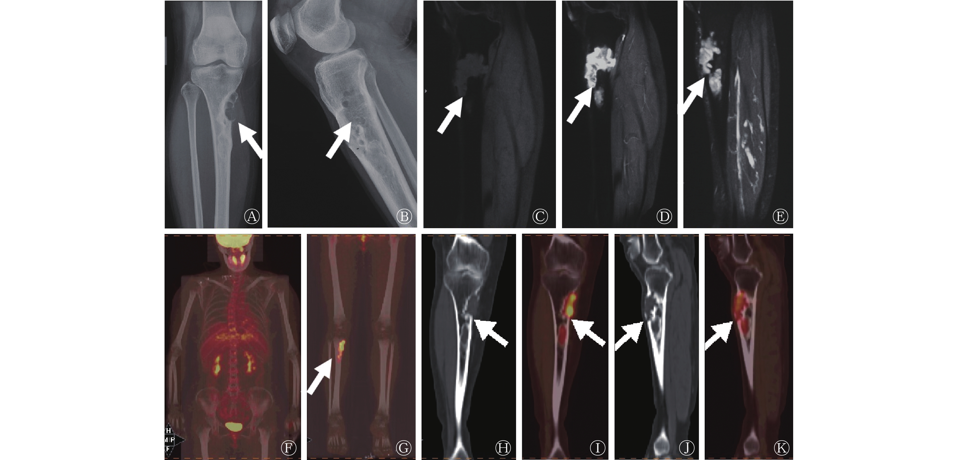
常规X线检查结果显示,位于右侧胫骨近端骨皮质有一个轮廓清晰的多囊状透亮区,边缘可见硬化边,内侧缘凸入髓腔呈花边样,病变长轴与骨干平行,无骨膜反应(图1A、B)。MRI检查结果显示,T1加权成像(weighted imaging,WI)图像(图1C)上呈不均匀的低信号,周围可见环形低信号硬化边,T2WI图像上呈中高信号(图1D),静脉注射造影剂Gd-DTPA后呈均匀强化(图1E)。

图 1 非骨化性纤维瘤患者(女性,44岁)的X线、MRI和18F-FDG PET/CT显像图
Figure 1. 18F-FDG PET/CT, X rays and MRI images of a patient with non-ossifying fibroma (female, 44 years old)
因该患者右肺有磨玻璃结节,为了进一步明确病灶的良恶性及全身情况,该患者在行常规X线和MRI检查后一周进行了18F-FDG PET/CT显像(图1F、G),显示右胫骨近端出现偏心性多房样密度减低区,范围约3.4 cm×1.8 cm×9.0 cm,边缘可见硬化边,病变长轴与胫骨长轴平行,并伸入髓腔,无骨膜反应,呈不均匀18F-FDG摄取增高,SUVmax为8.1(图1H~K)。患者因担心病理性骨折而进行手术,经组织病理学检查结果确诊为NOF。
-
NOF是一种界限清楚的良性孤立性纤维骨组织增生性病变,男性比女性更常见,可能发生于多达35%的儿童中,且多发生于青少年的长骨干骺端处,一般无明显临床症状。
NOF的常规X线、CT和MRI特征已被广泛研究[2-4]。X线片上,NOF表现为位于长骨干骺端近骨骺板处的边界清晰的偏心透亮区,呈多房性外观,边缘常见硬化,部分内可见骨嵴,一般没有骨膜反应或骨折。CT较X线能更好地显示骨皮质变薄及骨髓腔受侵犯程度,表现为皮质或皮质下的密度减低,常偏于一侧生长,病灶长轴与骨干平行,大部分累及骨髓腔,硬化边界清晰如扇贝,未见明显骨膜反应及软组织肿块影。根据病变的不同阶段,MRI会显示出不同的信号强度。病变最初在T1WI上呈低信号,在T2WI上呈中高信号,周边低信号边缘与硬化边界相对应。当病变成熟和骨化时,所有序列上的信号都变低。病灶一般无骨外延伸,也没有邻近的软组织异常,且其余的骨髓信号强度正常。放射性核素骨显像上NOF可显示轻至中度或无99Tcm-MDP摄取增加[5-6]。轻至中等程度的99Tcm-MDP摄取表明病变处于愈合阶段,但较高的摄取可能提示伴有病理性骨折[6]。18F-FDG PET/CT显像已在临床上发挥了重要的作用,具有比常规影像更高的诊断价值。尽管本病例的X线和MRI影像征象比较典型,但NOF通常发生于青少年男性,很少见于成年人。本例患者为中年女性,伴有右膝关节疼痛病史,且右肺磨玻璃结节倾向于腺癌,故进一步行18F-FDG PET/CT显像排除转移性病变。通常骨转移瘤首先表现为髓腔内的18F-FDG摄取增高,摄取比较局限,边界较清晰,后期可伴有溶骨性骨质破坏或软组织肿块形成,并呈明显的18F-FDG摄取增高。然而,18F-FDG不是肿瘤特异性示踪剂。由于激活的粒细胞和巨噬细胞可能表现出显著的葡萄糖摄取增加,例如感染、应用粒细胞集落刺激因子(G-CSF)治疗、骨折以及多种良性肿瘤等,也可以表现为18F-FDG摄取增加[7]。NOF的18F-FDG摄取分为以下几类:病变的18F-FDG摄取≤周围软组织的1.5倍,视为轻度;病变的18F-FDG摄取>周围软组织的1.5倍且病变的18F-FDG摄取<周围软组织的3倍,则视为中度;如病变的18F-FDG摄取≥周围软组织的3倍,则视为重度[8]。按照这一标准,笔者总结了目前国内外文献报告的NOF在18F-FDG PET显像上的代谢情况[8-13]。这6篇文献共纳入了17例NOF患者的18F-FDG PET/CT显像结果,NOF主要发生部位在股骨远端(58.8%,10/17),其次是胫骨远端(23.5%,4/17)和胫骨近端(17.7%,3/17);18F-FDG的摄取通常为轻度至中度(76.5%,13/17),SUVmax的范围为0.7~18.8。18F-FDG摄取增高的机制可能与血流量增加、成纤维细胞代谢活跃等有关。在本例患者中,病变呈重度不均匀18F-FDG摄取增高,组织学上,由轮辐状的梭形细胞、坚韧致密的纤维细胞(成纤维细胞)、多核巨细胞和组织细胞组成,并有散在的黄瘤细胞,这些都与18F-FDG的摄取明显增高有关。此外,Khalaf等[14]首次报道了1例NOF摄取68Ga-生长抑素受体的PET/CT显像结果,这种摄取可能与骨吸收后骨重建的成骨过程有关,而成骨细胞会表达生长抑素受体2 。
NOF临床上被认为是“不用处理的病变”,因此也不必要进行活检,主要是随访观察,当病变较大,存在骨折风险时再考虑手术治疗[15]。Goodin等[8]和Pagano等[10]认为18F-FDG PET/CT显像对NOF的代谢活性检测和特征分析更敏感,18F-FDG 摄取增高可能与急性骨折相关的血流量增加及成骨活跃有关,我们推断这部分18F-FDG摄取明显增高的NOF患者可能需要外科干预治疗,但尚需更多的病例观察积累。尽管NOF的影像特征通常比较典型,但该病发病率较低,影像医师对其认识不够,而且相关18F-FDG PET/CT征象的报道不多。充分认识NOF的影像表现,有助于获取更多的信息,做出更准确的诊断,并指导进一步的随访和治疗。
利益冲突 所有作者声明无利益冲突
作者贡献声明 房娜负责研究命题的提出、影像数据的分析、论文的撰写及修订;李超伟负责影像数据的分析;靳飞负责影像数据的处理、分析;姜雯雯负责影像数据的处理;王清负责影像数据的收集、处理;王艳丽负责影像数据的分析,论文的修订
非骨化性纤维瘤18F-FDG PET/CT显像1例及文献回顾
18F-FDG PET/CT imaging of non-ossifying fibroma: one case report and literature review
-
摘要: 笔者报道了1例发生在中年女性右侧胫骨近端的非骨化性纤维瘤(NOF)的X线、磁共振成像及PET/CT影像表现,并通过文献回顾总结了NOF的18F-氟脱氧葡萄糖(FDG) PET/CT影像表现。关于NOF的18F-FDG PET/CT检查的报道不多,充分认识其影像征象,有助于获取更多的信息,进而做出准确诊断。Abstract: The authors reported a case of non-ossifying fibroma (NOF) located in the cortical layer of the proximal tibia of a middle-aged woman. The characteristics of NOF were analyzed from X-ray, magnetic resonance imaging and 18F-fluorodeoxyglucose (FDG) PET/CT. The imaging features on 18F-FDG PET/CT were summarized through literature review. NOF can show mild-to-moderate 18F-FDG uptake on PET/CT. There are few reports on 18F-FDG PET/CT examination of NOF. So emphasize the awareness of this situation and get full understanding of its imaging signs are particularly important to get more information and make accurate diagnosis.
-
Key words:
-
[1] Jaffe HL, Lichtenstein L. Non-osteogenic fibroma of bone[J]. Am J Pathol, 1942, 18(2): 205−221. [2] Graham P. Nonossifying fibroma of the distal femur[J]. Orthop Nurs, 2020, 39(6): 416−417. DOI: 10.1097/NOR.0000000000000714. [3] 李长军, 申斌. 非骨化性纤维瘤的影像诊断[J]. 医学影像学杂志, 2017, 27(1): 131−134.
Li CJ, Shen B. Imaging diagnosis of non-ossifying fibroma[J]. J Med Imaging, 2017, 27(1): 131−134.[4] Goldin A, Muzykewicz DA, Dwek J, et al. The aetiology of the non-ossifying fibroma of the distal femur and its relationship to the surrounding soft tissues[J]. J Child Orthop, 2017, 11(5): 373−379. DOI: 10.1302/1863-2548.11.170068. [5] Sharma P, Singh H, Bal C, et al. Non-ossifying fibroma mimicking distant metastasis of osteosarcoma on 99mTc-methylene diphosphonate bone scintigraphy: diagnosis with single photon emission tomography/computed tomography[J]. Indian J Nucl Med, 2014, 29(3): 163−164. DOI: 10.4103/0972-3919.136573. [6] Sabaté-Llobera A, Notta PC, Pons-Escoda A, et al. Scintigraphic depiction of non-ossifying fibromas and the role of SPECT/CT[J]. Rev Esp Med Nucl Imagen Mol, 2015, 34(3): 181−184. DOI: 10.1016/j.remn.2014.10.004. [7] Cheung H, Yechoor A, Behnia F, et al. Common skeletal neoplasms and nonneoplastic lesions at 18F-FDG PET/CT[J]. Radiographics, 2022, 42(1): 250−267. DOI: 10.1148/rg.210090. [8] Goodin GS, Shulkin BL, Kaufman RA, et al. PET/CT characterization of fibroosseous defects in children: 18F-FDG uptake can mimic metastatic disease[J]. AJR Am J Roentgenol, 2006, 187(4): 1124−1128. DOI: 10.2214/AJR.06.0171. [9] von Falck C, Rosenthal H, Gratz KF, et al. Nonossifying fibroma can mimic residual lymphoma in FDG PET: additional value of combined PET/CT[J]. Clin Nucl Med, 2007, 32(8): 640−642. DOI: 10.1097/RLU.0b013e3180a1ad09. [10] Pagano M, Berta M, Postini AM, et al. Nonossifying fibroma: a possible pitfall in F18-FD-PET/CT imaging of Hodgkin's disease[J/OL]. Radiol Case Rep, 2015, 6(2): 271[2022-04-17]. https://www.sciencedirect.com/science/article/pii/S1930043315301369?via%3Dihub. DOI: 10.2484/rcr.v6i2.271. [11] Hetts SW, Hilchey SD, Wilson R, et al. Case 110: nonossifying fibroma[J]. Radiology, 2007, 243(1): 288−292. DOI: 10.1148/radiol.2431040427. [12] Iagaru A, Henderson R. PET/CT follow-up in nonossifying fibroma[J]. AJR Am J Roentgenol, 2006, 187(3): 830−832. DOI: 10.2214/AJR.05.0264. [13] Tsai LL, Drubach L, Fahey F, et al. [18F]-Fluorodeoxyglucose positron emission tomography in children with neurofibromatosis type 1 and plexiform neurofibromas: correlation with malignant transformation[J]. J Neurooncol, 2012, 108(3): 469−475. DOI: 10.1007/s11060-012-0840-5. [14] Khalaf A, Hirmas N, Anwer F, et al. 68Ga DOTA-TOC Uptake in non-ossifying fibroma: a case report[J]. Nucl Med Mol Imaging, 2020, 54(4): 199−203. DOI: 10.1007/s13139-020-00650-x. [15] Baghdadi S, Nguyen JC, Arkader A. Nonossifying fibroma of the distal tibia: predictors of fracture and management algorithm[J]. J Pediatr Orthop, 2021, 41(8): e671−e679. DOI: 10.1097/BPO.0000000000001882. -

 点击查看大图
图(1)
计量
- 文章访问数: 1908
- HTML全文浏览量: 1370
- PDF下载量: 18