


 下载:
下载:
-
卵巢颗粒细胞瘤(ovarian granulosa cell tumor, OGCT)是低度恶性性索间质肿瘤,该肿瘤可以产生雌激素,故通常在临床Ⅰ期时就能被诊断,预后相对较好[1]。腺泡软组织肉瘤(alveolar soft part sarcoma, ASPS)是一种罕见的软组织肿瘤,最常见于青少年和青壮年的下肢肌肉中,其血管丰富、生长缓慢,具有恶性倾向,肺转移较为常见[2]。目前关于OGCT和ASPS的影像学研究较少,但仍有一些影像学特征能够在术前提供参考。
-
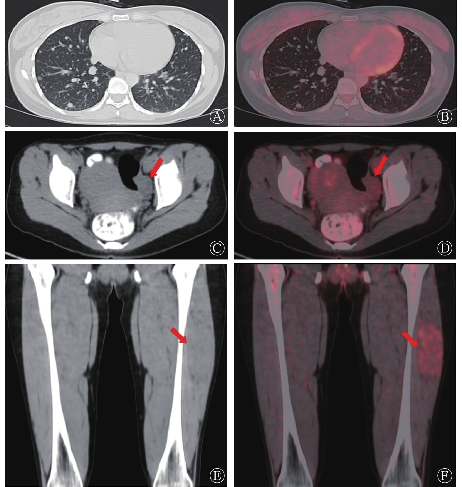
患者女性,25岁,无明显诱因出现咳嗽1月余,就诊外院行胸部CT检查,提示双肺多发结节。既往偶有咳嗽,无咳痰、咯血、发热等症状。之后患者就诊于海南省肿瘤医院,实验室检查结果:垂体泌乳素695.0 μIU/ml(非孕期女性:102.0~469.0 μIU/ml)、睾酮0.2(0.1~0.5) ng/ml、孕酮0.3(0.2~27.0) ng/ml、雌二醇46.1(12.0~498.0) pg/ml、促黄体生成素5.1(2.4~95.6) mIU/ml、卵泡生成素4.8(1.7~25.0) mIU/ml、血清胃泌素释放肽前体(ProGRP)74.3(0~63.0) pg/ml、癌胚抗原(CEA)0.4(0~4.7) ng/ml、细胞角蛋白19片段抗原(CYFRA21-1)2.3(0~3.3) ng/ml、鳞状细胞相关抗原1.3(0~1.5) ng/ml、神经元特异性烯醇化酶8.6(0~16.3) ng/ml、糖类抗原125(CA-125)18.6(0~35.0) U/ml、糖类抗原15-3(CA15-3)8.9(0~25.0)U/ml、人附睾蛋白4(HE-4)52.7(0~60.5) pmol/L。随后,患者行18F-FDG PET/CT全身显像检查(图1),结果显示,左侧附件区稍低密度结节,其内密度欠均匀,大小约为25 mm×26 mm,PET呈轻度放射性浓聚,SUVmax=1.9。双肺多发大小不等结节,PET上未见明显放射性浓聚。左侧大腿中段外侧稍低密度肿块,PET呈轻度放射性浓聚,SUVmax=2.7。

图 1 卵巢颗粒细胞瘤伴肺转移瘤合并左侧大腿腺泡状软组织肉瘤患者(女性,25岁)的18F-FDG PET/CT图
Figure 1. 18F-FDG PET/CT images of a ovarian granulosa cell tumor patient (female, 25 years old) with lung metastasis and alveolar soft part sarcoma of left thigh
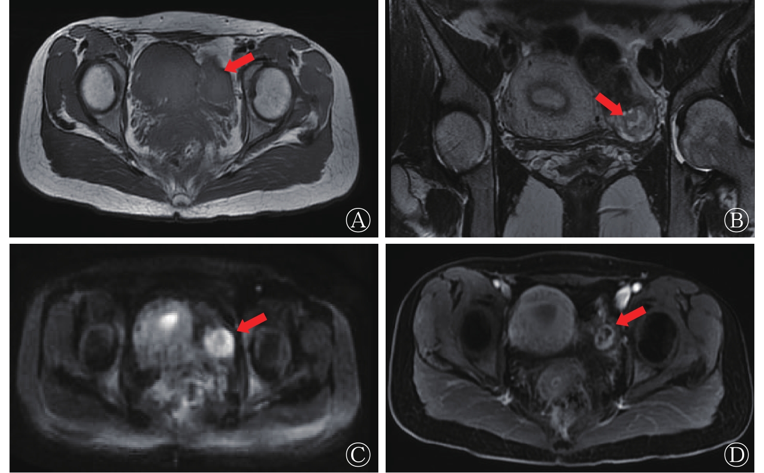
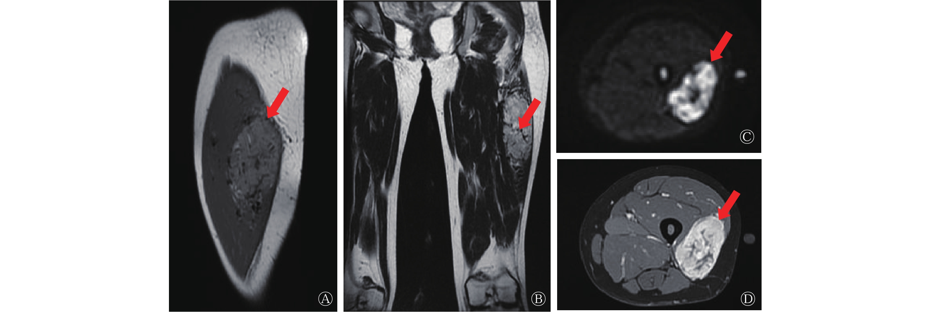
根据PET/CT结果,进一步明确病变的诊断,该患者行盆腔及大腿MRI检查。盆腔MRI表现(图2):左侧附件区类圆形占位,大小约为23 mm×26 mm×18 mm,T1WI呈等信号及稍高信号,T2WI呈等信号及高信号,内见多发小囊性灶,弥散加权成像(diffusion weighted imaging, DWI)呈不均匀高信号,MRI增强扫描病灶实性部分可见强化。左侧大腿MRI表现(图3):左侧大腿中段外侧肌层内见软组织肿块,大小约为34 mm×62 mm×105 mm,T1WI呈稍高信号,T2WI呈高信号,其内信号不均,DWI呈不均匀高信号,MRI增强扫描呈明显强化,其内及周围见多发血管影。

图 2 卵巢颗粒细胞瘤伴肺转移瘤合并左侧大腿腺泡状软组织肉瘤患者(女性,25岁)的盆腔MRI图
Figure 2. Pelvic MRI images of a ovarian granulosa cell tumor patient (female, 25 years old) with lung metastasis and alveolar soft part sarcoma of left thigh

图 3 卵巢颗粒细胞瘤伴肺转移瘤合并左侧大腿腺泡状软组织肉瘤患者(女性,25岁)的大腿MRI图
Figure 3. Thigh MRI images of a ovarian granulosa cell tumor patient (female, 25 years old) with lung metastasis and alveolar soft part sarcoma of left thigh
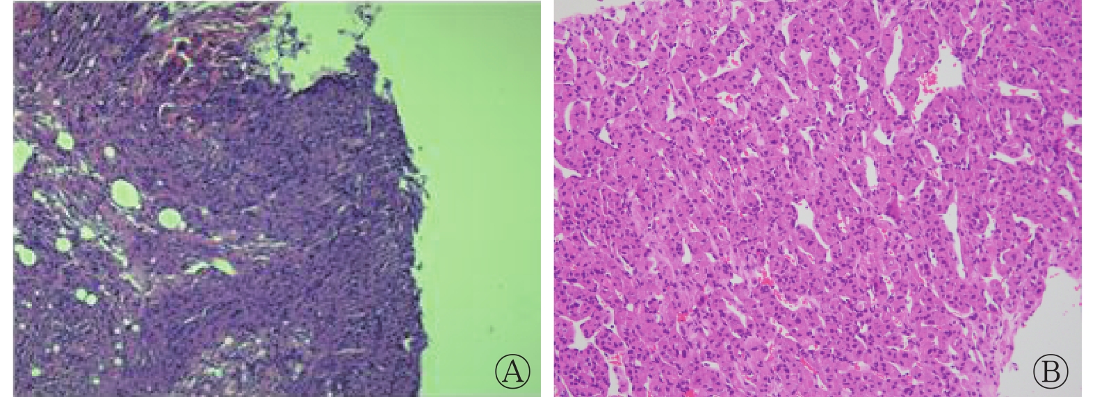
随后,患者行肺转移瘤及大腿病灶组织病理学活检,胸腔镜下右肺活检组织病理学检查示肿瘤细胞上皮样,异型性小,胞质丰富嗜伊红,核小、圆形、居中,核分裂难见,呈细梁状、片状分布,间质少(图4A)。免疫组织化学检查结果示:细胞增殖核抗原(Ki-67,3%)、神经元特异性烯醇化酶(NSE,+)、酸性钙结合蛋白(S-100,+)、突角素(Syn,+)、α抑制素(α-inhibin,+)、CK7(−)、CK20(−)、雌激素受体(ER,−)、上皮细胞膜抗原(EMA,−)、黑色素相关抗原(HMB45,−)、黑色素细胞抗原(MelanA,−)、肝细胞抗原1(Hep-1,−)、孕激素受体(PR,−)、细胞角蛋白(CK,−)、波形蛋白(Vimentin,−)。结合细胞学、组织学和免疫组织化学标记结果,符合多中心性颗粒细胞瘤。建议检查左右卵巢,排除OGCT转移。

图 4 卵巢颗粒细胞瘤伴肺转移瘤合并左侧大腿腺泡状软组织肉瘤患者(女性,25岁)的穿刺活检组织病理学检查图(苏木精-伊红染色,×100)
Figure 4. Histopatho logical examination chart of puncture biopsy of a ovarian granulosa cell tumor patient (female, 25 years old) with lung metastasis and alveolar soft part sarcoma of left thigh
左侧大腿肿物穿刺活检结合组织学镜下形态及免疫组化和特染结果,提示为ASPS(图4B)。免疫组化结果:CK(−)、原肌球蛋白调节蛋白(Myo-D1,−)、上皮细胞膜抗原(EMA,−)、α抑制素(α-inhibin,弱+)、CD99(+)、磷脂酰肌醇蛋白聚糖3(GPC-3,−)、CD34(−)、CD31(+)、肌肉组织肿瘤表达的结蛋白(Desmin,部分+)、肝细胞抗原(Hepatocyte,−)、AFP(+)、酸性钙结合蛋白(S-100,−)、嗜铬素A(CgA,+)、突触素(Syn,−)、CD56(个别细胞+)、转录因子E3(TFE3,+)、P53(30%弱+)、黑色素相关抗原(HMB45,−)、细胞增殖核抗原(Ki-67,5%+)。特殊染色(过碘酸雪夫染色法)结果示肿瘤细胞胞浆内见嗜酸性结晶体。
-
GCTO是一种罕见肿瘤,约占所有卵巢肿瘤的2%~5%;大部分患者在早期可被诊断,预后相对较好[3]。OGCT通常分为成人颗粒细胞瘤 (adult OGCT, AOGCT)和幼年颗粒细胞瘤( juvenile OGCT, JOGCT)[4]。AOGTC是一种低度恶性、晚期复发的卵巢肿瘤,主要见于老年女性。同时,JOGTC是一种较为罕见的OGCT,多见于青春期前及30岁以下的女性[5]。大多数OGCT伴有雌激素的升高,其原因可能是肿瘤通过增加雄烯二酮向雌二醇的转化而增加雌激素和(或)孕酮的产生[6]。但是本例患者雌激素水平并未升高,这可能与肿瘤大小有关,具体原因需更多研究证实。卵巢肿瘤转移的常见部位依次为肝、远处淋巴结、肺、骨骼等,但是OGCT的转移并不常见[7]。回顾文献并结合该患者的资料,总结卵巢原发肿瘤的影像学特征如下。(1)在CT上,OGCT表现为低密度实性肿块,偶尔有囊性成分,可出血,或伴有很多小叶,呈“葡萄串”状。CT增强扫描后实性成分可见强化[8];(2)在MRI上,从肿瘤成分看,OGCT呈纯实性至完全囊性肿块,形态各异,多数呈多房性囊性病变,似“海绵状”。T1WI多呈高信号或混杂信号,这可能与出血后血液吸收有关。MRI增强扫描后呈轻度至中度强化[6];(3)在18F-FDG PET/CT中,大多数OGCT表现为18F-FDG的摄取较低。这可能与肿瘤基因型和增殖活性有关[4]。OGCT的双肺转移瘤摄取18F-FDG的水平亦较低。OGCT应与卵巢囊腺癌、卵巢纤维瘤及纤维-卵泡膜细胞瘤及带蒂肌瘤相鉴别。(1)卵巢囊腺癌为卵巢囊实性肿块,形态不规则,边界不清,囊壁厚度不均匀。壁结节常突入腔内,表现为浸润性生长,通常可见腹水[9];(2)卵巢纤维瘤及纤维-卵泡膜细胞瘤中含有纤维成分,故T2WI上可见低信号,肿瘤内很少发生坏死和囊变,几乎不伴有出血,MRI增强扫描略有强化;(3)带蒂肌瘤与实性肿块的OGCT有一定相似度,带蒂肌瘤可以通过与子宫的连接与OGCT相鉴别,T1WI、T2WI序列上的低信号是非退行性带蒂肌瘤的特征,而OGCT在T1WI序列上表现为低信号,在T2WI序列上可见部分高信号。此外,带蒂肌瘤在增强扫描后T1WI图像中常常呈显著强化,而OGCT通常表现为轻度至中度强化[8]。总而言之,MRI可以较好地显示局部软组织病变,而PET/CT对全身的评估有着不可替代的作用。
既往的病例报道中,OGCT发生肺转移的概率很小,本例患者通过 18F-FDG PET/CT发现了附件区病灶,使得我们对双肺转移瘤有了新的思考。同时,PET/CT检查时发现左侧大腿肿物,SUVmax=2.7;穿刺后组织病理学检查结果为ASPS。这再一次体现了PET/CT全身评估的重要性。
ASPS是一种独特的恶性软组织肿瘤,其组织病理学特征为假腺泡结构,伴有丰富的窦状血管。15~35岁的年轻人是好发人群,女性发病率比男性略高[10]。回顾文献并结合该患者的资料,总结本例患者大腿病灶的影像学特征如下。(1)在MRI上,T1WI呈高信号和多发的流空血管影最为常见,这可能是由于肿瘤血管丰富和血管内血流缓慢[11];在DWI上,病灶表现为高信号,这可能与细胞质丰富、细胞密集排列,导致水分子扩散有限有关[12];(2)在18F-FDG PET/CT上,ASPS的18F-FDG摄取较高,与之前的研究结果相似[13]。PET/CT图像上18F-FDG摄取的增加有助于区分良、恶性肿瘤。18F-FDG PET/CT可以更容易区分肌间血管瘤和ASPS。ASPS应与脂肪肉瘤、滑膜肉瘤及肌间血管瘤相鉴别。(1)脂肪肉瘤等软组织肿瘤有很高的18F-FDG摄取,但MRI图像上的T1WI高信号及压脂后低信号的特点可能有助于与ASPS鉴别诊断;(2)在CT和PET/CT图像上很难将伴有钙化的ASPS与滑膜肉瘤相鉴别,但滑膜肉瘤在MRI上通常无血管流空效应,且滑膜肉瘤易出现出血、坏死和囊变[14];(3)与ASPS相似的肌间血管瘤在MRI的T1WI图像上可表现为高信号和明显强化,然而,其在DWI图像上通常表现为低信号,PET/CT图像上18F-FDG摄取一般较低[13]。因此,多模态影像学检查的应用可为ASPS的诊断及鉴别诊断提供更多依据。
综上所述,本例患者虽然未直接获得卵巢肿物组织病理学检查结果,但肺转移瘤活检结果足以证实卵巢病灶的性质。OGCT好发于老年女性或青春期前及30岁以下的女性,大多数伴有雌激素水平的升高;ASPS好发于中青年女性,临床相对无症状。ASPS转移比OGCT更为常见,但本例患者双肺转移瘤确是OGCT来源,具体原因仍需进一步研究证实。OGCT及ASPS均为罕见肿瘤,还未有病例报道二者同时发生,但二者在影像学上具有一定特点,利用多模态影像学检查方法可以让诊断更有依据,但组织病理学检查结果仍是最终诊断的“金标准”。
利益冲突 所有作者声明无利益冲突
作者贡献声明 潘登负责病例图片的整理与分析、论文的起草与撰写;林秀艳、龚伟负责研究命题的提出与理论的指导、论文的修订;李雪艳、王玉君负责病例图片的获取、文献的收集与分析;于丽娟负责论文的审核、最终版本的修订
卵巢颗粒细胞瘤合并大腿腺泡状软组织肉瘤影像学表现1例及文献复习
Imaging findings of ovarian granulosa cell tumor combined with alveolar soft part sarcoma of thigh: a case report and literature review
-
摘要: 卵巢颗粒细胞瘤(OGCT)及腺泡状软组织肉瘤(ASPS)在临床上相对少见,特别是2种肿瘤同时存在更为罕见。OGCT预后尚可,但ASPS预后并不乐观,且目前对于OGCT和ASPS的18F-氟脱氧葡萄糖(FDG)PET/CT的研究较少。笔者报道了1 例OGCT伴肺转移瘤合并左侧大腿ASPS患者的全身18F-FDG PET/CT 显像及MRI成像,分析其图像特点并进行文献复习,为该疾病的诊断提供更多参考。Abstract: Ovarian granulosa cell tumor (OGCT) and alveolar soft part sarcoma (ASPS) are relatively rare in clinical practice, especially in the presence of both tumors. The prognosis of OGCT is fair, but the prognosis of ASPS is not optimistic, and there are few reports of 18F-fluorodeoxyglucose (FDG) PET/CT of OGCT and ASPS at present. The author reported the whole body 18F-FDG PET/CT imaging and MRI of a patient with OGCT accompanied by lung metastasis and left thigh ASPS, analyzed the image characteristics and reviewed the literature to provide more reference for the diagnosis of this disease.
-
Key words:
-
图 1 卵巢颗粒细胞瘤伴肺转移瘤合并左侧大腿腺泡状软组织肉瘤患者(女性,25岁)的18F-FDG PET/CT图
Figure 1. 18F-FDG PET/CT images of a ovarian granulosa cell tumor patient (female, 25 years old) with lung metastasis and alveolar soft part sarcoma of left thigh
图 2 卵巢颗粒细胞瘤伴肺转移瘤合并左侧大腿腺泡状软组织肉瘤患者(女性,25岁)的盆腔MRI图
Figure 2. Pelvic MRI images of a ovarian granulosa cell tumor patient (female, 25 years old) with lung metastasis and alveolar soft part sarcoma of left thigh
图 3 卵巢颗粒细胞瘤伴肺转移瘤合并左侧大腿腺泡状软组织肉瘤患者(女性,25岁)的大腿MRI图
Figure 3. Thigh MRI images of a ovarian granulosa cell tumor patient (female, 25 years old) with lung metastasis and alveolar soft part sarcoma of left thigh
-
[1] Matsuki M, Numoto I, Suzuki A, et al. Magnetic resonance imaging of recurrent adult granulosa cell tumor of the ovary: a retrospective analysis of 11 cases[J]. J Comput Assist Tomogr, 2020, 44(6): 887−892. DOI: 10.1097/RCT.0000000000001096. [2] Gulati M, Mittal A, Barwad A, et al. Imaging and pathological features of alveolar soft part sarcoma: analysis of 16 patients[J]. Indian J Radiol Imaging, 2021, 31(3): 573−581. DOI: 10.1055/s-0041-1735501. [3] Khosla D, Dimri K, Pandey AK, et al. Ovarian granulosa cell tumor: clinical features, treatment, outcome, and prognostic factors[J]. N Am J Med Sci, 2014, 6(3): 133−138. DOI: 10.4103/1947-2714.128475. [4] Huang BS, Sun HD, Hsu YM, et al. Clinical presentation and outcome of adult-type granulosa cell tumors: a retrospective study of 30 patients in a single institute[J]. J Chin Med Assoc, 2014, 77(1): 21−25. DOI: 10.1016/j.jcma.2013.09.007. [5] Rustamadji P, Wiyarta E, Anggraeni TD, et al. Adult granulosa cell tumor with minor foci of juvenile granulosa cell tumor in postmenopausal woman: a rare case report[J/OL]. Int J Surg Case Rep, 2021, 88: 106531[2022-04-06]. https://www.ncbi.nlm.nih.gov/pmc/articles/PMC8536526/. DOI: 10.1016/j.ijscr.2021.106531. [6] Zhang H, Zhang HY, Gu SX, et al. MR findings of primary ovarian granulosa cell tumor with focus on the differentiation with other ovarian sex cord-stromal tumors[J/OL]. J Ovarian Res, 2018, 11(1): 46[2022-04-06]. https://www.ncbi.nlm.nih.gov/pmc/articles/PMC5989475/. DOI: 10.1186/s13048-018-0416-x. [7] Zhang C, Guo X, Peltzer K, et al. The prevalence, associated factors for bone metastases development and prognosis in newly diagnosed ovarian cancer: a large population based real-world study[J/OL]. J Cancer, 2019, 10(14): 3133−3139[2022-04-06]. https://www.ncbi.nlm.nih.gov/pmc/articles/PMC6603379/. DOI: 10.7150/jca.30335. [8] Elsherif S, Bourne M, Soule E, et al. Multimodality imaging and genomics of granulosa cell tumors[J]. Abdom Radiol (NY), 2020, 45(3): 812−827. DOI: 10.1007/s00261-019-02172-3. [9] 翁建辉, 邱淦滨, 李建辉. 卵巢颗粒细胞瘤的影像学表现[J]. 临床医学研究与实践, 2021, 6(14): 110−112. DOI: 10.19347/j.cnki.2096-1413.202114036.
Weng JH, Qiu GB, Li JH. Imaging manifestation of ovarian granulosa cell tumor[J]. Clin Res Pract, 2021, 6(14): 110−112. DOI: 10.19347/j.cnki.2096-1413.202114036.[10] Martínez R, Niklander S, Deichler J, et al. Alveolar soft-part sarcoma of the masseter and mandibular ramus: report of a case and review of the literature[J]. J Dent Sci, 2018, 13(1): 75−79. DOI: 10.1016/j.jds.2013.02.032. [11] Wu ZJ, Bian TT, Zhan XH, et al. Computed tomography, magnetic resonance imaging, and F-deoxyglucose positron emission computed tomography/computed tomography findings of alveolar soft part sarcoma with calcification in the thigh: a case report[J/OL]. World J Clin Cases, 2020, 8(15): 3349−3354[2022-04-06]. https://www.ncbi.nlm.nih.gov/pmc/articles/PMC7441268/. DOI: 10.12998/wjcc.v8.i15.3349. [12] Cui JF, Chen HS, Hao DP, et al. Magnetic resonance features and characteristic vascular pattern of alveolar soft-part sarcoma[J]. Oncol Res Treat, 2017, 40(10): 580−585. DOI: 10.1159/000477443. [13] Dong AS, Wang Y, Cheng C, et al. CT, MRI, and FDG PET/CT in a patient with alveolar soft part sarcoma[J]. Clin Nucl Med, 2014, 39(3): 265−267. DOI: 10.1097/RLU.0b013e3182817b09. [14] Li HM, Sun J, Ye J, et al. Magnetic resonance imaging and computed tomography features of alveolar soft-part sarcoma in the right deltoid muscle: a case report[J]. Oncol Lett, 2016, 11(4): 2857−2860. DOI: 10.3892/ol.2016.4290. -

 点击查看大图
计量
- 文章访问数: 2351
- HTML全文浏览量: 1872
- PDF下载量: 12